BY 115 Nervous Tissue & Brain
 Call Kai
 Learn
 Practice Test
 Spaced Repetition
 Match
 Flashcards
 Knowt Play
1/228

There's no tags or description
Looks like no tags are added yet.
229 Terms
Functions of the Nervous System
Sensory input, integration, motor output
Sensory input
Detects changes inside and outside the body
Integration
Processes and interprets information
Motor output
Produces responses through muscles and glands
Structural organization of the nervous system
Central nervous system (CNS: brain and spinal cord); peripheral nervous system (PNS: cranial nerves, spinal nerves, ganglia)
Sensory nervous system
Afferent (towards CNS); carries information from receptors to CNS; consists of somatic sensory system and visceral sensory system
Somatic sensory system
Touch, pain, pressure, vibration, proprioception (determining where the body is located/positioned in 3D)
Visceral sensory system
Internal organs, vessels
Motor nervous system
Efferent (away from CNS); carries information from CNS to effectors (muscles or glands); consists of somatic motor system and autonomic motor system
Somatic motor system
Skeletal muscle
Autonomic/visceral motor system
Cardiac muscle, smooth muscle, glands
Sympathetic nervous system
Flight, fight, or stress response
Parasympathetic nervous system
Rest and digest
Collection of neuronal cell bodies
Nucleus in CNS and ganglion in PNS
Bundle of axons
Tract in CNS and nerve in PNS
Gray matter
Primarily neuronal cell bodies, dendrites, synapses, and unmyelinated processes; forms cerebral cortex and nuclei (clusters of neuron cell bodies)
White matter
Composed primarily axons, many are myelinated, and tracts (axons within CNS organized into bundles)

ID 1
Gray matter

ID 2
White matter
Neurons
Electrically excitable; receive and transmit information
Glial cells
Support cells of nervous tissue, protect and nourish neurons, and maintain neural environment; generally do not generate action potentials; can undergo mitosis; 4 types in CNS and 2 types in PNS
Astrocytes
CNS glial cells; support blood-brain barrier; regulate extracellular environment; participate in scar formation

ID 1
Astrocyte
Action potential
Electrical change in a neuron that’s used for signaling
Cell body (soma)
Contains nucleus and nucleolus; major metabolic and integrative region of neuron—integrates incoming information; contains chromatophilic substance (Nissl bodies)
Nissl bodies
Clusters of endoplasmic reticulum and ribosomes; produces proteins
Axon hillock
Region where the axon begins

ID 1
Cell body

ID 2
Axon hillock

ID 1
Cell body
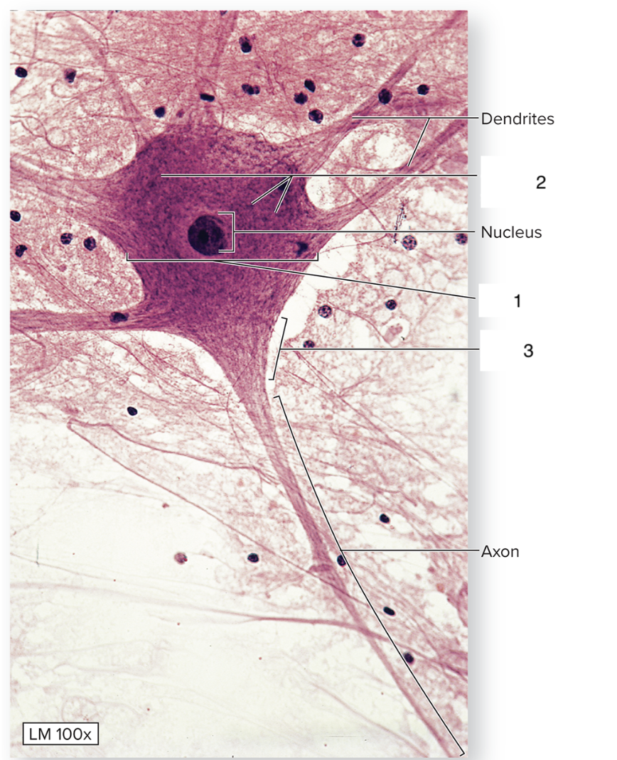
ID 2
Nissl bodies

ID 3
Axon hillock
Dendrites
Receive information and carry information toward the cell body
Axon
Carries information away from the cell body
Pathway of input and output in a neuron
Dendrites to cell body to axon to axon terminals

ID 3
Axon
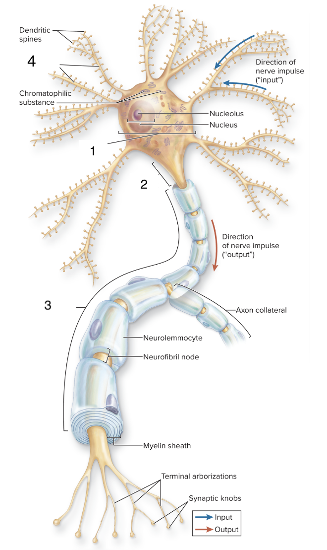
ID 4
Dendrites
Basic structure of a synapse
Presynaptic terminal, synaptic cleft, postsynaptic cell
Presynaptic terminal
Synaptic knob that contains vesicles with neurotransmitter
Synaptic cleft
Space between cells
Postsynaptic cell
Receives the chemical signal
Cell communication
Electrical signal arrives, neurotransmitter crosses the cleft, next cell receives the signal

ID 1
Synaptic cleft

ID 2
Presynaptic terminal

ID 3
Postsynaptic cell
Multipolar neurons
One axon, many dendrites; most common; most motor neurons and interneurons
Bipolar neurons
One axon, one dendrite; associated with special senses (retina and olfactory pathways)
Unipolar/pseudounipolar neurons
Single process that divides; most sensory neurons; cell bodies commonly located in sensory ganglia (dorsal root ganglion)

ID
Unipolar neuron

ID
Bipolar neuron
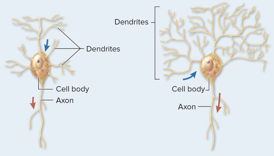
ID
Multipolar neuron
Interneurons
Located primarily in CNS; process and integrate information; connect neurons within CNS
Basic reflex arc
Receptor → sensory/afferent neuron → CNS (with possible participation from interneuron) → motor/efferent neuron → effector

ID 1
Skin receptors

ID 2
Sensory neuron

ID 3
Cell body of sensory neuron

ID 4
Posterior/dorsal root ganglion

ID 5
Interneuron
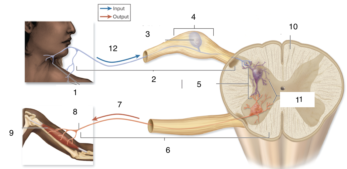
ID 6
Motor neuron

ID 7
Efferent transmission
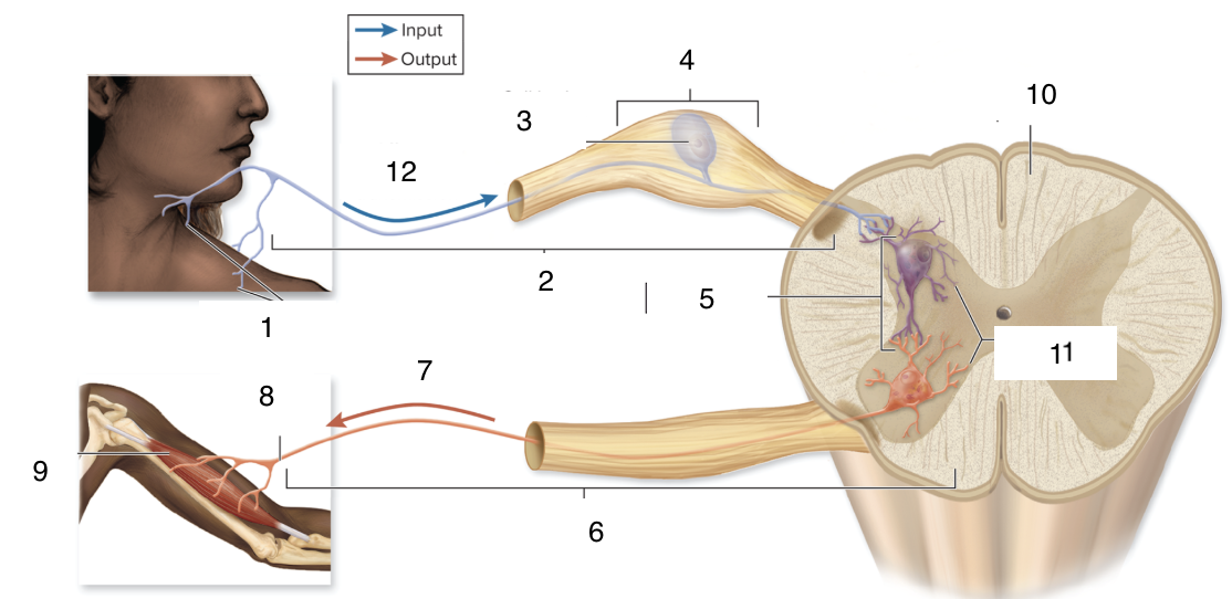
ID 8
Axon

ID 9
Skeletal muscle

ID 10
Spinal cord

ID 11
Dendrites
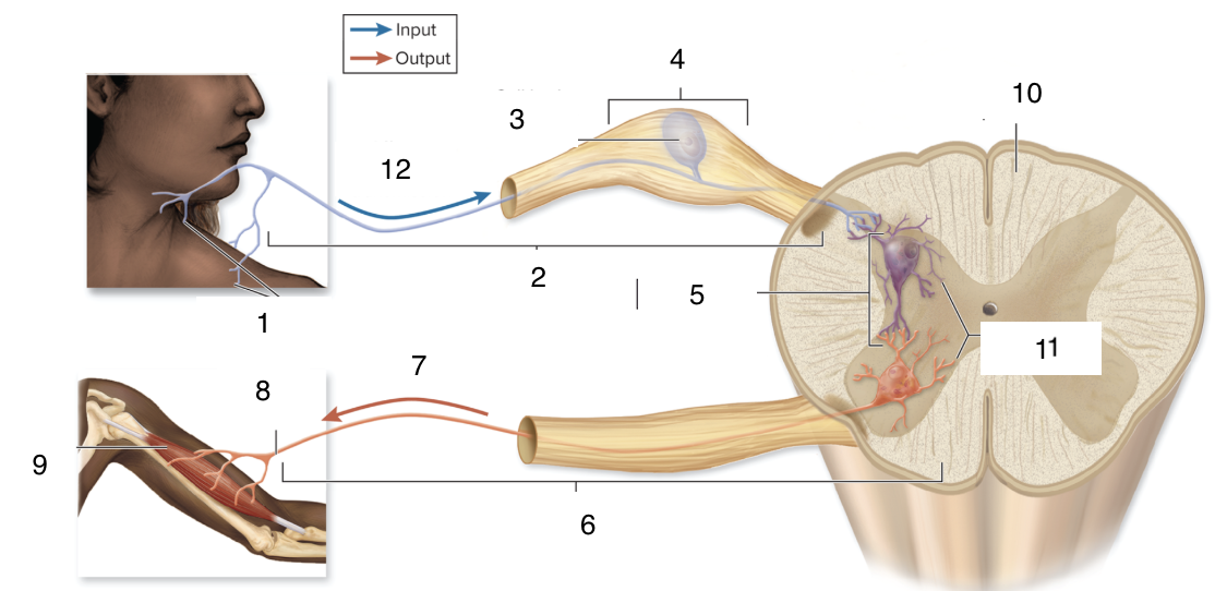
ID 12
Afferent transmission
Converging neural circuit
Many neurons become one neuron
Diverging neural circuit
One neuron becomes many neurons

ID
Converging neural circuit

ID
Diverging neural circuit
Ependymal cells
CNS glial cells; line ventricles and central canal; help form choroid plexus/produce cerebrospinal fluid (CSF)
Astrocytes and ependymal cells
Maintain the CNS environment

ID 1
Ependymal cells
Microglia
CNS glial cells; immune-defense cells that remove debris by phagocytosis
Oligodendrocytes
CNS glial cells; form myelin around axons; one cell can myelinate portions of multiple axons

ID 1
Oligodendrocytes/CNS myelination
Satellite cells
PNS glial cells; surround neuron cell bodies in ganglia; help regulate the local environment
Neurolemmocytes/Schwann cells
PNS glial cells; form myelin around axons; one cell myelinates one segment of one axon; support PNS axon regeneration

ID 1
Neurolemmocytes
Myelin
Layers of glial cell membrane around an axon; electrically insulates the axon and increases conduction speed (saltatory conduction)
Myelinated axons
Myelin interrupted by nodes of Ranvier/neurofibril nodes; conduct signals faster than unmyelinated axons
Saltatory conduction
Signal travels rapidly from node to node
CNS Axon regeneration
Extremely limited; oligodendrocytes and scar tissue from astrocytes inhibit regrowth
PNS Axon regeneration
More successful than in CNS; some damaged axons can regenerate; cell body must remain intact; neurolemmocytes help guide regrowth
Nerve
Bundle of parallel axons in the PNS; peripheral nerves contain neurolemmocytes, connective tissue, blood vessels; may contain sensory axons, motor axons, or both (mixed nerve)
Connective-tissue wrappings of a nerve
Endoneurium, epineurium, perineurium
Endoneurium
Surrounds axon; made of dense regular CT
Perineurium
Surrounds fascicle; made of dense irregular CT
Epineurium
Surrounds whole nerve; made of areolar CT
Brain regions
Cerebrum, diencephalon, midbrain, pons, medulla oblongata, cerebellum
Brainstem
Midbrain, pons, medulla oblongata; connects cerebrum, diencephalon, and cerebellum to spinal cord; contains ascending (sensory) tracts and descending (motor) tracts
Surficial brain anatomy
Outer surface is folded; ridges = gyri, depressions between ridges = sulci; deep sulci = fissures
Structures that protect the brain
Cranial bones (physical protection), cerebrospinal fluid (cushions and supports the brain), Blood-brain barrier (regulates movement of substances from blood into nervous tissue), meninges (connective tissue coverings)
Cranial meninges
Three connective tissue layers that enclose and protect blood vessels supplying brain; help contain and circulate cerebrospinal fluid
Pia mater
Innermost, adheres to brain surface; thin layer of areolar connective tissue
Arachnoid mater
Lies external to pia mater
Subarachnoid space
Contains cerebrospinal fluid
Subdural space
Lies between dura mater and arachnoid mater; can develop when bleeding separates the membranes; normally a potential space
Dura mater
Tough, outer membrane, made of dense irregular connective tissue in two layers: meningeal layer (deep) and periosteal layer (superficial)
Dural venous sinuses
Dural layers separate to form; drain venous blood from the brain via the jugular vein
Epidural space
Potential space between skull and dura; can form when bleeding separates dura from skull