Topic F - Conjugation
 Call Kai
 Learn
 Practice Test
 Spaced Repetition
 Match
 Flashcards
 Knowt Play
1/70

There's no tags or description
Looks like no tags are added yet.
71 Terms
Vertical and Horizontal Gene Transfer
Vertical Gene Transfer - Transfer of DNA from a parent cell to its progeny through cell division (ex; parent cell providing DNA to two daughter cells)
Horizontal Gene Transfer - Transfer of DNA between cells unrelated/distantly related (produces genetic diversity in a bacterial population)
Mechanisms of Horizontal Gene Transfer
Conjugation - Physical cell-to-cell contact required
-Donor cell creates pilus, grabbing onto recipient cell, transferring genetic information
Transformation - Exchange of genetic material via free-floating DNA material (donor cell dies releasing DNA, recipient cell uptakes the DNA)
Transduction - Transfer of genetic material between cells using bacteriophage
-Phage accidently package bacterial DNA, resulting in the transfer of bacterial DNA to foreign bacteria
Conjugation
Transfer of genetic material from one cell to another via direct cell-to-cell contact
-Pilus connecting donor/recipient cell is the “Conjugative Pilus”
History of Conjugation
First observed by Lederberg and Tatum in 1946, using 2 experiments
Experiment 1 - Conjugation
Mixing different auxotrophic strains and got prototrophs (WTs)
-Strain A - Auxotrophic for multiple nutrients when plated on MM = no colonies
-Strain B - Auxotrophic for a different set of nutrients when plated on MM = no colonies
Mixing Strain A + B and plating on MM = observed colonies
Experiment 2 - Conjugation
Separate cells to see if transfer occurs
-Needed to rule out factors in the medium that may have caused observed phenotypes (DNA, bacteriophage)
Two auxotrophic strains separated by filter allowing passage of nutrients, but NOT bacteria
-Vacuum applied to maintain movement of liquid
Observed no colonies on MM → Direct contact therefore needed for transfer of DNA
Bacterial Plasmids
Small, circular supercoiled dsDNA molecules
-Must contain origin of replication
Depends on host cell for most replication proteins (helicase, DNAP)
-Some encode special proteins for plasmid replication and segregation
Low Copy Number Plasmids
Plasmid replication coordinated with chromosome
-Once prior to cell division, segregated following division to ensure each daughter cell gets a copy
-Proteins used to move one copy to each end of thecell
High Copy Number Plasmids
Plasmid replication not linked to cell division
-Lots of replication to get lots of plasmid copies, regulation control maximum number of plasmids in the cell
Random segregation during division, high number ensures each daughter cell gets at least one copy
Plasmid Incompatibility
Inability of two plasmids to co-exist stably in the same cell, occurs if:
-Plasmids have the same replication mechanism; replication mechanism sees them as the same plasmid, making only 6 copies of the plasmid (rather than 6 copies of EACH plasmid), overtime one of the plasmids will be lost (ex; both plasmids having the same ori)
-Plasmids have the same partitioning mechanism
Role of Plasmids
Carry genes that are dispensable for growth (don’t have essential genes)
-may be lost (cured) with no negative effect
But provide selective advantage under some conditions
Ex; Tol Plasmid - Toluene degradation (energy source)
Ex; Ti plasmid - tumor inducing (bacteria infects plant-host cells, causing tumor via conjugation)
Ex; R plasmid - antibiotic resistance
Ex; F plasmid - sex pili + conjugation
Addiction Systems
Some plasmids contain addiction systems
-Encode a stable toxin and unstable anti-toxin
If plasmid is lost, the cell will die
-Allows plasmid to be maintained in a bacterial population, even in non-selective conditions

Mechanism of Addiction Systems
When the anti-toxin is expressed, it binds to the toxin preventing it from killing the bacterial cell
-If the bacteria cell divides and only one daughter cell receives the plasmid, the remaining unstable anti-toxin in cytoplasm will become degraded resulting in cell death
Plasmid and Conjugation
Some plasmids carry genes for plasmid transfer by conjugation between bacteria
-Includes “tra” genes or “mob” genes
F Plasmid of E.Coli
F (fertility) plasmid, has a low copy number (1 per cell), consists of:
1. OriV - Origin of replication
2. OriT - Origin of Transfer (for Conjugation)
3. Tra genes - Encodes proteins involved in conjugation
4. IS (Insertion Sequence) Elements - Transposons

Transfer of Plasmid DNA
Some plasmids are self-transmissible
-Encode all functions needed for conjugation from cell-to-cell
Donor Strain - Bacterium that carries a self-transmissible plasmid
Recipient Strain - Bacterium lacking self-transmissible plasmid, receives DNA by conjugation
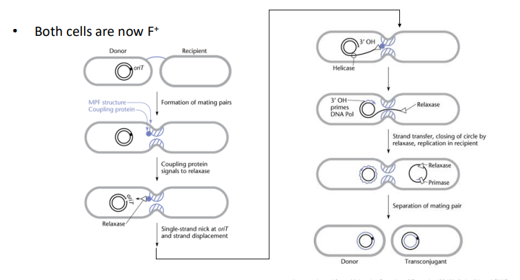
Five-Step Process of Plasmid Transfer
Formation of Mating Pairs
Preparation for DNA Transfer
DNA Transfer
Restoration of Double-Stranded plasmid in donor
Formation of replicative plasmid in recipient
Formation of Mating Pairs
Requires cell-to-cell contact between donor and recipient;
-Mediated by the conjugative pilus (F pilus or sex pilus); polymer of a single protein called “Pilin”
a) Pilus tip binds to receptor on recipient cell surface to initiate mating pair formation
b) Pilus contracts to pull the two cells into close contact
c) Conjugation Bridge forms between cells, consists of pore complex formed by proteins
-Passageway to allow DNA transfer
Preparation for DNA Transfer
Requires relaxase enzyme, F Plasmid-encoded protein
Recognizes unique DNA sequence of the oriT
-Nicks one strand of plasmid DNA at oriT

DNA Transfer
Cut strand of plasmid is transferred to recipient via conjugation bridge
Transfer starts at 5’ end of nick at oriT
Transferred strand is linear/single-stranded
Enabled by protein complex (relaxosome) that binds at oriT
Helicase separates two strands of plasmid
-Other DNA strand stays in donor cell as intact circle
Restoration of Double-Stranded Plasmid in Donor
ssDNA remaining in donor is replicated to regenerate double-stranded plasmid
-Primed by 3’ end of nicked strand of plasmid
Synthesis occurs during plasmid transfer
Formation of Replicative Plasmid in Recipient
ssDNA in recipient recircularizes:
-Nick is sealed by relaxase, transferred into recipient with plasmid DNA
Second strand produced by replication to give double-stranded plasmid
-Primase enzyme (from recipient cell) synthesizes RNA primer to initiate DNA replication
Both cells are now F+
-DNA synthesis only occurs after DNA transfer is complete
Diagram of Transfer
tra Genes
Conjugation functions are encoded by tra genes
-Two main types of genes:
MPF - Encode mating pair formation proteins
DTR - Encode DNA transfer and replication
Are trans-acting; act on another plasmid in the same cell, as long as it has an oriT
-oriT is “Cis-acting”; plasmid must have an oriT in its sequence to transfer
-Plasmid MUST have an oriT in its sequence to transfer (oriT in one plasmid, can’t act on another plasmid)

Self-Transmissible Plasmid
Transfer between cells without help
-has MPF and DTR genes
MPF Gene - Pilin subunit, assembly of pilus, stabilization of mating pairs, inhibition of mating between two F+ cells
DTR Gene - Helicase, initiation of plasmid transfer, Relaxase
Conjugative Plasmid
Can form pilus and mating pair, but can’t be transferred by conjugation
-Has MPF genes only, no DTR genes or oriT
Mobilizable Plasmid
Two types:
Has DTR + oriT, no MPF
When in cell with conjugative/self-transmissible plasmid, it can be transferred by conjugation
Only has oriT, no MPF/DTR
Can be transferred by conjugation only when in a cell with self-transmissible plasmid (has MPF and DTR)
Plasmid Interaction Example
Two Plasmids:
-F Plasmid - Self-transmissible
-Other Plasmid - Mobilizable
Genes encoding proteins for transfer are encoded on a different plasmid than the one being transferred
-recipient does not become F+
Hfr Strains
“High Frequency of Recombination” strains
-Formed when F-plasmid integrates into the bacterial chromosome
-Can enable transfer of chromosomal DNA to a recipient by conjugation
Hfr Formation - Recombination
Integration occurs via recombination between homologous IS (insertion sequence) elements
-Found on both the F plasmid and chromosome, have conserved sequences
F-Plasmid Integration
Can integrate in one of two ways, and at any one of more than 20 sites in the E.coli chromosome
-only ONE F-plasmid is allowed per genome since cell becomes F+ (no additional conjugation an occur)
Hfr Formation - Direction of Integration
Homologous sequence on plasmid will align in the direction of the homologous sequence in the chromosome
-Different direction of integration produces a different direction of transfer during conjugation
-Transfer occurs as if arrow is being shot into the recipient cell
DNA behind the oriT (the “arrow”) will be transferred first during conjugation

Hfr Transfer - Recombination
When donor DNA from an Hfr strain enters the recipient cell, it recombines with the recipient chromosome
-Requires sequence similarity
-Recipient genes are replaced with donor genes
Many recombinants are formed
Hfr Transfer - Integration
Upon integration, F-plasmid behaves as part of the chromosome
-Mediates DNA transfer
If cells get seperated; transfer stops and recipient only gets a linear chromosome fragment
-Rare for cells to stay in contact long enough to transfer entire chromosome (100 minutes)
Mechanism of Hfr Transfer
Same as plasmid conjugation
-Conjugative bridge is formed, bringing the two cells close together
OriT nicked by relaxase in one strand
Transfer of ssDNA starts at 5’ end of nick
ssDNA moves through conjugation bridge
In donor, free 3’ end used to primer for DNA replication to restore ds chromosome
In recipient, primase enzymes initiates DNA replication to produce dsDNA
-Conjugative bridge breaks leaving behind a linear chromosome, won’t recircularize
Hfr Transfer - Crossovers
If new linear fragment contains similar sequences to recipient chromosome, crossovers can occur
-Need 2 crossovers to replace recipient DNA with donor DNA
-Recipient becomes a transconjugant
Two crossovers are required to ensure bacterial chromosome remains circular (one crossover results in a linear chromosome)
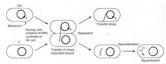
Hfr Transfer - F Sequence Not Incorporated
F-sequence does not get incorporated into recipient chromosome during Hfr transfer
-Therefore, recipient does not become F+
Hfr Transfer vs Plasmid Conjugation
Complete transfer takes longer
Transfer almost never completed
Transferred chromosome does not recircularize and replicate in recipient
Recombinants are produced
Recipient cell remains F-
Complete Transfer Takes Longer
Constant transfer rate
-100 min for chromosome vs 2 minutes for F plasmid
Transfer Almost Never Completed
Mating pair usually breaks before entire chromosome transfer
-F-plasmid transfer almost always completes
Transferred Chromosome Does Not:
Recircularize
Replicate in recipient
-Chromsome is too large
F-plasmid always recircularizes
Recombinants are Produced
Recombination between donor DNA fragments and recipient chromosome
-Recombinants not produced from normal F-plasmid transfer
Recipient Cell Remains F-
In normal F plasmid transfer, recipient alwayas becomes F+
Detecting Transconjugants
Selection
Counterselection
Selection for Transconjugant Recipients
Selectable marker gene located on the donor chromosome (ex; Trp+ = prototrophic)
-Recipient lacks marker phenotype (Ex; Trp- = auxotrophic)
Select for recipient that has undergone recombination, plate on MM and look for prototrophs (Trp- —> Trp+)
Recipient that hasn’t received transferred DNA will die
Donors (Trp+) will also survive, so we have to counterselect
Counterselection to Prevent Growth of Donor
Ex; Antibiotic resistance marker
-Str = streptomycin resistance
-Donor = Str(s) - Sensitive
-Recipient = Str(R) - Resistant
Marker gene must be located far from oriT, and therefore not likely to transfer to recipient
-Donor will be sensitive and die
All recipients will be resistant and live, both transconjugant and non-recombinant
Detecting Transconjugants - Example
Recipient can make arginine, but not tryptophan
But donors can make tryptophan, but not arginine
-Through conjugation and recombination, Trp+ recombines with Trp- in the recipient, producing a recipient containing Trp+ and Arg+ (recipient is now prototrophic)
Selecting and Counterselecting Transconjugants
What will the Selectable Marker be?
-Trp+ (Selects for recipient that has undergone recombination, but also donor)
Counter selectable Marker
-Arg+ (Since donor does not have it)
What Medium will be Used to Isolate Transconjugants
-Minimal medium since donor is auxotrophic
Number of OriT in Hfr Strains
Hfr strains have ONE oriT
-Only one F-plasmid is allowed per cell
Genes on the chromosome will be transferred from Donor to Recipient in the order they occur
Ex: First Line: 10, IS2 are transferred before 92, IS2
-Second Line: 10, IS2 are transferred before 92, IS2

Steps to Determine Time a Locus is Transferred
1. Initiate mating between Hfr donor and F- recipient
2. Break mating pairs at different time points using a blender
3. Count the number of transconjugants at each time point
-Requires selection and counterselection
Determining the Time of Entry
Earliest time during transfer at which breakage no longer prevents recombinant formation
-Mating pair breaks but transconjugants still form
-Determining time of entry allows us to know the position of the selected gene relative to the oriT
Mapping with Hfr Example - Mating
pro+/str(R) recombinants are plated out and counted after various times of mating between Hfr pro+/str(S) and F- pro-/str(R)
-pro+ = prototroph, pro- = auxotroph
-str(s) = sensitive to str, str(R) is resistance
Mapping with Hfr Example - Selection/Counterselection
-Selection is using MM, recipients that received DNA from donor will grow
-Counterselection is using streptomycin, kills off the donor leaving the transconjugants
-What Medium to Count Transconjugants - Minimal Medium and Strep Resistance
Results:
0-15 minutes = no transconjugants
20 minutes = transconjugants are detected; 20 minutes of incubation allows pro+ DNA to move into the recipient and recombine

Mapping with Hfr Example - Entrance Time of Pro Gene
Time is graphed on the X axis and the “Number of Transconjugants" on the Y axis
-Extrapolate back to zero transconjugants to determine time of entry of the pro+ gene
(Conjugation occurs at a constant rate)
Graph shows the Pro+ gene entered at around 15 minutes

Mapping Genes Relative to Each Other
Examining several different types of transconjugants, genes can be mapped relative to one another
Ex: Gene A enters at 6 minutes, Gene B at 36, and Gene C at 42, and Gene D at 20
-Gene Order is A – D – B – C
This method requires:
All genes must be selectable
Each time is relative to oriT
Genetic Distance in Hfr Mapping
Called “minute”
-One minute contains 30-50 genes
-Not equivalent to a map unit
Example: E-coli chromosome = 100 minutes
Determining Gene Location With a Single Time Point
Hfr donor with oriT near the gene of interest and in the correct direction will transfer the WT gene to the recipient at high frequency
-Produces lots of recombinants
Hfr donor with oriT is far from the gene of interest or in the wrong direction will form few recombinants
Single Time Point Example - Mating
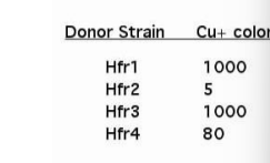
Induce 4 different mating pairs between cu-/str(R)/kanaS mutants and cu+/str(S)/kanaR
Each Hfr will have oriT at different position of the chromosome
Hfr 1 x mutant, Hfr 2 x mutant, Hfr 3 x mutant, Hfr 4 x mutant
Single Time Point Example - Selecting/Counterselecting
Select for transconjugants by plating on kanamycin-containing media, then kill the donor by plating on streptomycin
-Replicate plate onto medium containing copper; mutants are copper sensitive so if they did not receive they’ll die
-But those that did receive the WT copper gene will be copper resistant, so they grow
Single Time Point Example - Analysis of Colonies
If cu+ is near oriT, lots of recipient will be cu+
-if it’s located further, less recipients will be cu+
Same amount = between the two Hfr strains
Mutation is located between Hfr1 and 3 because large number of colonies produced indicates the WT copper is close to the oriT in each strain
-Hfr4 only produced 80 colonies, requires more DNA transference to produce WT
R Plasmids
Most multidrug resistant bacteria isolated from patients contain resistance genes on R plasmids
-Encode:
Enzymes that modify and inactivate the drug
Proteins that prevent uptake of drug or actively pump it out of the cell
Resistance genes will affect all drugs belonging to the same structural class
Examples: penicillins, aminoglycosides, etc.
-These plasmids also contain tra genes = self-transmissible
Location of R Plasmids in the Wild
R plasmids commonly found in soil bacteria, before clinical antibiotic use
-Some bacteria produce antibiotics to kill competitors
-But antibiotic-producing organisms (Streptomyces, Penicillium fungi) live in the soil
So they have resistance genes for antibiotics they make
Purpose of R Plasmids
Genes on R plasmids may be derived from resistance mechanisms or these producers
-R plasmids may provide selective advantage for survival in soil environments containing antibiotics
-Transfer between organisms in soil by conjugation
Evidence for the Origin of Antibiotic Resistance
Evidence One: Bacteria never exposed to clinical antibiotics are resistant to most antibiotics
Evidence Two: Tribes in South America have antibiotic resistant bacteria on their skin/digestive tract
How Does Antibiotic Resistance Occur
Use of antibiotics provides selective conditions for spread of R plasmids to pathogens
How Does Antibiotic Resistance Occur
1. Few bacteria in large populations are resistant
2. Antibiotics kill those who are sensitive (including good bacteria)
3. Resistant bacteria have preferred conditions to grow
4. Bacteria can then transfer drug resistance to other bacteria
Agriculture - Antibiotic Resistance
-Growth promoting agents
-Prophylactic additive to prevent disease
-Antibiotic released into the environment
-Selects for antibiotic resistance in food-borne organisms and water
Human Medicine - Antibiotic Resistance
-Prescriptions for non-bacterial infections
-Inappropriate doses/duration of treatment
-Patient non-compliance
Largely due to “inapproriate” use by doctors/patients
Veterinary Medicine - Antibiotic Resistance
Inapprorpiate use
Antimicrobial Consumer Products - Antibiotic Resistance
Triclosans (found in hand-soaps, toothpaste, blankets)
Methods for the Spread of Resistance
1. Agriculture use selects for R plasmid transfer from soil organisms to food-borne organisms
2. Resistant bacteria enter food and water supply
3. Exposure to organisms introduces them to our systems
4. Clinical use of antibiotics select for:
-Increased population of resistant organisms in our bodies
-Spread of R plasmids to pathogens by conjugative transfer
Clinical Use of Antibiotics
Paralleled by emergence of bacteria resistant to them
-Ex; Penicillin use began in 1943, resistance emerged in 1960