oral anatomy and embryology EXAM 2
 Call Kai
 Learn
 Practice Test
 Spaced Repetition
 Match
 Flashcards
 Knowt Play
1/123

Earn XP
Description and Tags
chapters 3, 4, 5, 6, 12, 13, and 14
124 Terms
embryology
the study of prenatal development
primordium
earliest recognizable structure during stages of development of organ or tissue during prenatal development
karotype
oraganized profile of an individual’s chromosomes
phenotype
an observable characteristic or trait (thin/thick gingival phenotype)
endometrium
innermost lining of the uterus on its posterior wall
trophoblast layer
layer of peripheral cells — involved in forming prenatal support tissue
embryoblast layer
small inner mass of embryonic cells — involved in forming embryo during embryonic period
stomodeum
promotive mouth of future embryo, beginning of digestive tract
preimplantation period
1st week after fertilization (females ovum penetrated by and united with males sperm which then forms the zygote (fertilized egg))
second week of prenatal development during the embryonic period
blastocyst has been implanted (trophoblast and embryoblast layers)
proliferation, differentiation, and morphogenesis are all occuring
embryonic cell layers form within blastocyst
bilaminar embryonic disc has 2 layers:
superior epiblast layer - composed of high columnar cells
inferior hypoblast layer - composed of small cuboidal cells
third week of prenatal development during the embryonic period
primitive streak is formed and mesoderm is created between epiblast layer and endoderm
bilaminar disc becomes trilaminar disc
top of epiblast layer becomes ectoderm —> skin epidermis, central nervous system and other structures
mesoderm is created between endoderm and ectoderm —> formation of connective tissue like cartilage, bone, blood, and muscle
bottom of epiblast layer becomes endoderm
head (cephalic end) and tail (caudal end) are not visible
fourth week of prenatal development during the embryonic period
trilaminar disc undergoes cephalic embryonic folding — places tissues into correct position/location for further development
endoderm ends up inside ectoderm with mesoderm between most areas — creates a tubular embryo
inner tube is lined by endoderm
tube divides into:
foregut - anterior portion of tube-forms primitive throat
midgut and hindgut - eventually develops into the rest of the digestive system
face and neck begin to develop and primitive eyes, ears, nose, oral cavity, jaw areas become present
main events that occur during neck development
neck development begins 4th week and completed during fetal period
neck forms from primitive pharynx and pharyngeal apparatus
primitive pharynx formed by hollow tube of embryo derived from anterior part of foregut
cleft palate
failure of fusion of palatal shelves with primary palate and/or with each other
cleft uvula
least complicated type of cleft palate
palatal development
starts 5th week of development — formed from the primary palate and secondary palate
begins as intermaxillary segment — formed as result of fusion of 2 medial nasal processes internally within embryo
primary palate forms from this moving posteriorly, stopping where incisive foramen will be
secondary palate forms 6th week of prenatal development — will form posterior 2/3 of hard palate, soft palate, uvula, and contain maxillary canines and premolars
palate completion - primary and secondary palates fuse
final palate includes hard and soft palate and separates nasal cavity from oral cavity and process is completed by 12th week
main events that occur during nasal cavity and nasal septum development
nasal cavity and septum begin forming at same time as palate (5th to 12th week)
tissues that form nasal septum grow inferiorly and deep to the median nasal proccesses
main events that occur during tongue development
tongue develops 4th-8th week
body of tongue - develops from 1st pharyngeal arch
begins with proliferation in 1st pharyngeal arch forming tuberculum impar
2 lateral lingual swellings form and fuse together forming the body of the tongue
base of tongue - develops from 2nd, 3rd, and 4th pharyngeal arches
1st swelling - copula - forms between 2nd and 3rd pharyngeal arch
2nd swelling - hypopharyngeal eminence - forms from the 3rd pharyngeal arch, overgrows copula — eventually forms mature base of tongue
3rd welling - epiglottic swelling — forms from the 4th pharyngeal arch, forms the epiglottis
lingual papilla begins to appear 8th week
odontogenesis
process of tooth development
dentition
natural teeth in jaws
primary dentition
20 teeth, develop during embryonic and fetal periods
permanent dentition
32 teeth, develop mostly during fetal period
adontia (hypodontia)
absence of a tooth or teeth — caused by a failure of induction within dental lamina at this stage
supernumerary teeth (hyperdontia)
development of 1 or more extra teeth — caused by extra teeth being initiated
microdontia
atypical finding of small tooth — peg laterals or peg molars
macrodontia
atypical finding of large teeth — caused by disturbance in proliferation process
enamel organ
leads to enamel
dental papilla
leads to dentin and pulp
dental sac
leads to periodontium (cementum, periodontal ligament, alveolar process)
enamel knot
will form first cusp and signal formation of secondary enamel knots
dens en dente
enamel organ may mistakenly undergo invagination by growth into dental papilla — tooth within a tooth

gemination
single tooth germ tries to divide into 2 tooth germs — 1 pulp, 1 root, but 2 crowns

fusion
union of 2 adjacent tooth germs during cap stage — 2 separate and distinct pulp cavities and united enamel, dentin, and pulp

tubercles
small round enamel extensions forming extra cusp — could be from enamel knots or enamel organ
outer enamel epithelium (OEE)
serves as protective barrier and nutritional supply for rest of enamel organ during enamel production
inner enamel epithelium (IEE)
differentiate by phases into ameloblasts (enamel-secreting cells)
dental core
area between OEE and IEE that supports future enamel matrix production — contains stellate reticulum (star shaped cells) and stratum intermedium (flat cuboidal cells)
enamel hypoplasia
caused by reduction in QUANTITY of enamel matrix production
enamel hypocalcification
caused by reduction in QUALITY of enamel matrix production — teeth appear more opaque, yellower, or browner within because of intrinsic staining of enamel
amelogenesis imperfecta
abnormal formation of dentin — because of lack of supportive structure, enamel is weaker, more brittle
cervical loop
most cervical part of the enamel organ — responsible for root development
hertig epithelial root sheath (HERS)
determines the number of roots and all the characteristics of them like size and shape
cementogenesis
appositional growth of cementum in the root area
cementoblasts
at dentin areas producing cementoid (cementum matrix)
cementocytes
mature cementoblasts that have become embedded in cementum they produced
dentinocemental junction (DCJ)
junction between dentin and cementum formed during root formation
concrescence
excessive cementum produced causing 2 or more teeth to be joined by the cementum only
taurodontism
teeth that have larger pulp chambers
enamel pearl
misplaced ameloblasts migrate to root surface causing enamel to grown on cemental root surface — pretty common and visible during x-rays
dilaceration
either distorted roots or severe associated crown angulation in formed tooth

periodontal ligament (PDL)
fibrous joint that anchors the root of the tooth to the alveolar bone socket
alveolar process
tooth-bearing part of each jaw containing alveoli
sharpey’s fibers
ends of these collagen fibers that insert into cementum and alveolar bone
reduced enamel epithelium (REE)
layers of enamel organ become compressed
osteoclasts
cells that resorb or breakdown bone
fibroclasts
cells that resorb or breakdown collagen fibers
odontoclasts
cells that resorb or breakdown dentin and cementum
stages of tooth development
initiation, bud, cap, bell
initiation stage of tooth development
odontogenesis of primary dentition begins 6-7th week of development; oral epithelium consists of 2 horseshoe-shaped bands of tissue (1 for each future arch) and is induced to produce a dental lamina
bud stage of tooth development
8th week of development for primary dentition; extensive proliferation is occurring at site of each dental placode creating oval masses (buds) — 10 buds on each arch and each bud develops into a tooth germ
cap stage of tooth development
9-10th week of development — morphogenesis is the main process at this stage — earliest forms of tooth tissues form; primordium of tooth forms at this stage
bell stage of tooth development
11-12th week of development for primary dentition; process of proliferation, differentiation, morphogenesis are ongoing — outer cells of dental papilla become odontoblasts and central cells of dental papilla become primordium of pulp
process of root development
occurs long after crown development and beginning of eruption
cervical loops grows deeper into dental sac, elongating and moving away from crown forming heritage epithelial root sheath (HERS)
dental papilla cells are induced to become odontoblasts which then begin producing predentin
HERS disintegrate allowing undifferentiated cells in dental sac to contact dentin induced those cells to become cementoblasts
periodontal ligament and alveolar process development
dental sac cells have differentiated into: collagen fibers to form PDL and osteoblasts to form alveoli (alveolar process forming tooth sockets)
events that occur during tooth eruption
eruption occurs in chronological order in both dentitions
formation of REE indicates primary tooth is ready to begin eruption
active eruption begins where enzymes are related from REE and the REE fuses with oral epithelium for oral epithelium begins to disintegrate
then coronal fused epithelial tissue peels back from crown, leaving primary junctional epithelium
when root formation is complete, primary JE is replaced by definitive JE
amelogenesis
enamel production
dental fluorosis
hypo mineralization due to excessive systemic fluoride levels
neonatal line
line of retizus that marks trauma to ameloblasts at time of birth
attrition
wear due to tooth to tooth contact, seen as wear facets (grinding)

abrasion
wear due to friction (toothbrushing)

abfraction
flexing of tooth structure due to compressive and tensile forces causes enamel to chip away from dentin beginning at cervical area (clenching)

erosion
wear due to chemical acids not involving cariogenic bacteria (bulimia and sucking on acidic foods like lemons)
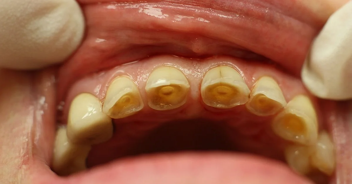
caries
breakdown of enamel due to cariogenic bacteria that causes demineralization eventually leading to a cavitated lesion
enamel properties
hardest material tissue in the body — 96% inorganic or mineralized material and 1% organic material and 3% water
mature enamel consists of mostly calcium hydroxyapatite
protects tooth from physical, thermal, and chemical forces — can withstand 20-30 lbs of pressure per tooth
enamel production
ameloblasts secrete initial enamel matrix through Tomes Process
initial enamel matrix made up of proteins, carbohydrates, and only small amount of calcium hydroxyapatite crystals
begins on masticatory surface of future crown (incisal or occlusal) — appositional growth continues, forming DEJ, moving cervical to CEJ
process of enamel completing mineralization from initial enamel matrix (30%) to full mineralization level (96%)
histology of enamel
enamel rod (enamel prism) - crystalline structural unit of enamel — each rod is made up of ribbons of crystals and lie perpendicular to denitn
lines of Retzius - incremental lines that seen to transverse the enamel rods — associated with imbrication lines (raised bumps) and perikymata (grooves)
systemic remineralization
water fluoridation, supplements — impact teeth that are going to erupt
topical remineralization
water fluoridation, dental fluoride treatment — impact only erupted teeth
globular dentin
areas where both primary and secondary mineralization have occurred — more mineralized = harder
interglobular dentin
areas where only primary mineralization has occurred — less mineralized = softer
dentinal tubules
long tubes in dentin that extend from DEJ in crown area or DCJ in root area to outer wall of the pulp = only sensation is to pain / not to cold or heat just pain
what do dentinal tubules contain
cell bodies of odontoblasts that remain in the pulp after maturation and odontoblastic processes
resorption
inflammation and loss of dentin structure within the tooth
internal resorption
resorption starting from the inside out
external resorption
resorption starts from the outside
dentin hypersensitivity
exposed dentin tubules lead to pain sensation
dentin-pulp complex
not clinically visible if tooth and supporting periodontium are health
dentin = 70% inorganic or mineralized material, 20% organic material, 10% water
mature dentin consists of mostly calcium hydroxyapatite and determines tooth shade
dentinoenamel junction (DEJ)
junction between dentin and enamel tissues — formed by the contact of predentin and the enamel matrix
dentinogenesis
production of dentin, begins before production of enamel
apposition and maturation of dentin
odontoblasts begin appositional growth of predentin (dental matrix)
ameloblasts are lost after eruption
odontoblasts are not and will continue to produce dentin
mineralization of predentin begins shortly after appositional growth and occurs in 2 phases
primary mineralization
calcium hydroxyapatite crystals forms as globules in predentin
secondary mineralization
another layer of crystals form, layered on the first, but don’t fuse everywhere
peritubular dentin
dentin that creates a wall around dentinal the tubule — highly mineralized after dentin maturation
intertubular dentin
dentin found between tubules — high level of mineralization
mantel dentin
first predentin produced found along dentinoenamel junction (DEJ) below enamel — creates CEJ
circumpulpal dentin
forms deep to mantle dentin and surrounds pulp, making up most of the dentin in the tooth — produces and matures after mantle dentin
primary dentin
produced within the tooth BEFORE completion of apical foramen of the root — most dentin is produced during this time
secondary dentin
formed AFTER completion of apical foramen — continues to be produced throughout life of the tooth and produced more slowly than primary dentin
tertiary dentin
forms in response to localized injury to exposed dentin, produced quickly in local regions
reactionary tertiary dentin
produced by existing odontoblasts