muscle histology and cytology
 Call Kai
 Learn
 Practice Test
 Spaced Repetition
 Match
 Flashcards
 Knowt Play
1/85

There's no tags or description
Looks like no tags are added yet.
86 Terms
muscle cells are called
fibers
myoblasts
muscle fiber stem cells
different muscle tissues are distinguished by
shape of muscle fibers and striations/lack of striations
striations
alternate dark and light bands found on skeletal and cardiac muscle

muscle tissue is mostly made up of
muscle fibers
muscle tissue is generally intermixed with
connective tissue
types of muscle
smooth, cardiac, skeletal
smooth muscle

appearance of smooth muscle fibers
non-striated, spindle shaped, single nucleus
what control does smooth muscle have
involuntary control
important organelles/cytoskeleton/junctions in smooth muscle
numerous gap junctions, actin and myosin placed in multiple planes
what are gap junctions used for
communication
why does smooth muscle line blood vessels
to maintain blood pressure, direct blood flow
why does smooth muscle line gastrointestinal/digestive tract
to move digested materials
cardiac muscle
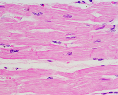
appearance of cardiac muscle fibers
striated, cylindrical and branched, single nucleus
what control do cardiac muscles have
involuntary control
example of involuntary control in cardiac muscles
heart rate can't be controlled
important organelles/cytoskeleton/junctions in cardiac muscle
gap junctions, desmosomes, highly organized actin and myosin, mitochondria take up 20% of fiber volume
functions of cardiac muscle
To pump blood and maintain blood pressure
skeletal muscle

appearance of skeletal muscle fibers
striated, long cylinders, multiple nuclei (multinucleate)
what control does skeletal muscle have
voluntary control by motor neurons
important organelles/cytoskeleton/junctions in skeletal muscle
highly organized actin and myosin, mitochondria take up 2% of fiber volume, sarcoplasmic reticulum
function of skeletal muscle
movement, posture, joint stability, heat generation
important structures of the skeletal muscle fiber
sarcolemma, multinucleate, sarcoplasmic reticulum
general structure of skeletal muscle
long cylinders
sarcolemma
cell membrane of muscle fibers
parts of sarcolemma
motor end plate, transverse tubules ( T-tubules)
Motor end plate of sarcolemma
specialized region where motor neuron interacts with the muscle fiber, part of neuromuscular junction

structure of motor end plate
folds, acetylcholine receptors

transverse tubules (T-tubules) of sarcolemma
cylindrical indentation of sarcolemma

function of transverse tubules
brings action potential/electrical stimulus into the fiber's interior
multinucleate
many nuclei
why do muscles need a lot of nuclei
muscles have a lot of protein, nuclei have genetic material that encode for them
sarcoplasmic reticulum
located near ends of the transverse tubules
function of sarcoplasmic reticulum
stores calcium
important molecules for atp production
glycogen, myoglobin, creatine phosphate
myoglobin
protein only found in muscle
what kind of protein is myoglobin
globular protein
what kind of protein is actin
fibrous protein
globular protein function
functional
fibrous protein function
structural
what does myoglobin bind to
oxygen (O2)
why does myoglobin bind to oxygen
so it can be stored in muscle
Creatine Phosphate
storage form for ATP
is creatine phosphate quicker than producing ATP in the mitochondria?
yes
function of creatine phosphate
can donate a P to ADP, making ATP
major proteins
myofilaments, proteins associated with actin
types of myofilaments
thin filaments and thick filaments
thick filament
myosin
myosin structure
fibrous tails, globular heads
myosin tails
fibrous proteins bundled into a rope
myosin head
globular
what do globular and fibrous portions of myosin indicate
myosin is both structural and functional
thin filament
actin
what type of protein is actin
globular
actin structure
arranged into two long filaments wrapped around each other

proteins associated with actin
tropomyosin and troponin
what kind of protein is tropomyosin
fibrous
what kind of protein is troponin
globular
tropomyosin structure
thin filament wound around the actin

tropomyosin function
blocks actin-myosin binding sites
troponin structure
linked to tropomyosin

troponin function
calcium binding site
what happens when troponin is bind to?
it changes shape
sarcomere
functional unit of skeletal muscle fiber
what is sarcomere banding pattern determined by
myosin and actin
sarcomere function
gives striated appearance of muscle fibers
sarcomere location
between two Z lines

z line (z disc)
ends of sarcomeres

light band
split in half by a Z-line, one half sits in one muscle unit and the other half sits in the next
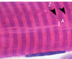
dark band
width defined by thick filaments

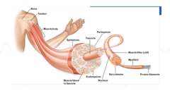
order of skeletal muscle gross anatomy
muscle fiber, fascicle, gross muscle
muscle fiber
muscle cells
fascicle (fasciculus)
group of muscle fibers
gross muscle
several fascicles
connective tissue sheaths
epimysium, perimysium, endomysium, fascia
composition of connective tissue sheaths
loose/areolar connective tissue, each layer is continuous/interconnected with the others
Endomysium
surrounds individual muscle fiber
Perimysium
surrounds a fascicle
what does perimysium contain
blood vessels and nerves run in perimysium in between fascicles
Epimysium
outer layer of connective tissue, surrounds all fascicles/muscle
epimysium also serves as
bone attachments (indirect and direct attachments)
Fascia
connective tissue that forms a sheet of tissue under the skin
what can fascia also surround
individual muscles or muscle groups