Anatomy Unit C: Lab Practical: Axial Skeleton
 Call Kai
 Learn
Learn Practice Test
 Spaced Repetition
 Match
Match Flashcards
 Knowt Play
1/104

There's no tags or description
Looks like no tags are added yet.
Name | Mastery | Learn | Test | Matching | Spaced | Call with Kai | Chat |
|---|
No analytics yet
Send a link to your students to track their progress
105 Terms
Frontal bone (Question answer not definition)
Which bone form the forehead?
Parietal bones (Question answer not definition)
Which bones form the superior and lateral skull?
Temporal bone (Question answer not definition)
Which bone lies inferior to the parietal bone?
External acoustic meatus (Question answer not definition)
What opening leads to the eardrum?
Mastoid process (Question answer not definition)
Which projection is the rough bump behind the ear?
Styloid process (Question answer not definition)
Which thin, needle like projection is below the ear canal?
Foramen magnum (Question answer not definition)
What large opening lets the spinal cord pass through the skull?
Occipital condyles (Question answer not definition)
Which skull projections articulate with C1?
Sphenoid bone (Question answer not definition)
Which bone contains the sella turcica?
Sella turcica (Question answer not definition)
What structure houses the pituitary gland?
Ethmoid bone (Question answer not definition)
Which bone forms the upper nasal septum and roof of the nasal cavity?
Crista Gali (Question answer not definition)
What vertical ethmoid projection attaches to the dura mater?
Cribriform plates (Question answer not definition)
Through which plates do olfactory fibers pass?
Mandible (Question answer not definition)
What is the lower jaw bone?
Maxillae (Question answer not definition)
Which bones form the upper jaw?
Zygomatic bone (Question answer not definition)
Which bone forms the cheekbone?
Zygomatic process (Question answer not definition)
Which projection of the temporal bone helps form the zygomatic arch?
Nasal bones (Question answer not definition)
Which paired bones form the bridge of the nose?
Vomer (Question answer not definition)
Which bone forms most of the nasal septum?
Lacrimal bones (Question answer not definition)
Which small bones help form the medial orbit and provide a tear passageway?
Palatine bones (Question answer not definition)
Which paired bones form the posterior hard plate?
Inferior nasal conchae (Question answer not definition)
Which bones project into the nasal cavity from its lateral walls?
Sagittal suture (Question answer not definition)
Which structure joins the two parietal bones superiorly?
Coronal suture (Question answer not definition)
Which structure joins the frontal and parietal bones?
Squamous suture (Question answer not definition)
Which structure joins the parietal and temporal bones?
Lambdoid suture (Question answer not definition)
Which structure joins the parietal and occipital bones?
Hyoid bone (Question answer not definition)
Which horseshoe-shaped bone lies in the throat above the larynx?
Intervertebral discs (Question answer not definition)
What pads between vertebrae absorb shock?
Body (Question answer not definition)
What is the rounded anterior portion of a vertebra?
Vertebral foramen (Question answer not definition)
What opening in a vertebra allows the spinal cord to pass?
Transverse processes (Question answer not definition)
Which paired vertebral projections extend laterally?
Spinous process (Question answer not definition)
What single vertebral projection extends posteriorly?
Superior and inferior articular processes (Question answer not definition)
Which projections allow adjacent vertebrae to articulate?
Atlas (C1) (Question answer not definition)
Which vertebra supports the skull and allows you to nod “yes”?
Axis (C2) (Question answer not definition)
Which vertebra allows head rotation and contains the dens?
Dens (Question answer not definition)
What projection of C2 acts as a pivot for head rotation?
Thoracic vertebrae (Question answer not definition)
Which vertebrae articulate with the ribs?
Lumbar vertebrae (Question answer not definition)
Which vertebrae have the largest, block like bodies?
Sacrum (Question answer not definition)
Which bone forms from the fusion of five vertebrae?
Alae (Question answer not definition)
What are the winglike projections of the sacrum called?
Coccyx (Question answer not definition)
What is the human tailbone called?
Manubrium, body, and xiphoid process (Question answer not definition)
What are the three parts of the sternum, from superior to inferior?
Manubrium (Question answer not definition)
Which part of the sternum articulates with the clavicles?
Body of the sternum (Question answer not definition)
Which part forms most of the sternum?
Xiphoid process (Question answer not definition)
Which part lies at the inferior end of the sternum?
12 pairs (Question answer not definition)
How many pairs of ribs are in the human thoracic cage?
True ribs (1-7) (Question answer not definition)
Which ribs attach directly to the sternum through their own costal cartilage?
False ribs (8-12) (Question answer not definition)
Which ribs do not attach directly to the sternum?
Floating ribs (11-12) (Question answer not definition)
Which ribs have no sternal attachment?

Frontal bone
Forms the forehead and the upper portions of the eye sockets
Located at the front of the cranium

Parietal bones
Two large, flat bones forming most of the top and upper sides of the skull.
Meet along the sagittal suture.

Temporal bones
Located on both sides of the skull around the ears, inferior to the parietal bones.
Contain bone, forming part of the cheek arch.

Zygomatic process
A thin, arching projection of the temporal bones forming most that extends forward to join the zygomatic bone
Forming part of the cheek arch.

External acoustic meatus
The round or oval opening on the temporal bone that leads inward toward the eardrum.

Styloid process
A thin, needle like projection extending downward from the temporal bone.
Located just below the ear canal.
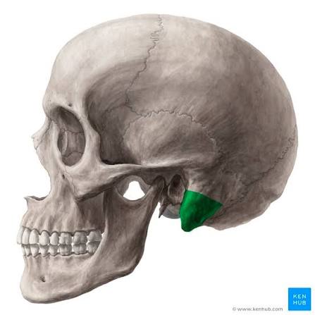
Mastoid process
A thick, rounded, rough projection behind and below the external acoustic meatus.
You can feel this bump behind your ear.

Occipital bone
Forms the back and base of the skull.
Recognize it by the large foramen magnum near its center.

Foramen magnum
The very large opening at the base of the skull where the brainstem connects to the spinal cord.

Occipital condyles
Two smooth, rounded projections on either side of the foramen magnum.
They articulate with the atlas (C1)

Sphenoid bone
A bat-shaped bone across the base of the skull, behind the eyes.
It’s broad wings extend laterally.
Contains the sella turcica.

Sella turcica
A saddle shaped depression in the center of the sphenoid bone.
It houses the pituitary gland.
Ethmoid bone
An irregular, delicate bone between the eye sockets.
Forms part of the roof of the nasal cavity, upper nasal septum, and medial orbital walls.

Crista galli
A small, upright, midline projection on the ethmoid bone, rising above the cibriform plates.

Cibriform plates
Thin, perforated horizontal areas of the ethmoid bone on either side of the crista galli.
Tiny holes allow olfactory nerve fibers to pass through.

Mandible
The lower jawbone.
Has a horizontal body and two upright rami
It is the movable bone of the skull

Maxillae
The paired upper jawbones that hold the upper teeth
Form part of the hard palate, floor of the orbits, and central face

Palatine bones
Paired L-shaped bones at the back of the hard plate
Form its posterior portion and contribute to the nasal cavity and orbit

Zygomatic bone
The prominent cheekbone on the outer side of each orbit
Connects with the temporal bone to help form the zygomatic arch

Lacrimal bones
Tiny, thin bones on the medial (inner) walls of the eye sockets
Near the tear drainage passage

Nasal bones
Two small, rectangular bones joined at the bridge of the nose
Located between the frontal bone and the upper nasal cartilages

Vomer
A thin, plow-shaped of blade-shaped bone in the middle of the nasal cavity.
Forms the inferior and posterior part of the nasal septum

Inferior nasal conchae
Two curved, scroll-like bones projecting inward from the walls of the nasal cavity
They are separate bones from the ethmoid

Sagittal suture
Runs along the midline of the skull
Separating the left and right parietal bones

Coronal suture
Runs from side to side across the top of the skull
Separating the frontal bone from the two parietal bones

Squamous suture
Located on each side of the skull Separating
Where parietal bones meets the temporal bone

Lambdoid suture
Located at the back of the skull
Separating the parietal bones from the occipital bone

Hyoid bone
A small, horseshoe-shaped bone in the front of the neck
Located above the larynx
Has a central body and two pairs of projections called horns (cornua)
It does not directly articulate with another bone

Cervical vertebrae (C1-C7)
Seven vertebrae in the neck
Generally smaller than other vertebrae
Many have openings in their transverse processes called transverse foramina

Thoracic vertebrae (T1-T12)
Twelve vertebrae in the upper and middle back
Have rib-articulating facets and usually long spinous bodies and thick processes

Lumbar vertebrae (L1-L5)
Five large, sturdy vertebrae in the lower back
Recognize them by their massive, block like bodies and thick processes

Intervertebral discs
Pads of fibrocartilage between the bodies of adjacent vertebrae
Visible as cushioning spaces or discs between vertebral bodies

Body
The thick, rounded or block like anterior part of a vertebra
It bears most of the body’s weight

Vertebral foramen
The central opening surrounded by the vertebral body and arch
When vertebrae are stacked, these openings form the vertebral canal for the spinal cord

Transverse processes
Two projections extending laterally, one to each side of the vertebral arch
Often serve as muscle attachment points

Spinous process
The single posterior projection extending backward from the vertebral arch
Often palpable along the midline of the back

Superior and inferior articular processes
Paired projections with smooth joint surfaces that connect neighboring vertebrae Visible
Look near the vertebral arch, above and below

Atlas (C1)
The first cervical vertebra, directly beneath the skull
Ring-shaped and lacks a vertebral body
Its superior surfaces receive the occipital condyles
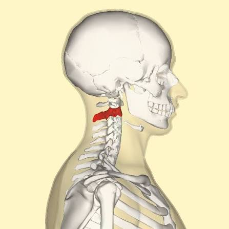
Axis (C2)
The second cervical vertebra
Identified by the dens, which projects upward and serves as a pivot for head rotation

Dens
A toothlike vertical projection rising from C2
Fits into the atlas and allows the head to rotate from side to side

Sacrum
A large, triangular bone at the base of the spine
Formed from five fused vertebrae Visible
Recognize its paired sacral foramina and broad upper base
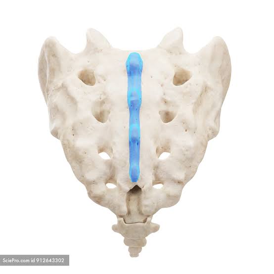
Median sacral crest
A raised ridge running down the posterior midline of the sacrum
Represents fused remnants of the spinous processes

Alae of the sacrum
The broad, winglike expansions on the upper sides of the sacrum
Connect with the hip bones at the spinous joints

Sacral foramina
Paired openings on the anterior and posterior surfaces of the scrum
They allow nerves and blood vessels to pass through

Coccyx
The small, pointed tailbone inferior to the sacrum
Formed from three to five fused, reduced vertebrae

Thoracic cage
The protective framework of the chest, made up of the sternum, 12 pairs of ribs, costal cartilages, and thoracic vertebrae

Sternum
The flat breastbone in the anterior midline of the chest
Consists of the Manubrium, body, and xiphoid process

Manubrium
The broad, superior part of the sternum
Has a jugular notch at the top and articulates with the clavicles and first rib pair

Body of the sternum
The long, central, longest portion of the sternum
Articulates with costal cartilages from several ribs

Xiphoid process
The small, inferior tip of the sternum
May be pointed, rounded, or slightly forked in models

Ribs (12 pairs)
Curved bones forming the sides of the thoracic cage
Attach posteriorly to thoracic vertebrae
Their anterior attachments vary by rib group