Heart Anatomy and Physiology
 Call Kai
 Learn
 Practice Test
 Spaced Repetition
 Match
 Flashcards
 Knowt Play
1/32

There's no tags or description
Looks like no tags are added yet.
33 Terms
Location of the Heart
Thoracic cavity in the mediastinum, obliquely situated.
Anterior Wall
Front portion containing the right atrium and both ventricles.

Inferior Wall
Bottom portion of both ventricles resting on the diaphragm.
Posterolateral Wall
Back portion, greatest portion of left ventricle; largest muscular wall.
Right vs. Left Side of Heart
Right side has oxygen-poor blood; left side has oxygenated blood.

Cycle of Blood Flow (First Half)
Right Atrium -> Right Ventricle -> Pulmonary Arteries -> Lungs.
Cycle of Blood Flow (Second Half)
Pulmonary Veins -> Left Atrium -> Left Ventricle -> Aorta.

Atrioventricular Sulcus
Groove that separates the atria from the ventricles.
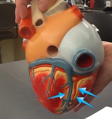
Interventricular Sulcus
Grooves separating ventricles; contain interventricular arteries.

Fibrous Pericardium
Tough, dense outer sac attaching the heart to the diaphragm.

Serous Pericardium
Inner layer with parietal and visceral portions.

Pericardial Space
Space containing serous fluid between pericardial layers to reduce friction.

Pericarditis
Inflammation of the pericardium causing chest pain and breath shortness.

Epicardium
Outermost layer composed of fibrous connective tissue and adipose tissue.

Myocardium
Middle layer of the heart composed of cardiac muscle.

Endocardium
Innermost layer composed of simple squamous epithelium.

Superior and Inferior Vena Cava
Superior carries upper body blood; inferior carries lower body blood.

Pulmonary Veins
Four veins delivering oxygenated blood from lungs to left atrium.

Aorta
Transports oxygenated blood from the left ventricle to the body.

Pulmonary Trunk
Transports deoxygenated blood from the heart to the lungs.

Function of Heart Valves
Gates that prevent backflow to ensure one-way blood flow.

Bicuspid Valve
Left AV valve that prevents backflow into the left atrium.

Tricuspid Valve
Right AV valve that prevents backflow into the right atrium.

Aortic Semilunar Valve
Valve between left ventricle and aorta preventing backflow.

Pulmonary Semilunar Valve
Valve between right ventricle and pulmonary trunk.

Systole
Contraction phase of the cardiac cycle where AV valves close and blood is ejected.

Diastole
Relaxation phase of the cardiac cycle.

Papillary Muscles
Structures that prevent valve inversion and stabilize valve leaflets.

Chordae Tendineae
Connect papillary muscles to valve leaflets to keep them touching when they close.

Sinoatrial (SA) Node
Pacemaker of the heart that initiates the heartbeat and makes atria contract.

Atrioventricular (AV) Node
Receives action potential from the SA node and passes it to the ventricles.

Myocardial Infarction
Condition where a coronary vessel gets blocked by a thrombus or embolus blood clot.

Heart Sounds ("Lubb" and "Dubb")
"Lubb" is closure of AV valves and ventricular contraction; "Dubb" is closure of semilunar valves and ventricular relaxation.