10 Acute Kidney Injury
 Call Kai
 Learn
 Practice Test
 Spaced Repetition
 Match
 Flashcards
 Knowt Play
1/53

There's no tags or description
Looks like no tags are added yet.
54 Terms
Define acute kidney injury
1. increase in serum creatinine by 26.5umol/l within 48 hours.
2. increase in serum creatinine to >1.5x baseline (known to have occured within prior 7 days).
3. urine volume
subsets of AKI
pre-renal, renal and post renal
aki mortality vs non aki mortality
aki has 23% higher mortality
risk factors for AKI
polypharmacy,elderly, diabetes, heart falure, liver cirrhosis, dehydration, infection, renal stones, begin prostatic hyperplasia, chronic kidney disease
classification of AKI according to KDIGO
stage 1: 1.5-1.9x baseline/>0.3mg/dl increase in serum creatinine. with urine output 0.5ml/kg/h for 6-12 hours
stage 2: 2.0-2.9x baseline and
stage 3: 3x baseline with urine output less than 0.3ml/kg/h for more than 24 hours or anuria for more than 12 hours. increase in serum creatinine to >4.0mg/dl or initiation of renal replacement therapy ot in pts

AKI classifications
KDIGO, rifle and akin
RIFLE classification of AKI

AKIN classification of AKI

Prerenal AKI causes
autogregulation of glomerulus interruption
chronic heart and liver failure causing decreased effective circ vol, decreased cardiac volume, hypovolaemia
or
NSAIDS, ARBs, ACE inhibitors, cyclosporine impair renal autoregulation
Majority of AKI causes are...
pre-renal
intrinsic acute kidney injury can be broken down into
tubular (majority), interstitial, glomerular, vascular
tubular causes of intrinsic AKI
acute tubular necrosis by ischaemia (eg pre-renal blood loss), nephrotoxic drugs, myeloma, myoglobin
interstitial (10%) causes of intrinsic aki
pyelonephritis, nephrotoxic drugs, autoimmune eg SLE, infiltration with lymphoma/infection/tumour lysis syndrome
how does multiple myeoloma cause intrinsic aki
due to deposition of light chains
myoglobin in intrinsic aki
v nephrotxic. damages tubules
glomerular causes of AKI
glomerular nephritis (nephrotic and nephritic), haemolytic uraemic syndrome
haemolytic uraemic syndrome
often young children with e.coli infections . thrombocytopoenia, haemolytic anaemia and uraemia. bruising, low platelets, cell breakdown and urea

vascular causes of intrinsic aki
vasculitis, malignant hypertension
Nephritic syndrome
Hematuria, hypertension, and some prt uria. due to damage of glomerular basement membrane.

nephrotic syndrome
characterized by massive proteinuria caused by glomerular damage.
what is acute tubular necrosis pathophysiology
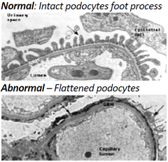
acute tubules have normal epithelia with their adhesion molecules. in damage they lose their brush border and polarity. lose adhesion molecules triggers apoptosis and necrosis which block off lumen with dead cells. worsening leads to end stage RF
what does acute tubular necrosis cause
decrease in GFR and urine output, increase in urea and creatinine. metabolic acidosis, fluid retention and electrolyte imbalance resulting in intrinsic renal AKI
coca cola urine
Rhabdomyolysis
nephrotoxins
aminoglycosides (gentamicin, amikacin), diuretics. bacterial endotoxins eg e.coli. IV contrast. tumour lysis syndrome
tumour lysis syndrome
cancer cells die rapidly causing hyperuricaemia as uric acid, potassium and phosphate is released. precipitation into renal tubules, renal vasoconstriction, impaired autoregulation, decreased renal blood flow and inflammation. hyperphosphtaemia as calcium phosphate deposits in renal tubules
cancers that cause tumour lysis syndrome
leakemia, lymphoba bc high tumour burden.
post renal aki (10-25%)
most common places for kidney stones to obstruct
ureteropelvic junction obstruction, ureteral stricture, ureterovesicle junction
post renal obstruction causes
luminal, mural and extrinsic
luminal causes of PRO
renal stones, blood clots, sloughed tissues
mural causes of PRO
strictures due to infection or post TURP/TURBT in males; cancer
TURP (urinary) as a mural cause of PRO
transurethral resection of the prostate can cause strictures in urethra
TURBT (transurethral resection of bladder tumor)
as a mural cause of PRO
causing stricture in uterovesicle junction
extrinsic causes of PRO
benign prostatic hyperplasia pressing on urethra, cancer (eg of bladder, pelvic organs, ovaries, cervix), pregnancy, retroperitoneal fibrosis
pregnancy as PRO extrinsic compression
bladder anterior to vagina and uterus. growing uterus presses on ureter and bladder
retroperitoneal fibrosis
scarring due to radiotherapy. treated by steroids and stenting
not treating post obstructive renal causes of aki can cause
hydroureter leading to hydronephrosis

clinical features of renal failure
nausea, lethargy. decreased urine output!!, fluid overload (oedema, orthopnoea), electrolyte abnormalities, acid-base disturbance
long lie injury
lying down for a long time causing rhabdomyolysis so intra renal causes of aki
prerenal symptoms of aki
hypovolaemia, oedema if intrinsic renal
intra renal symptoms of aki
often precipitating cause. drug history, immune disease, toxins, falls/crush injury/ long lie
post renal symptoms of AKI
voiding problems, enlarged bladder, red flag cancer signs (weight loss, frank haematuria)
most at risk in AKI
elderly, CKD, cardiac failure, liver disease, diabetes, vascular disease, nephrotoxic medications
acute STOP in aki
sepsis and hypoperfusion, toxicity, obstruction, parenchymal kidney disease
a-e approach for AKI
obs, asses fluid status and for sepsis give abx
serum K and aki
because of dehydration, aki pts will have hyperkalaemia. give insulin + dextrose infusion and +/- salbutamol
investigations with pt with aki
establish baseline: refer to previous tests of urea and creatinine
urinalysis: proteinuria +/- haematuria. casts, crystals, osmorality
blood tests: FBC, LFTs, ABGs, CRP, ESR, CK, blood cultures
ESR blood test
raised in vasculitis and SLE
LFT in aki
liver cirrhosis can cause aki
ck in aki blood test
rhabdomyolysis
serum electrophoresis in aki
in multiple myeloma sees breakdown of cells
blood test when suspecting vasculitis or SLE
free light chains, c3/c4 complement depletion, autoAb ANA, ANCA, anti-GBM
indications for renal replacement therapy
AEIOU
a: acidaemia, severe metabolic acidosis and depleted bicarb
E: uncontrolled hyperkalaemia
I: ingested toxins (lithium, ethylene glycol)
o: severe overload oedema
u: symptomatic uraemia
uraemic frost
late stage uraemia. uraemic salts leak from skin to head. present with chronic anorexia, blood nitrogen.
