Lecture 7 & 8 (first half)
 Call Kai
 Learn
 Practice Test
 Spaced Repetition
 Match
 Flashcards
 Knowt Play
1/21

There's no tags or description
Looks like no tags are added yet.
22 Terms
Multi-substrate enzyme interactions
ternary complex
random: all substrates bind to enzyme in random order forming the ternary complex
ordered: substrates bind in an ordered fashion, 1 after the other
Ping-pong mechanism
subtrate 1 binds and releases product forming an enzyme intermediate for substrate B to pick up and form a different product
no ternary complex formed
ex: hexokinase (ATP and Glucose)
each substrate has a Km
Types of reversible inhibition
competitive
uncompetitive
non competitive (mixed)
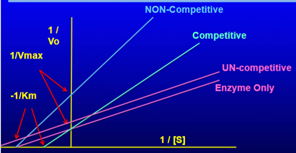
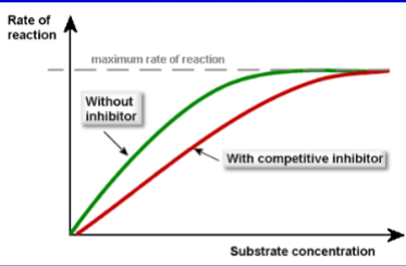
competitive inhibition
inhibitor competes with ONLY active site → NO catalysis
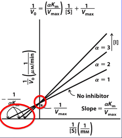
forms E1 complex
Km INCREASES, Vmax CONSTANT
alphaKm is the Km observed with comp. inhibitor is present

uncompetitive inhibition
inhibitor binds in spot OTHER than active site (allosteric site)
conformational change renders enzyme INACTIVE
forms ES1 complex
Km DECREASES, Vmax DECREASES
alpha’Km is the Km observed when uncomp. inhibitor is present

mixed inhibition
inhibitor binds in spot OTHER than active site
either E or ES complex (has bias toward one)
Km INCREASES, Vmax DECREASES
kinetics impacted by alpha and alpha ‘

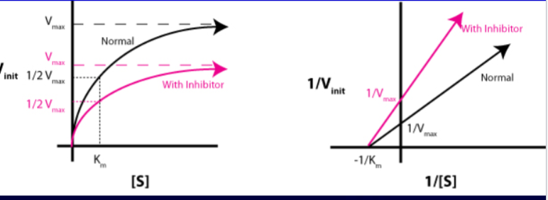
non-competitive inhibition
a special case of MIXED inhibition
alpha = alpha’
can bind either E or ES complex
Km CONSTANT, Vmax DECREASES
irreversible inhibition
suicide inhibitors - form covalent bonds with their targets
structurally similar to the normal substrate so the enzyme treats it like a substrate catalyzing the mechanism
permanently bonded to the active site
forms a reactive intermediate covalently traps a catalytics residue in the active site
ex: penicilin acts as a suicide inhibitor of bacterial glycopeptide transpeptidase
Regulatory enzyme function
enzymes positioned at key points in metabolic pathways that regulate catalytic activity in response to cellular signals
does not obey michaelis-menten kinetics
Feedback inhibition
regulatory mechanism where the end product of a multi-step metabolic pathways acts as a negative allosteric modulator (inhibitor) of the first enzyme in the pathway
saves cellular resources and energy

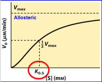
allosteric modulation
when binding of a molecule on one part of a protein affects the shape and behavior of the protein
have an active site and regulatory site (BOTH specific)
positive modulators: stabilize the active site, high affinity R state, lowers the K0.5, INCREASES catalytic rate
negative modulators: stabilize the T state, raises K0.5 and DECREASES catalytic activity

heterotropic modulator
enzymes are when the modulator is different from substrate binds outside active site
positive modulators: stabilize the active site, high affinity R state, lowers the K0.5, INCREASES catalytic rate (can shift curve to left - hyperbolic like)
negative modulators: stabilize the T state, raises K0.5 and DECREASES catalytic activity (can shift the curve to the right)

homotropic modulator
enzymes are when the substrate is the modulator (active site is regulatory site)
Types of reversible covalent modifications
methylation: addition of methyl group
ADP-ribosylation: addition of ribose
acetylation: addition of acetyl group
adenylation: addition of an AMP to tyrosine
myristoylation:
Prenylation: addition of lipid
ubiquitination: addition of ubiquitin to lysine
phosphorylation: addition of phosphate groups
importance of phosphorylation
thermodynamically favorable & rapid
kinase: adds a phosphoryl group to a protein (takes it off ATP first) (recognize specific sequences)
phosphatase: removes a phosphoryl group from an enzyme
neg charge introduced electrostatic interactions or steric clashes that either active or inactive an enzyme
forms the basis of kinase cascades that relay extracellular info throughout the cell

Amino Acid targets of phosphorylation
all have OHs (nucleophilic)
Serine
Thereonine
Tyrosine
Low Activity → High Activity
2 ATP → 2 ADP
2 OH Phosphorylase b → 2 P Phosphorylase a
High Activity → Low Activity
2 H2O → PP1 → 2 Pi
2 P Phosphorylase → 2 OH Phosphorylase b
Low Blood sugar → High Blood sugar
glucagon
ATP → PKA → ADP
phosphorylase kinase (P_
High Blood sugar → low blood sugar
insulin
H2O → PP1 → Pi
phosphorylase kinase (OH)

irreversible activation
occurs by proteolytic cleavage of a zymogen (precursor)
cleavage triggots a localized conformational rearrangement opening up a properly aligned catalytics residues → converts zymogen into fully active enzyme
trypsin: a protease that is activated by proteolytic cleavage - breaks down proteins in the gut
zymogen: trypsinogen
Proteases
peptidases are enzymes that catalyze the hydrolysis of peptide bonds linking amino acids in proteins
uses:
nutritional digestion (pepsin, trypsin)
blood coagulation (fibrin)
programmed cell death
protein turnover
classes:
serine
cysteine
aspartyl (HIV- protease)
metallo-
Serine Proteases Mechanism (Chymotrypsin, Trypsin)
substrate binding: polypeptide chain is positioned near active site with the polypeptide chain near serine
nucleophilic attack (acylation): catalytic triad (Ser-His-Asp) polarizes serine. serine OH attacks the carbonyl carbon of the peptide bond, forming a high energy TETRAHEDRAL intermediate (stabilized by oxyanion hole)
bond cleavage & amine release: tetrahedral intermediate collapses, breaking the peptide bond, N-term departs forming acyl-enzyme intermediate
deacylation: catalytic water molecule enters active site and nucleophilically attacks the acyl-enzyme bond with His stabilization
product: second tetrahedral intermediate collapses, releasing the c-term peptide and regenerating serine
Beta-lactamases
nucleophilic attack: serine OH attacks the carbonyl of the strained Beta-lactam ring of penicillin
Beta-lactam ring opens covalently forming a stable acyl-enzyme intermediate trapping the enzyme
hydrolysis: beta-lactamase hydrolyzes the covalent bond with the addition of catalytic water resulting in an opened, inactive antibiotic molecule
Chymotrypsin vs Trypsin
substrate specificity pockets differ
chymotrypsin: deep, hydrophobic substarte pocket lined with non polar amino acids (Gly & Ala)
accommodates and binds bulky, hydrophobic aromatic amino acid side chains (Phe, Tyr, Trp)
Trypsin: neg charged (asp)
attracts and forms strong salt bridges with pos charged basic side chains (Lys, Arg)
AA based on pH vs Activity curves
rising phase: catalytic residue must be deprotonated (inflection point on this slope corresponds to pka)
falling phase: second catalytic must be protonated (inflection point on this slope gives pka)
at high pH: salt bridge btw alpha amino group and Asp is broken → closes active site
low pH: his is protonated, cannot interact in catalysis