DEN 105 Lecture #5
 Call Kai
 Learn
 Practice Test
 Spaced Repetition
 Match
 Flashcards
 Knowt Play
1/54

There's no tags or description
Looks like no tags are added yet.
Name | Mastery | Learn | Test | Matching | Spaced | Call with Kai | Chat |
|---|
No analytics yet
Send a link to your students to track their progress
55 Terms
What is a gland?
it is a structure that produces a chemical secretion necessary for normal bodily functions
Glandular tissue in the head and neck includes…
Lacrimal
Salivary
Thyroid
Parathyroid
Thymus
Classification of glands
2 major types
Exocrine
Endocrine
Salivary glands- exocrine glands
produce saliva and are controlled by the autonomic nervous system (ANS)
Saliva
is a watery secretion with a pH of around 7. It is referred to as the “mouth’s bloodstream”
What role does saliva play?
plays an important role in bolstering and maintaining the health of oral soft and hard tissues
What is saliva made up as?
Salivary amylase
Lingual lipase
Mucus
lysozyme
Immunoglobulin A (IgA)
Electrolytes
Salivary amylase
an enzyme that begins starch digestion in the mouth
Lingual lipase
an enzyme that is activated by stomach acid and digest fat after the food is swallowed
Mucus
bind and lubricates the food mass and aids in swallowing
Lysozyme
an enzyme that kills bacteria and other pathogens
Immunoglobulin A (IgA)
an antibody that inhibits bacteria growth
Electrolytes
including sodium, potassium, chloride, phosphate, and bicarbonate
Function of saliva
Helps prepare the bolus of food for easier swallowing
Helps to keep the mucous membranes lubricated
Helps prevent dehydration; dry mouth stimulus to drink water
pH buffer
Breaks down starches (carbohydrates)
Cleanses teeth
Neutralizes acids to prevent tooth decay
Aids in taste
development of salivary gland
arise from a cord or epithelium
Grows to form a tube
end of tube is either round or tubelike clusters
Acini- cluster of secretory cells forms at the end of the tube
What are acini?
Secretory end pieces
Secrete proteins
There are three types of secretory units (acini)
Three types of acini
Serous acini
Mucous acini
Seromucous acini
Types of secretions
Serous
Mucous
Seromucous
Serous
Secretion is thin, watery, protein/enzyme-rich secretion
Less viscous
Mucous
Secretion is more viscous and mucin-rich
99% water
Inorganic ions (e.g., sodium, potassium, and chloride)
Minor amounts of amylase
Has mucins: Key characteristic in their ability to form gels
Seromucous
has characteristics of both types of secretions
Major and Minor salivary glands
are divided by size
both major and minor salivary glands are exocrine glands and thus have ducts associated with them
These ducts help drain the saliva directly into the oral cavity where the saliva can function
Major salivary glands
large paired glands and have maned ducts associated with them
Three major salivary glands
Parotid glands
Submandibular glands
Sublingual glands
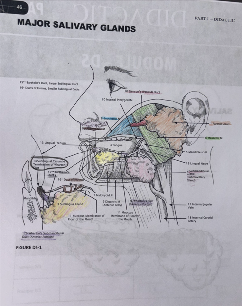
Parotid salivary gland
is the largest encapsulated major salivary gland
the gland is divided into two lobes: the superficial and deep lobes
Produces approximately 25% of saliva
Predominantly serous secretions

Parotid salivary gland: Innervation
The parotid salivary gland is innervated by the efferent (parasympathetic) nerves of the otic ganglion of the ninth cranial or glossopharyngeal nerve by way of the lesser petrosal nerve, as well as by the afferent nerve from the auriculotemporal nerve of the mandibular nerve of the fifth cranial or trigeminal nerve.
However, the seventh cranial or facial nerve and its branches travel through the parotid salivary gland between its superficial and deep lobes to serve as a divider but are not involved in its innervation
Parotid salivary gland location
The parotid salivary gland occupies the parotid space, an area posterior to the mandibular ramus, anterior and inferior to the ear
Parotid salivary gland: Ducts
the duct associated with the parotid salivary gland is the parotid duct or Stenson’s duct

Parotid salivary gland duct location
This long duct emerges from the anterior border of the gland, superficial to the masseter muscle.
The duct pierces the buccinator muscle, and then opens up into the oral cavity on the inner surface of the buccal mucosa of the cheek, usually opposite the maxillary second molar

Parotid papilla
is a small elevation of tissue that marks the opening of the parotid duct on the inner surface of the cheek
Mumps
When the parotid salivary gland becomes enlarged and tender
This contagious viral infection usually involves inflammatory enlargement or parotitis of both glands, first one side and then the other, giving the characteristics “chipmunk” cheeks
Submandibular gland
is the second larges encapsulated major salivary gland
Produces approximately 60-65% of saliva
Mucous and serous secretions
Posterior to the sublingual gland
Wraps around the mylohyoid muscle

Submandibular salivary gland location
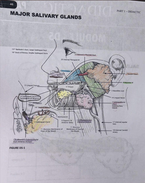
the submandibular salivary gland occupies the submandibular fossa in the submandibular space mainly in its posterior part
Submandibular salivary gland: duct
the duct associated with this gland is the submandibular duct or Wharton’s duct

Submandibular salivary gland: Wharton’s duct location
This duct travels along the anterior floor of the mouth then opens into the oral cavity at the sublingual caruncle, a small papilla near the midline of the floor of the mouth on each side of the lingual frenum

Submandibular salivary gland: innervation
it is innervated by the efferent (parasympathetic) fibers of the chorda tympani and the submandibular ganglion of the seventh cranial or facial nerve
Sublingual salivary gland
is the smallest most diffuse, and only unencapsulated major salivary gland
Approximately 10% of the total salivary volume
Mixed serous and mucous secretion


Sublingual salivary gland: location
this gland occupies the sublingual fossa in the sublingual space at the floor of the mouth
Sublingual salivary gland: duct
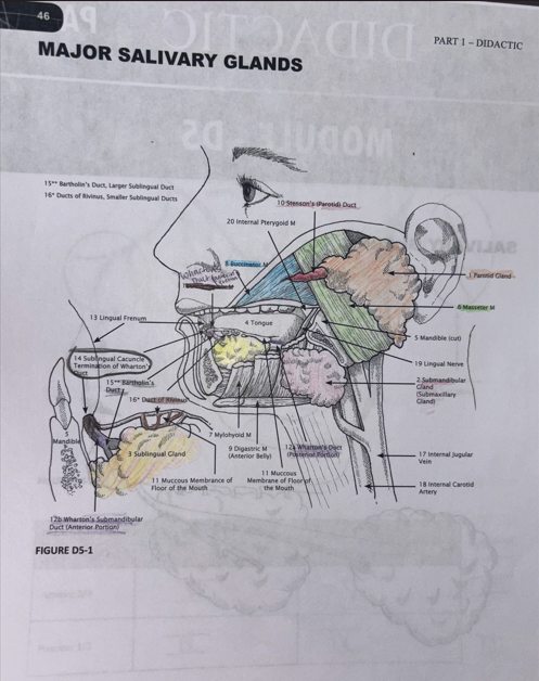
the short ducts associated with the gland sometimes combine to for the sublingual duct or Bartholin’s duct
The sublingual duct then opens directly into the oral cavity through the same opening as the submandibular duct, the sublingual caruncle

Sublingual caruncle
is a small papilla near the midline of the floor of the mouth on each side of the lingual frenum
Ducts of Rivinus
other smaller ducts of the sublingual gland open along the sublingual fold

Sublingual salivary gland: innervation
has the same innervation as the submandibular salivary gland, which is by the efferent (parasympathetic) fibers of the chorda tympani and the submandibular ganglion of the seventh cranial or facial nerve
Minor salivary glands
are smaller than the larger major salivary glands but are more numerous in number; however, they are not individually encapsulated by surrounded by connective tissue within the submucosa
Minor salivary glands: Location
Most of the minor salivary glands are mainly scattered throughout the oral cavity in the buccal, labial, and lingual mucosa, the soft palate, the posterior part of the lateral zones of the hard palate, and the floor of the mouth.
What are the minor salivary glands, von Ebner’s glands associated with?
the base of the large circumvallate lingual papillae on the posterior part of the tongue’s dorsal surface
Where can the minor salivary glands also be found in?
found in lesser numbers along the mucosa of the tonsils, supraglottis region, and paranasal sinuses as well as between the muscle fibers of the tongue
They are not found in the gingival tissue and the anterior part of the lateral zones of the hard palate or its medial zone
Minor salivary glands: duct
Each minor salivary gland has a single duct that secretes saliva directly into the oral cavity since they are exocrine glands like the major salivary glands.
But the unnamed ducts of the minor salivary glands are shorter than those of the major salivary glands.
All the minor salivary glands have a mainly mucous secretion with some serous secretion except for the glands of von Ebner which have only a serous secretion
Minor salivary glands: innervation
The minor salivary glands are innervated by various nerves depending on location
Lesions
Salivary glands may become enlarged, tender, and possibly firmer due to various disease processes. They may also become involved in the formation of a salivary stone (or sialolith) within the gland, blocking the drainage of saliva from the duct, especially with the submandibular gland.
Salivary gland obstruction may result in swelling and tenderness. Mucoceles (associated with minor salivary glands) and Ranulas (associated with the floor of the mouth) are salivary lesions caused by accumulation of mucus.
Salivary stones
Salivary stones are uncomfortable but not dangerous, and can involve one or more enlarged, tender salivary glands. The clinician may also be able to palpate the stony hard salivary stone(s) during an examination.
Facial radiographs or computed tomography (CT) can confirm the diagnosis. Salivary stones are usually removed manually or with minor surgery
Sialoliths
is a minor salivary gland on the floor of the mouth
Sialolithiasis
When a salivary stone becomes lodged within the duct, causing a blockage of saliva. The gland usually swells and feels painful, especially around meal times, and infection may follow.
Small stones may sometimes pass spontaneously or with conservative measures such as hydration, salivary stimulation, and gland massage.
Antibiotics may be indicated when bacterial infection is present.
Persistent stones may require minimally invasive removal or surgical treatment
Xerostomia
(DRY MOUTH) can result in increased trauma to a nonprotected oral mucosa, increased cervical caries, problems in eating, speech and mastication, and bad breath or halitosis.
Extensive cervical caries is a frequent complication of dry mouth. In addition to the lack of saliva, patients may attempt to stimulate salivary flow with sweets or chewing gums.
hyposalivation
reduced production of saliva
Treatment for dry mouth
Being very vigilant about good oral hygiene through brushing and flossing
Using a high-fluoride toothpaste (Prevident 5000)
Sip water frequently
Eat soft moist foods that are not overly salty
Avoid rinses that contain alcohol
Use products to moisturize your mouth including prescription or over-the- counter mouth rinses, artificial saliva or moisturizers to lubricate your mouth.
Mouthwashes designed for dry mouth, especially ones with xylitol. Ex. Biotene or ACT mouthwashes
Prescribe medication that stimulates saliva. Ex. pilocarpine (Salagen) to stimulate saliva production.