BDS1_BSC1.AHN.06 The skull part 1

0.0(0)
Studied by 0 people
call kaiCall Kai
learnLearn
examPractice Test
spaced repetitionSpaced Repetition
heart puzzleMatch
flashcardsFlashcards
GameKnowt Play
Card Sorting

1/39

flashcard set

Earn XP

Description and Tags

Define and identify the principal parts of the skull. Describe and identify the bones which make up the skull and cranio-facial skeleton. Describe the embryological origin of the skull. Describe and identify the main features of the skull in its principal views (posterior, superior, lateral, anterior and inferior).

40 Terms

1
New cards

What is the Neurocranium vs viscerocranium? Which bones are in each? 

  • Neurocranium: The bony case covering the brain. Has a roof (calvaria) and a floor/base (basicranium) 

  • Viscerocranium = the facial skeleton


<ul><li><p class="Paragraph SCXW138576991 BCX8" style="text-align: left;"><span style="line-height: 18.5333px;">Neurocranium: The bony case covering the brain. Has a roof (calvaria) and a floor/base (basicranium)&nbsp;</span></p></li></ul><ul><li><p class="Paragraph SCXW138576991 BCX8" style="text-align: left;"><span style="line-height: 18.5333px;">Viscerocranium = the facial skeleton</span></p></li></ul><p></p>
2
New cards

What 5x bones are in the neurocranium and which 10x bones are in the viscerocranium?

  • 5 Neurocranium bones = 2x Occipital, 2x parietal, 2x temporal, 1x frontal and 1x sphenoid.

  • 10x viscerocranium = 2x sphenoid, 2x ethmoid, 2x lacrimal, 2x nasal, 2x zygomatic, 2x maxilla, 1x mandible, 1x vomer, 2x inferior nasal conchae, 2 x palatine,

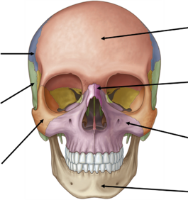
<ul><li><p><span>5 Neurocranium bones = 2x Occipital, 2x parietal, 2x temporal, 1x frontal and 1x sphenoid.</span></p></li><li><p class="MsoListParagraphCxSpLast"><span>10x viscerocranium = 2x sphenoid, 2x ethmoid, 2x lacrimal, 2x nasal, 2x zygomatic, 2x maxilla, 1x mandible, 1x vomer, 2x inferior nasal conchae, 2 x palatine,</span></p></li></ul><p></p>
3
New cards
<p><span style="font-family: &quot;Times New Roman&quot;; line-height: normal; font-size: 7pt;"> </span><span>Label the 7 anterior facial bones.</span></p>

Label the 7 anterior facial bones.

knowt flashcard image
4
New cards
<p><span style="font-family: &quot;Times New Roman&quot;; line-height: normal; font-size: 7pt;"> </span><span>What are the following 3 regions and what structures do they transmit?</span></p>

What are the following 3 regions and what structures do they transmit?

  • Supraorbital margin: 1/3rd along is located the supraorbital Notch or Foramen which transmits the supraorbital nerve (branch of CN5 V1), supraorbital artery (branch of the ophthalmic artery which is from the ICA), and supraorbital vein, which supply sensation and blood flow to the forehead and upper eyelid

  • Infraorbital margin - sits directly above the infraorbital foramen. The infraorbital foramen transmits the infraorbital nerve (terminal branch CN5 V₂, along with the infraorbital artery (branch of the maxillary artery – ECA) and vein.

  • Anterior nasal aperture


<ul><li><p><span>Supraorbital margin: 1/3rd along is located the supraorbital Notch or Foramen which transmits the supraorbital nerve (branch of CN5 V1), supraorbital artery (branch of the ophthalmic artery which is from the ICA), and supraorbital vein, which supply sensation and blood flow to the forehead and upper eyelid</span></p></li><li><p class="MsoListParagraphCxSpMiddle"><span>Infraorbital margin - sits directly above the infraorbital foramen. The infraorbital foramen transmits the infraorbital nerve (terminal branch CN5 V₂, along with the infraorbital artery (branch of the maxillary artery – ECA) and vein.</span></p></li><li><p class="MsoListParagraphCxSpLast">Anterior nasal aperture</p></li></ul><p></p>
5
New cards
<p><span>What are the 7 bones of the lateral view of the skull?</span></p>

What are the 7 bones of the lateral view of the skull?

knowt flashcard image
6
New cards
<p><span>What are the structures/regions in this lateral view?</span></p>

What are the structures/regions in this lateral view?

  • Superior temporal line: The attachment for the fascia associated with the temporalis muscle

  • Inferior temporal line: The upper limit of the attachment of the temporalis muscle

  • Zygomatic arch: Formed by the temporal process of the zygomatic bone and the zygomatic process of the temporal bone.

  • Temporal fossa: A shallow depression between the superior temporal line and the zygomatic arch. Contains Temporalis muscle. Deep temporal arteries and nerves. Zygomaticotemporal nerve. The floor of the temporal fossa features the pterion, a weak junction of bones. Directly underneath runs the anterior division of the middle meningeal artery; trauma to this area can cause a dangerous epidural hematom

  • Infratemporal fossa: A space inferior to the temporal fossa and deep to the ramus of the mandible.Includes the pterygoid and temporalis muscles, branches of the mandibular nerve and otic ganglion, and the maxillary artery with the pterygoid venous plexus


<ul><li><p><span>Superior temporal line: The attachment for the fascia associated with the temporalis muscle</span></p></li><li><p class="MsoListParagraphCxSpMiddle"><span style="font-family: &quot;Times New Roman&quot;; line-height: normal; font-size: 7pt;"> </span>Inferior temporal line: The upper limit of the attachment of the temporalis muscle</p></li><li><p class="MsoListParagraphCxSpMiddle">Z<span>ygomatic arch: Formed by the temporal process of the zygomatic bone and the zygomatic process of the temporal bone.</span></p></li><li><p class="MsoListParagraphCxSpMiddle"><span>Temporal fossa: A shallow depression between the superior temporal line and the zygomatic arch. Contains<strong> Temporalis muscle. Deep temporal arteries and nerves. Zygomaticotemporal nerve.</strong> The floor of the temporal fossa features the <strong>pterion</strong>, a weak junction of bones. Directly underneath runs the anterior division of the middle meningeal artery; trauma to this area can cause a dangerous epidural hematom</span></p></li><li><p class="MsoListParagraphCxSpMiddle"><span>Infratemporal fossa: A space inferior to the temporal fossa and deep to the ramus of the mandible.Includes the<strong> pterygoid and temporalis muscles, branches of the mandibular nerve and otic ganglion, and the maxillary artery with the pterygoid venous plexus</strong></span></p></li></ul><p class="MsoListParagraphCxSpLast"></p>
7
New cards

How can odontogenic infection spread from Infratemporal fossa to temporal fossa?

  • The temporal fossa and the infratemporal fossa communicate with one another through the gap between the zygomatic arch and the zygomatic bone (deep to the zygomatic arch).

  • Infection can therefore spread: (1) From the infratemporal fossa.  (2) Superiorly, deep to the zygomatic arch. (3) Into the temporal fossa, where it can spread around the temporalis muscle.


<ul><li><p><span style="font-family: &quot;Times New Roman&quot;; line-height: normal; font-size: 7pt;"> </span>The temporal fossa and the infratemporal fossa communicate with one another through the gap between the zygomatic arch and the zygomatic bone (deep to the zygomatic arch).</p></li><li><p class="MsoListParagraphCxSpLast"><span>Infection can therefore spread: (1) From the infratemporal fossa.&nbsp; (2) Superiorly, deep to the zygomatic arch. (3) Into the temporal fossa, where it can spread around the temporalis muscle.</span></p></li></ul><p></p>
8
New cards

How can odontogenic infection spread from Infratemporal fossa to cavernous sinus? And which teeth are often responsible?

  • Venous drainage rather than along tissue planes.

  • Infection reaches the pterygoid region within the Infratemporal fossa à Enters the pterygoid venous plexus (venous network within the infratemporal fossa). The pterygoid venous plexus communicates with the cavernous sinus through emissary veins. Emissary veins pass through skull-base foramina, particularly foramen ovale. Once infection is within the cavernous sinus it can cause a cavernous sinus thrombosis

9
New cards
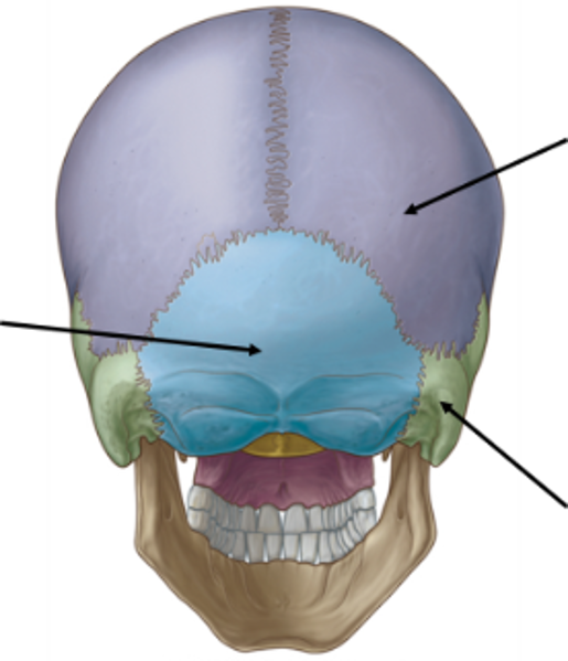
<p><span style="font-family: &quot;Times New Roman&quot;; line-height: normal; font-size: 7pt;"> </span><span>What are the three bones in this posterior view?</span></p>

What are the three bones in this posterior view?

knowt flashcard image
10
New cards
<p><span>What are the following 5 bones?</span></p>

What are the following 5 bones?

Vomer- forms the posteroinferior (back and lower) part of the nasal septum

<p><span>Vomer- forms the posteroinferior (back and lower) part of the nasal septum</span></p>
11
New cards

What are skull sutures and when do they fuse?

  • Sutures are a form of fibrous joint that are only found in the skull.

  • They are immovable in adults and fuse completely by roughly 20 years of age.


12
New cards
<p><span>Whare are the following sutures?</span></p>

Whare are the following sutures?

knowt flashcard image
13
New cards

Define a craniometric point and label the following.

Craniometric points are used radiographically to make cranial measurements, compare and describe the shape the cranium and note variations.

  • Pterion: H-shaped junction of the sphenoid, temporal, frontal and parietal bones. It sits in the temporal fossa on the side of the head, roughly 2 to 4 cm above the zygomatic arch and behind the temple. Weakest part of the skull and the middle meningeal artery (maxillary artery branch, therefore ECA) lies directly underneath. A fracture in this area can tear the artery and lead to a dangerous extradural hematoma

  • Bregma: Junction of the coronal and sagittal sutures. Site of the anterior fontanelle in neonates (closes at 18 months).

  • Lambda: Junction of the lambdoid and sagittal sutures.  Site of the posterior fontanelle in neonates (closes at 2 months)


14
New cards

Which type of haematoma forms if there is significant force/damage to the pterion?

The pterion is indicated by H-shaped formation of sutures. It is found at the point which unites the frontal, parietal, sphenoid and temporal bones. The pterion overlies the middle meningeal artery. A fracture in this area by cause injury to this vessel. If this vessel is injury and blood accumulates between the skull and the dura mater it results in an extradural haematoma.

<p><span>The pterion is indicated by H-shaped formation of sutures. It is found at the point which unites the frontal, parietal, sphenoid and temporal bones. The pterion overlies the middle meningeal artery. A fracture in this area by cause injury to this vessel. If this vessel is injury and blood accumulates between the skull and the dura mater it results in an extradural haematoma.</span></p>
15
New cards
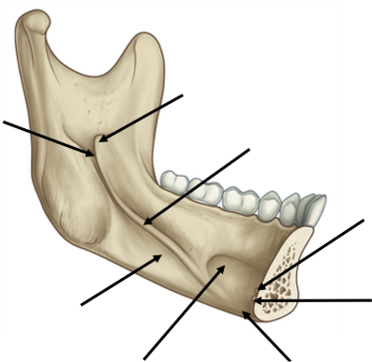
<p><span style="font-family: &quot;Times New Roman&quot;; line-height: normal; font-size: 7pt;"> </span><span>Label the following components of the mandible:</span></p>

Label the following components of the mandible:

knowt flashcard image
16
New cards

Muscle attachments and mandible:

Region

Muscles attached

Function

Body

6


Ramus (lateral)

1


Ramus (medial)

1


Coronoid process

1


Condylar neck

1


Region

Muscles attached

Function

Body

Mylohyoid, geniohyoid, genioglossus, digastric (ant.), platysma, buccinator

Oral floor support, tongue movement, jaw depression

Ramus (lateral)

Masseter

Elevation, clenching

Ramus (medial)

Medial pterygoid

Elevation, side-to-side movement

Coronoid process

Temporalis

Elevation, retraction

Condylar neck

Lateral pterygoid

Protrusion, depression


<table style="min-width: 75px;"><colgroup><col style="min-width: 25px;"><col style="min-width: 25px;"><col style="min-width: 25px;"></colgroup><tbody><tr><td colspan="1" rowspan="1" style="width: 106.1pt; border: 1pt solid black; padding: 0cm 5.4pt;"><p class="MsoNormal"><span>Region</span></p></td><td colspan="1" rowspan="1" style="width: 184.25pt; border-width: 1pt 1pt 1pt medium; border-style: solid solid solid none; border-color: black black black currentcolor; border-image: none; padding: 0cm 5.4pt;"><p class="MsoNormal"><span>Muscles attached</span></p></td><td colspan="1" rowspan="1" style="width: 160.45pt; border-width: 1pt 1pt 1pt medium; border-style: solid solid solid none; border-color: black black black currentcolor; border-image: none; padding: 0cm 5.4pt;"><p class="MsoNormal"><span>Function</span></p></td></tr><tr><td colspan="1" rowspan="1" style="width: 106.1pt; border-width: medium 1pt 1pt; border-style: none solid solid; border-color: currentcolor black black; border-image: none; padding: 0cm 5.4pt;"><p class="MsoNormal"><span>Body</span></p></td><td colspan="1" rowspan="1" style="width: 184.25pt; border-width: medium 1pt 1pt medium; border-style: none solid solid none; border-color: currentcolor black black currentcolor; padding: 0cm 5.4pt;"><p class="MsoNormal"><span>Mylohyoid, geniohyoid, genioglossus, digastric (ant.), platysma, buccinator</span></p></td><td colspan="1" rowspan="1" style="width: 160.45pt; border-width: medium 1pt 1pt medium; border-style: none solid solid none; border-color: currentcolor black black currentcolor; padding: 0cm 5.4pt;"><p class="MsoNormal"><span>Oral floor support, tongue movement, jaw depression</span></p></td></tr><tr><td colspan="1" rowspan="1" style="width: 106.1pt; border-width: medium 1pt 1pt; border-style: none solid solid; border-color: currentcolor black black; border-image: none; padding: 0cm 5.4pt;"><p class="MsoNormal"><span>Ramus (lateral)</span></p></td><td colspan="1" rowspan="1" style="width: 184.25pt; border-width: medium 1pt 1pt medium; border-style: none solid solid none; border-color: currentcolor black black currentcolor; padding: 0cm 5.4pt;"><p class="MsoNormal"><span>Masseter</span></p></td><td colspan="1" rowspan="1" style="width: 160.45pt; border-width: medium 1pt 1pt medium; border-style: none solid solid none; border-color: currentcolor black black currentcolor; padding: 0cm 5.4pt;"><p class="MsoNormal"><span>Elevation, clenching</span></p></td></tr><tr><td colspan="1" rowspan="1" style="width: 106.1pt; border-width: medium 1pt 1pt; border-style: none solid solid; border-color: currentcolor black black; border-image: none; padding: 0cm 5.4pt;"><p class="MsoNormal"><span>Ramus (medial)</span></p></td><td colspan="1" rowspan="1" style="width: 184.25pt; border-width: medium 1pt 1pt medium; border-style: none solid solid none; border-color: currentcolor black black currentcolor; padding: 0cm 5.4pt;"><p class="MsoNormal"><span>Medial pterygoid</span></p></td><td colspan="1" rowspan="1" style="width: 160.45pt; border-width: medium 1pt 1pt medium; border-style: none solid solid none; border-color: currentcolor black black currentcolor; padding: 0cm 5.4pt;"><p class="MsoNormal"><span>Elevation, side-to-side movement</span></p></td></tr><tr><td colspan="1" rowspan="1" style="width: 106.1pt; border-width: medium 1pt 1pt; border-style: none solid solid; border-color: currentcolor black black; border-image: none; padding: 0cm 5.4pt;"><p class="MsoNormal"><span>Coronoid process</span></p></td><td colspan="1" rowspan="1" style="width: 184.25pt; border-width: medium 1pt 1pt medium; border-style: none solid solid none; border-color: currentcolor black black currentcolor; padding: 0cm 5.4pt;"><p class="MsoNormal"><span>Temporalis</span></p></td><td colspan="1" rowspan="1" style="width: 160.45pt; border-width: medium 1pt 1pt medium; border-style: none solid solid none; border-color: currentcolor black black currentcolor; padding: 0cm 5.4pt;"><p class="MsoNormal"><span>Elevation, retraction</span></p></td></tr><tr><td colspan="1" rowspan="1" style="width: 106.1pt; border-width: medium 1pt 1pt; border-style: none solid solid; border-color: currentcolor black black; border-image: none; padding: 0cm 5.4pt;"><p class="MsoNormal"><span>Condylar neck</span></p></td><td colspan="1" rowspan="1" style="width: 184.25pt; border-width: medium 1pt 1pt medium; border-style: none solid solid none; border-color: currentcolor black black currentcolor; padding: 0cm 5.4pt;"><p class="MsoNormal"><span>Lateral pterygoid</span></p></td><td colspan="1" rowspan="1" style="width: 160.45pt; border-width: medium 1pt 1pt medium; border-style: none solid solid none; border-color: currentcolor black black currentcolor; padding: 0cm 5.4pt;"><p class="MsoNormal"><span>Protrusion, depression</span></p></td></tr></tbody></table><p></p>
17
New cards

What are the four muscles of mastication?

Elevators: Masseter, temporalis, medial pterygoid.

Depressors/Protruders: Lateral pterygoid (primary).

18
New cards

Attachment, function and innervation of Mylohyoid.

o    Mylohyoid – suprahyoid muscle: attaches along the mylohyoid line to the hyoid bone.

o   Forms floor of oral cavity (with geniohyoid and digastric and stylohyoid), elevates hyoid bone and floor of mouth, depresses mandible

o    Innervated by inferior alveolar nerve – CN5 V3 branch.

<p><span style="font-family: &quot;Courier New&quot;;">o</span><span style="font-family: &quot;Times New Roman&quot;; line-height: normal; font-size: 7pt;">&nbsp;&nbsp;&nbsp; </span><span>Mylohyoid – suprahyoid muscle: attaches along the mylohyoid line to the hyoid bone.</span></p><p class="MsoListParagraphCxSpMiddle"><span style="font-family: &quot;Courier New&quot;;">o</span><span style="font-family: &quot;Times New Roman&quot;; line-height: normal; font-size: 7pt;">&nbsp;&nbsp;&nbsp;</span><span>Forms floor of oral cavity (with geniohyoid and digastric and stylohyoid), elevates hyoid bone and floor of mouth, depresses mandible</span></p><p class="MsoListParagraphCxSpLast"><span style="font-family: &quot;Courier New&quot;;">o</span><span style="font-family: &quot;Times New Roman&quot;; line-height: normal; font-size: 7pt;">&nbsp;&nbsp;&nbsp; </span><span>Innervated by inferior alveolar nerve – CN5 V3 branch.</span></p>
19
New cards

Attachment, function and innervation of: Geniohyoid.

o    Geniohyoid – pair short suprahyoid muscle.

o    Attaches to the inferior genial tubercles à to hyoid bone.

o    Elevates and draws hyoid bone anteriorly; shortens the mouth floor; widens pharynx

o    Innervated: anterior ramus of spinal nerve C1 via hypoglossal nerve.

<p><span style="font-family: &quot;Courier New&quot;;">o</span><span style="font-family: &quot;Times New Roman&quot;; line-height: normal; font-size: 7pt;">&nbsp;&nbsp;&nbsp; </span><span>Geniohyoid – pair short suprahyoid muscle.</span></p><p class="MsoListParagraphCxSpMiddle"><span style="font-family: &quot;Courier New&quot;;">o</span><span style="font-family: &quot;Times New Roman&quot;; line-height: normal; font-size: 7pt;">&nbsp;&nbsp;&nbsp; </span><span>Attaches to the inferior genial tubercles à to hyoid bone.</span></p><p class="MsoListParagraphCxSpMiddle"><span style="font-family: &quot;Courier New&quot;;">o</span><span style="font-family: &quot;Times New Roman&quot;; line-height: normal; font-size: 7pt;">&nbsp;&nbsp;&nbsp; </span><span>Elevates and draws hyoid bone anteriorly; shortens the mouth floor; widens pharynx</span></p><p class="MsoListParagraphCxSpLast"><span style="font-family: &quot;Courier New&quot;;">o</span><span style="font-family: &quot;Times New Roman&quot;; line-height: normal; font-size: 7pt;">&nbsp;&nbsp;&nbsp; </span><span>Innervated: anterior ramus of spinal nerve C1 via hypoglossal nerve.</span></p>
20
New cards

Attachment, function and innervation of Genioglossus.

o   Genioglossus – 1 of 4 extrinsic tongue muscles.

o     Origin: Superior mental spine of mandible. Insertion: Entire length of dorsum of tongue/lingual aponeurosis, body of hyoid bone.

o    Function: Bilaterally: Depress and protrude tongue.  Unilaterally: Contralaterally deviate tongue

o    Innervation: hypoglossal nerve (CN XII)

<p><span style="font-family: &quot;Courier New&quot;;">o</span><span style="font-family: &quot;Times New Roman&quot;; line-height: normal; font-size: 7pt;">&nbsp;&nbsp;&nbsp;</span><span>Genioglossus – 1 of 4 extrinsic tongue muscles. </span></p><p class="MsoListParagraphCxSpMiddle"><span style="font-family: &quot;Courier New&quot;;">o</span><span style="font-family: &quot;Times New Roman&quot;; line-height: normal; font-size: 7pt;">&nbsp;&nbsp;&nbsp; </span><span>&nbsp;Origin: Superior mental spine of mandible. Insertion: Entire length of dorsum of tongue/lingual aponeurosis, body of hyoid bone.</span></p><p class="MsoListParagraphCxSpMiddle"><span style="font-family: &quot;Courier New&quot;;">o</span><span style="font-family: &quot;Times New Roman&quot;; line-height: normal; font-size: 7pt;">&nbsp;&nbsp;&nbsp; </span><span>Function: Bilaterally:&nbsp;Depress and protrude tongue.&nbsp; Unilaterally:&nbsp;Contralaterally deviate tongue</span></p><p class="MsoListParagraphCxSpLast"><span style="font-family: &quot;Courier New&quot;;">o</span><span style="font-family: &quot;Times New Roman&quot;; line-height: normal; font-size: 7pt;">&nbsp;&nbsp;&nbsp; </span><span>Innervation:&nbsp;hypoglossal nerve (CN&nbsp;XII)</span></p>
21
New cards

Attachment, function and innervation of anterior belly of digastric.

o    Digastric is a paired suprahyoid muscle.

o   Its anterior belly originates at the digastric fossa and attaches to the body of hyoid. (The posterior belly originates at the mastoid notch of the temporal bone and attaches to the body of hyoid).

o    Function: depresses mandible. Elevates hyoid bone during chewing, swallowing.

o    Innervation: Anterior belly: mylohyoid nerve (of inferior alveolar nerve) (CN V3). Posterior belly: digastric branch of facial nerve (CN VII)

<p><span style="font-family: &quot;Courier New&quot;;">o</span><span style="font-family: &quot;Times New Roman&quot;; line-height: normal; font-size: 7pt;">&nbsp;&nbsp;&nbsp; </span><span>Digastric is a paired suprahyoid muscle.</span></p><p class="MsoListParagraphCxSpMiddle"><span style="font-family: &quot;Courier New&quot;;">o</span><span style="font-family: &quot;Times New Roman&quot;; line-height: normal; font-size: 7pt;">&nbsp;&nbsp;&nbsp;</span><span>Its anterior belly originates at the digastric fossa and attaches to the body of hyoid. (The posterior belly originates at the mastoid notch of the temporal bone and attaches to the body of hyoid). </span></p><p class="MsoListParagraphCxSpMiddle"><span style="font-family: &quot;Courier New&quot;;">o</span><span style="font-family: &quot;Times New Roman&quot;; line-height: normal; font-size: 7pt;">&nbsp;&nbsp;&nbsp; </span><span>Function: depresses mandible. Elevates hyoid bone during chewing, swallowing.</span></p><p class="MsoListParagraphCxSpLast"><span style="font-family: &quot;Courier New&quot;;">o</span><span style="font-family: &quot;Times New Roman&quot;; line-height: normal; font-size: 7pt;">&nbsp;&nbsp;&nbsp; </span><span>Innervation: Anterior belly: mylohyoid nerve (of inferior alveolar nerve) (CN V3). Posterior belly: digastric branch of facial nerve (CN VII)</span></p>
22
New cards

Attachment, function and innervation of Platysma.

o    Platysma – thin sheet-like muscle that lies superficially within anterior neck.

o    Origin: covers pec muscles and ascends superomedially to attached to the mandible, skin and lower face (lower lip, modiolus, orbicularis oris muscle).

o    Function: Depresses mandible and angle of mouth, tenses skin of lower face and anterior neck

o   Innervation: Cervical branch of facial nerve (CN VII)


<p><span style="font-family: &quot;Courier New&quot;;">o</span><span style="font-family: &quot;Times New Roman&quot;; line-height: normal; font-size: 7pt;">&nbsp;&nbsp;&nbsp; </span><span>Platysma – thin sheet-like muscle that lies superficially within anterior neck.</span></p><p class="MsoListParagraphCxSpMiddle"><span style="font-family: &quot;Courier New&quot;;">o</span><span style="font-family: &quot;Times New Roman&quot;; line-height: normal; font-size: 7pt;">&nbsp;&nbsp;&nbsp; </span><span>Origin: covers pec muscles and ascends superomedially to attached to the mandible, skin and lower face (lower lip, modiolus, orbicularis oris muscle).</span></p><p class="MsoListParagraphCxSpMiddle"><span style="font-family: &quot;Courier New&quot;;">o</span><span style="font-family: &quot;Times New Roman&quot;; line-height: normal; font-size: 7pt;">&nbsp;&nbsp;&nbsp; </span><span>Function: Depresses mandible and angle of mouth, tenses skin of lower face and anterior neck</span></p><p class="MsoListParagraphCxSpLast"><span style="font-family: &quot;Courier New&quot;;">o</span><span style="font-family: &quot;Times New Roman&quot;; line-height: normal; font-size: 7pt;">&nbsp;&nbsp;&nbsp;</span><span>Innervation: Cervical branch of facial nerve (CN VII)</span></p><p></p>
23
New cards

Attachment, function and innervation of buccinator.

o    Buccinator main muscle of the cheek.

o    It compresses the cheek against the molar teeth, which is important to keep the food bolus central in the oral cavity, and to prevent the cheeks from being bitten during mastication.

o    Insertion modiolus à to Superior part: Alveolar process of maxilla. Inferior part: Alveolar part of mandible Posterior part: Pterygomandibular raphe, buccinator crest of mandible

o    Innervation: buccal branch of CN7.

<p><span style="font-family: &quot;Courier New&quot;;">o</span><span style="font-family: &quot;Times New Roman&quot;; line-height: normal; font-size: 7pt;">&nbsp;&nbsp;&nbsp; </span><span>Buccinator main muscle of the cheek.</span></p><p class="MsoListParagraphCxSpMiddle"><span style="font-family: &quot;Courier New&quot;;">o</span><span style="font-family: &quot;Times New Roman&quot;; line-height: normal; font-size: 7pt;">&nbsp;&nbsp;&nbsp; </span><span>It compresses the cheek against the molar teeth, which is important to keep the food bolus central in the oral cavity, and to prevent the cheeks from being bitten during mastication.</span></p><p class="MsoListParagraphCxSpMiddle"><span style="font-family: &quot;Courier New&quot;;">o</span><span style="font-family: &quot;Times New Roman&quot;; line-height: normal; font-size: 7pt;">&nbsp;&nbsp;&nbsp; </span><span>Insertion modiolus à to Superior part:&nbsp;Alveolar process of maxilla. Inferior part:&nbsp;Alveolar part of mandible Posterior part:&nbsp;Pterygomandibular raphe, buccinator crest of mandible</span></p><p class="MsoListParagraphCxSpLast"><span style="font-family: &quot;Courier New&quot;;">o</span><span style="font-family: &quot;Times New Roman&quot;; line-height: normal; font-size: 7pt;">&nbsp;&nbsp;&nbsp; </span><span>Innervation: buccal branch of CN7.</span></p>
24
New cards

Attachment, function and innervation of superior pharyngeal constrictor.

o    Superior pharyngeal constrictor – one of three pharyngeal constructor muscles important in facilitating swallow.

o    Attached to pharyngeal tubercle of the occipital bone à then has 4 parts/attachments Pterygoid hamulus, pterygomandibular raphe, posterior end of mylohyoid line of mandible, the tongue.

o    Innervation: Branches of pharyngeal plexus (CN X)

<p><span style="font-family: &quot;Courier New&quot;;">o</span><span style="font-family: &quot;Times New Roman&quot;; line-height: normal; font-size: 7pt;">&nbsp;&nbsp;&nbsp; </span><span>Superior pharyngeal constrictor – one of three pharyngeal constructor muscles important in facilitating swallow.</span></p><p class="MsoListParagraphCxSpMiddle"><span style="font-family: &quot;Courier New&quot;;">o</span><span style="font-family: &quot;Times New Roman&quot;; line-height: normal; font-size: 7pt;">&nbsp;&nbsp;&nbsp; </span><span>Attached to pharyngeal tubercle of the occipital bone à then has 4 parts/attachments Pterygoid hamulus, pterygomandibular raphe, posterior end of mylohyoid line of mandible, the tongue.</span></p><p class="MsoListParagraphCxSpLast"><span style="font-family: &quot;Courier New&quot;;">o</span><span style="font-family: &quot;Times New Roman&quot;; line-height: normal; font-size: 7pt;">&nbsp;&nbsp;&nbsp; </span><span>Innervation: Branches of pharyngeal plexus (CN X)</span></p>
25
New cards

Attachment, function and innervation of Masseter

o Masseter – paired muscle of mastication.

o Origin from zygomatic arch à attaches to the lateral surface of the ramus and angle.

o  Function: Elevates and protrudes mandible

o  Innervation: masseteric nerve of CN5 V3.

26
New cards

Attachment, function and innervation of temporalis

o    Temporalis – paired thin fan shaped muscle of mastication.

o   Originates from Temporal fossa (up to inferior temporal line), Temporal fascia à inserts on the coronoid process and anterior border of the ramus.

o    Function: Anterior fibres: Elevates mandible. Posterior part: Retracts mandible

o    Innervation: deep temporal branches (of mandibular nerve (CN V3))

<p><span style="font-family: &quot;Courier New&quot;;">o</span><span style="font-family: &quot;Times New Roman&quot;; line-height: normal; font-size: 7pt;">&nbsp;&nbsp;&nbsp; </span><span>Temporalis – paired thin fan shaped muscle of mastication.</span></p><p class="MsoListParagraphCxSpMiddle"><span style="font-family: &quot;Courier New&quot;;">o</span><span style="font-family: &quot;Times New Roman&quot;; line-height: normal; font-size: 7pt;">&nbsp;&nbsp;&nbsp;</span><span>Originates from Temporal fossa (up to inferior temporal line), Temporal fascia à inserts on the coronoid process and anterior border of the ramus. </span></p><p class="MsoListParagraphCxSpMiddle"><span style="font-family: &quot;Courier New&quot;;">o</span><span style="font-family: &quot;Times New Roman&quot;; line-height: normal; font-size: 7pt;">&nbsp;&nbsp;&nbsp; </span><span>Function:</span> <span>Anterior fibres: Elevates mandible. Posterior part: Retracts mandible</span></p><p class="MsoListParagraphCxSpLast"><span style="font-family: &quot;Courier New&quot;;">o</span><span style="font-family: &quot;Times New Roman&quot;; line-height: normal; font-size: 7pt;">&nbsp;&nbsp;&nbsp; </span><span>Innervation: deep temporal branches (of mandibular nerve (CN V3))</span></p>
27
New cards

Attachment, function and innervation of pterygoids

o    Medial and lateral pterygoids are 2 of the 4 muscles of mastication.

o    Innervated by branches of the mandibular division of the trigeminal nerve (CN V3),

o    Medial pterygoid – attaches to the medial surface of the ramus and angle.

o    Lateral pterygoid – attaches to the neck of the condyle.

o    Function: Lateral pterygoid: Protrusion, depression, medial movement of mandible. Medial pterygoid: Protrusion, elevation, medial movement of mandible

<p><span style="font-family: &quot;Courier New&quot;;">o</span><span style="font-family: &quot;Times New Roman&quot;; line-height: normal; font-size: 7pt;">&nbsp;&nbsp;&nbsp; </span><span>Medial and lateral pterygoids are 2 of the 4 muscles of mastication.</span></p><p class="MsoListParagraphCxSpMiddle"><span style="font-family: &quot;Courier New&quot;;">o</span><span style="font-family: &quot;Times New Roman&quot;; line-height: normal; font-size: 7pt;">&nbsp;&nbsp;&nbsp; </span><span>Innervated by branches of the mandibular division of the trigeminal nerve (CN V3),</span></p><p class="MsoListParagraphCxSpMiddle"><span style="font-family: &quot;Courier New&quot;;">o</span><span style="font-family: &quot;Times New Roman&quot;; line-height: normal; font-size: 7pt;">&nbsp;&nbsp;&nbsp; </span><span>Medial pterygoid – attaches to the medial surface of the ramus and angle.</span></p><p class="MsoListParagraphCxSpMiddle"><span style="font-family: &quot;Courier New&quot;;">o</span><span style="font-family: &quot;Times New Roman&quot;; line-height: normal; font-size: 7pt;">&nbsp;&nbsp;&nbsp; </span><span>Lateral pterygoid – attaches to the neck of the condyle.</span></p><p class="MsoListParagraphCxSpLast"><span style="font-family: &quot;Courier New&quot;;">o</span><span style="font-family: &quot;Times New Roman&quot;; line-height: normal; font-size: 7pt;">&nbsp;&nbsp;&nbsp; </span><span>Function: Lateral pterygoid: Protrusion, depression, <strong>medial movement</strong> of mandible. Medial pterygoid: Protrusion, elevation, <strong>medial movement</strong> of mandible</span></p>
28
New cards

·         What are the Suprahyoid Muscles and what is their function?

o The suprahyoid muscles primarily elevate the hyoid bone and floor of the mouth, assist in swallowing and speaking, and can depress the mandible to help open the mouth.

o Mylohyoid, geniohyoid, digastric and stylohyoid.

<p><span style="font-family: &quot;Courier New&quot;;">o </span><span>The suprahyoid muscles primarily elevate the hyoid bone and floor of the mouth, assist in swallowing and speaking, and can depress the mandible to help open the mouth.</span></p><p class="MsoListParagraphCxSpLast"><span style="font-family: &quot;Courier New&quot;;">o</span><span style="font-family: &quot;Times New Roman&quot;; line-height: normal; font-size: 7pt;"> </span><span>Mylohyoid, geniohyoid, digastric and stylohyoid.</span></p>
29
New cards
<p><span>Label the following 6 structures of the external view of the mandible</span></p>

Label the following 6 structures of the external view of the mandible

knowt flashcard image
30
New cards
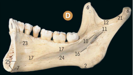
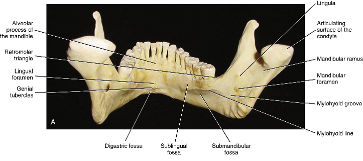
<p><span>Label the following 8 structures of this internal view of a mandible and<strong> what is the function of each?</strong></span></p>

Label the following 8 structures of this internal view of a mandible and what is the function of each?

  1. Mandibular foramen: just inferior to mandibular notch. It is an opening located on the inner (medial) surface of the ramus of the mandible. Contains inferior alveolar nerve (V3 of CN5), artery (branch maxillary artery) and nerve.

  2. Lingula: a sharp, tongue-shaped or triangular bony projection located on the inner (medial) surface of the mandibular ramus, immediately anterior/superior to the mandibular foramen.

  3. Mylohyoid line: a prominent, oblique bony ridge located on the internal (medial) surface of the mandible (from symphysis to 3rd molar). Attachments: mylohyoid muscle, superior pharyngeal constrictor muscle and pterygomandibular raphe.

  4. Submandibular fossa (superficial portion of the submandibular salivary gland.)

  5. Sublingual fossa: contains sublingual salivary gland).

  6. Digastric fossa: attachment site for the origin of the anterior belly of the digastric muscle.

  7. Superior genial tubercles: attachment of genioglossus muscle --> protrudes, elevates, and retracts parts of the tongue,

  8. Inferior genial tubercles:  attachment for geniohyoid muscle--> elevates the hyoid bone, helps pull it forward, and assists in lowering the epiglottis during swallowing to protect and close the larynx


<ol><li><p><span>Mandibular foramen: just inferior to mandibular notch. It is an opening located on the inner (medial) surface of the ramus of the mandible. Contains inferior alveolar nerve (V3 of CN5), artery (branch maxillary artery) and nerve.</span></p></li><li><p><span>Lingula: a sharp, tongue-shaped or triangular bony projection located on the inner (medial) surface of the mandibular ramus, immediately anterior/superior to the mandibular foramen.</span></p></li><li><p><span>Mylohyoid line: a prominent, oblique bony ridge located on the internal (medial) surface of the mandible (from symphysis to 3rd molar). Attachments: mylohyoid muscle, superior pharyngeal constrictor muscle and pterygomandibular raphe.</span></p></li><li><p><span>Submandibular fossa (superficial portion of the submandibular salivary gland.)</span></p></li><li><p><span>Sublingual fossa: contains sublingual salivary gland).</span></p></li><li><p><span>Digastric fossa: attachment site for the origin of the anterior belly of the digastric muscle.</span></p></li><li><p><span>Superior genial tubercles: attachment of genioglossus muscle --&gt; protrudes, elevates, and retracts parts of the tongue,</span></p></li><li><p><span>Inferior genial tubercles:&nbsp; attachment for geniohyoid muscle--&gt; elevates the hyoid bone, helps pull it forward, and assists in lowering the epiglottis during swallowing to protect and close the larynx</span></p></li></ol><p></p>
31
New cards
<p><span>Label: Frontal bone, Nasal bone, Maxilla, Zygomatic bone, Mandible:</span></p>

Label: Frontal bone, Nasal bone, Maxilla, Zygomatic bone, Mandible:

Frontal bone (3)

Nasal bone (18)

Maxilla (14)

Zygomatic bone (26)

Mandible (2)

<p><span>Frontal bone (3)</span></p><p class="MsoListParagraphCxSpMiddle"><span>Nasal bone (18)</span></p><p class="MsoListParagraphCxSpMiddle"><span>Maxilla (14)</span></p><p class="MsoListParagraphCxSpMiddle"><span>Zygomatic bone (26)</span></p><p class="MsoListParagraphCxSpLast"><span>Mandible (2)</span></p>
32
New cards
<p><span style="font-family: &quot;Times New Roman&quot;; line-height: normal; font-size: 7pt;"> </span><span>Identify the number that marks the following structures on the superior view of the skull: Frontal bone. Parietal bone. Coronal suture. Sagittal suture. Bregma. Foramina for emissary veins.</span></p>

Identify the number that marks the following structures on the superior view of the skull: Frontal bone. Parietal bone. Coronal suture. Sagittal suture. Bregma. Foramina for emissary veins.

  • Frontal bone (3)

  • Parietal bone (7)

  • Coronal suture (2)

  • Sagittal suture (10)

  • Bregma (1)

  • Foramina for emissary veins (9) = Emissary veins are valveless blood vessels that pass through small openings (foramina) in the skull. They connect the blood vessels outside the skull (such as the scalp and face) to the dural venous sinuses (the large blood channels inside the skull that drain the brain)


<ul><li><p><span>Frontal bone (3)</span></p></li><li><p><span>Parietal bone (7)</span></p></li><li><p><span>Coronal suture (2)</span></p></li><li><p><span>Sagittal suture (10)</span></p></li><li><p><span>Bregma (1)</span></p></li><li><p><span style="font-family: &quot;Times New Roman&quot;; line-height: normal; font-size: 7pt;"> </span><span>Foramina for emissary veins (9) = Emissary veins are valveless blood vessels that pass through small openings (foramina) in the skull. They connect the blood vessels outside the skull (such as the scalp and face) to the dural venous sinuses (the large blood channels inside the skull that drain the brain)</span></p></li></ul><p></p>
33
New cards
<p><span>Identify the number that marks the following structures on the posterior view of the skull: Lambda . Lambdoid suture. Occipital bone.</span></p>

Identify the number that marks the following structures on the posterior view of the skull: Lambda . Lambdoid suture. Occipital bone.

  • Lambda (3)

  • Lambdoid suture (4)

  • Occipital bone (9)


<ul><li><p><span style="font-family: &quot;Times New Roman&quot;; line-height: normal; font-size: 7pt;"> </span>Lambda (3)</p></li><li><p><span>Lambdoid suture (4)</span></p></li><li><p><span>Occipital bone (9)</span></p></li></ul><p></p>
34
New cards
<p><span><strong>Identify the number that marks the following structures on the lateral view of the skull: </strong>Temporal bone. Greater wing of sphenoid. Zygomatic bone. Zygomatic arch. Superior temporal line. Temporal fossa.</span></p>

Identify the number that marks the following structures on the lateral view of the skull: Temporal bone. Greater wing of sphenoid. Zygomatic bone. Zygomatic arch. Superior temporal line. Temporal fossa.

  • Temporal bone (31)

  • Greater wing of sphenoid (14)

  • Zygomatic bone (36)

  • Zygomatic arch (35)

  • Superior temporal lines (33)

  • Temporal fossa (superior temporal line to the zygomatic arch)
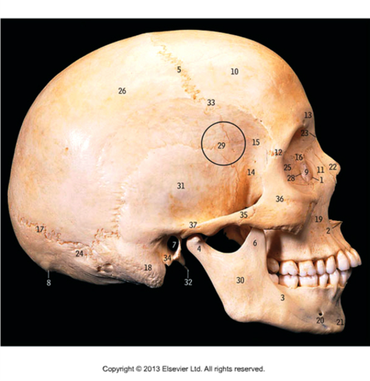

<ul><li><p><span>Temporal bone (31)</span></p></li><li><p><span>Greater wing of sphenoid (14)</span></p></li><li><p><span>Zygomatic bone (36)</span></p></li><li><p><span>Zygomatic arch (35)</span></p></li><li><p><span>Superior temporal lines (33)</span></p></li><li><p><span style="font-family: &quot;Times New Roman&quot;; line-height: normal; font-size: 7pt;"> </span><span>Temporal fossa (superior temporal line to the zygomatic arch)</span></p></li></ul><p></p>
35
New cards
<p><span style="font-family: &quot;Times New Roman&quot;; line-height: normal; font-size: 7pt;"> </span><span><strong>I</strong>dentify the number that marks the following structures on the mandible Body. Ramus. Angle. Condylar process. Coronoid process. Mandibular notch . Mental foramen.</span></p>

Identify the number that marks the following structures on the mandible Body. Ramus. Angle. Condylar process. Coronoid process. Mandibular notch . Mental foramen.

  • Body (5)

  • Ramus (22)

  • Angle (2)

  • Condylar process (8)

  • Coronoid process (6)

  • Mandibular notch (12)

  • Mental foramen (13)


<ul><li><p><span style="font-family: &quot;Times New Roman&quot;; line-height: normal; font-size: 7pt;"> </span><span>Body (5)</span></p></li><li><p><span>Ramus (22)</span></p></li><li><p><span>Angle (2)</span></p></li><li><p><span>Condylar process (8)</span></p></li><li><p><span>Coronoid process (6)</span></p></li><li><p><span style="font-family: &quot;Times New Roman&quot;; line-height: normal; font-size: 7pt;"> </span><span>Mandibular notch (12)</span></p></li><li><p><span style="font-family: &quot;Times New Roman&quot;; line-height: normal; font-size: 7pt;"> </span><span>Mental foramen (13)</span></p></li></ul><p></p>
36
New cards
<p><span>Identify the number that marks the following structures on the mandible: Mandibular foramen . Lingula. Mylohyoid line. Submandibular fossa. Sublingual fossa. Digastric fossa. Superior and inferior genial tubercles (mental spines).</span></p>

Identify the number that marks the following structures on the mandible: Mandibular foramen . Lingula. Mylohyoid line. Submandibular fossa. Sublingual fossa. Digastric fossa. Superior and inferior genial tubercles (mental spines).

  • Mandibular foramen (11)

  • Lingula (10)

  • Mylohyoid line (17)

  • Submandibular fossa (24)

  • Sublingual fossa (23)

  • Digastric fossa (7)

  • Superior and inferior genial tubercles (mental spines) (25)
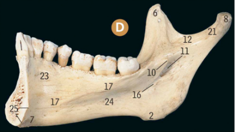

<ul><li><p><span>Mandibular foramen (11)</span></p></li><li><p><span>Lingula (10)</span></p></li><li><p><span>Mylohyoid line (17)</span></p></li><li><p><span>Submandibular fossa (24)</span></p></li><li><p><span>Sublingual fossa (23)</span></p></li><li><p><span>Digastric fossa (7)</span></p></li><li><p>Superior and inferior genial tubercles (mental spines) (25)</p></li></ul><p></p>
37
New cards
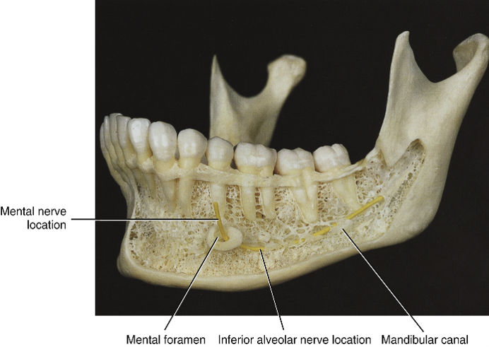
<p><span>·</span><span style="font-family: &quot;Times New Roman&quot;; line-height: normal; font-size: 7pt;">&nbsp;&nbsp;&nbsp;&nbsp;&nbsp;&nbsp;</span><br><span>Label the following structures &nbsp;</span></p>

·      
Label the following structures  

knowt flashcard image
38
New cards
<p>Label the structures </p>

Label the structures

knowt flashcard image
39
New cards
<p>Label the structures </p>

Label the structures

knowt flashcard image
40
New cards
<p>Label the structures </p>

Label the structures

knowt flashcard image