neural communication
 Call Kai
 Learn
 Practice Test
 Spaced Repetition
 Match
 Flashcards
 Knowt Play
1/32

There's no tags or description
Looks like no tags are added yet.
33 Terms
what are sensory receptors, location
Sensory receptors are specialised cells that detect specific types of stimuli or changes in the environment.
located in sensory organs, for example thermoreceptors in the skin.
Sensory receptors are specific to one type of stimulus; for example, rod and cone cells in the eye respond to changes in light. This ensures the brain can accurately identify and interpret different types of stimuli.
Sensory receptors act as transducers, converting a stimulus into an electrical signal (a nerve impulse). Neurones transmit electrical impulses to the CNS (central nervous system) where the brain coordinates a response via an effector (e.g., muscle).
1
Sensory receptors act as … and transmit impulse to…
Sensory receptors act as transducers = converting a stimulus into an electrical signal (a nerve impulse).
Neurones transmit electrical impulses to the CNS (central nervous system) where the brain coordinates a response via an effector (e.g., muscle).
1
explain pacinain corpuscles function, anatomy,(basic) physiology
Pacinian Corpuscles are mechanoreceptors (pressure receptors) that detect pressure changes in the skin.
They are made up of rings of connective tissue and gel wrapped around the end of a sensory neurone.
Changes in pressure deform the rings of connective tissue, which compress the nerve ending, generating a nerve impulse.

The mechanism of action of a Pacinian corpuscle is:
Pressure applied to the skin deforms the layers of gel and connective tissue surrounding the sensory neurone ending.
This deformation stretches the membrane of the sensory neurone, causing stretch–mediatedsodium ion channels to open.
Sodium ions diffuse into the neurone, causing depolarisation(Depolarisation means the inside of the neurone becomes less negatively charged compared with the outside.) of the membrane and production of a generator potential.
If the generator potential reaches the threshold, voltage–gated sodium channels open.
An action potential is generated and travels along the sensory neurone to the central nervous system.
nerve cells called… their phisioslgy is
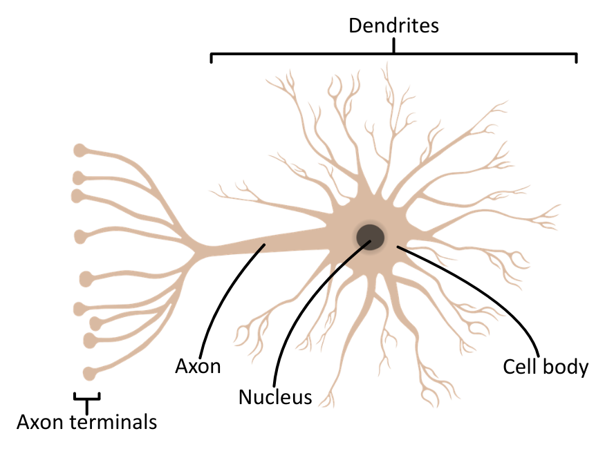
Nerve cells are called neurones.
They have a cell body,
dendrites which carry nerve impulses towards the cell body,
axon that carries nerve impulses away from the cell body.
Nerves are bundles of axons.

anatomy of sensory neuron
carry impulses from receptors→ central nervous system, where they synapse with relay or motor neurones.
long dendrons that carry an impulse from the receptor to the cell body.
short axon transmits an impulse to the CNS. the order is sensory neuron, relay in CNS,motor neuron,effector, response
order is stimulus,

anatomy of relay neron
Relay neurones connect sensory neurones to motor neurones within the CNS.
They have short dendrites that carry nerve impulses towards the cell body
short axon that carries nerve impulses away from the cell body.
Their short length allows rapid transmission of nerve impulses over the short distances within the CNS
anatomy of motor neutron
Motor neurones carry impulses from relay neurones (and sometimes directly from sensory neurones) to effectors such as muscles or glands.
They have many short dendrites that carry nerve impulses towards the cell body.
They have a single long axon that carries nerve impulses away from the cell body in the CNS to an effector.

describe function os Schwann cells
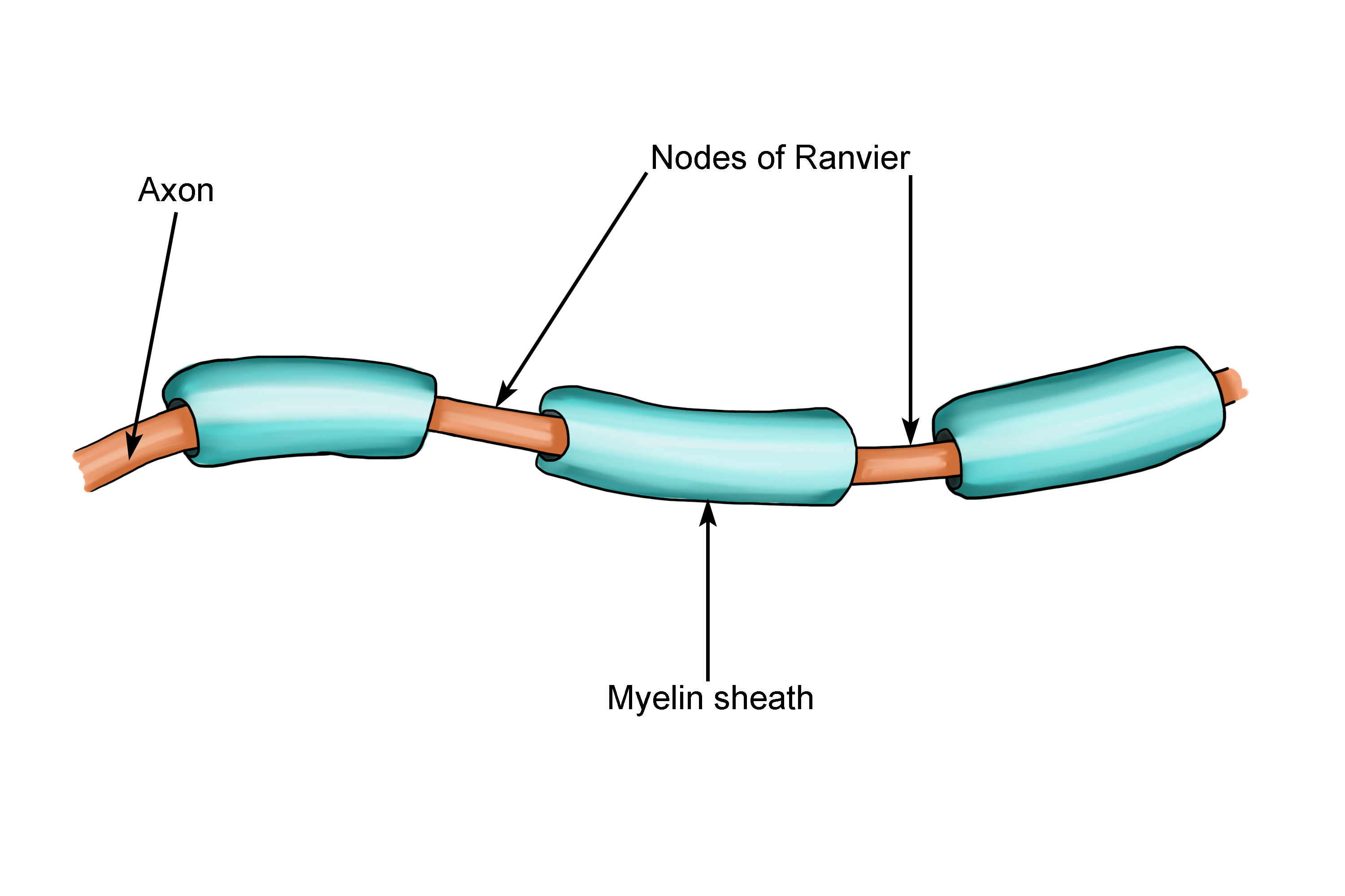
The myelin sheath around a neurone’s axon is made up of Schwann cell membranes. They insulate the axon and speed up impulse transmission.
Myelin (layers of plasma membrane) is an electrical insulator.
Myelinated neurones conduct up to 100× faster than neurones that are unmyelinated.
what are nodes of ranvier
Nodes of Ranvier are gaps between the Schwann cells making up the myelin sheath.
Electrical impulses jump between nodes, which speeds up signal transmission. This is known as saltatory conduction.
what is saltatory conduction
Nodes of Ranvier are gaps between the Schwann cells making up the myelin sheath.
Electrical impulses jump between nodes, which speeds up signal transmission. This is known as saltatory conduction.
how do electrical impulses move along a neurone
Electrical impulses move along a neurone by changing the voltage across its membrane.
The membrane is in one of two possible states, either resting potential or action potential.
0
describe resting potential, what occurs In it
When a neurone is not transmitting an action potential, it is at its resting potential.
Neurones maintain a resting potential of approximately -70 mv
In this state, the membrane is polarised.
The outside of the neurone membrane is more positively charged than the inside of the membrane.
The outside of the neurone membrane is more positively charged than the inside of the membrane. explain why
Na ion channels are gated, and others are always open across the cell membrane of neurones.
The sodium–potassium pump uses ATP to move 3 Na ions out for every 2 K ions pumped in,
resulting in more Na+ outside the cell and more inside the cytoplasm.
K ions also diffuse out of the neurone down the electrochemical gradient,
but Na ions cannot diffuse back in because the gated sodium ion channels are closed.
More positively charged ions are found outside the axon, so the inside of the neurone is negativelycharged relative to the outside at resting potential.
they only open when Stimulus — a receptor detects something. Sodium channels open — Na⁺ ions diffuse into the neurone.Depolarisation — the inside becomes less negative.Threshold reached — if the membrane reaches threshold, voltage-gated sodium channels open.Action potential — more Na⁺ enters rapidly, causing the inside to become positive relative to the outside.
what is depolarisation
Depolarisation is when sodium ions (Na⁺) diffuse into a neurone, making the inside of the membrane less negative and eventually positive relative to the outside.
The stimulus changes the shape of voltage-gated ion channels, causing the membrane potential to rise to
40 mv
how is depolarisation achieved
1. A stimulus causes sodium channels to open.
2. (Na⁺) diffuse into the axon down their electrochemical gradient.
3. This causes more voltage-gated sodium channels to open.
This is positive feedback because sodium entering causes even more sodium channels to open.
4. Depolarisation happens.
The inside of the axon becomes positive relative to the outside, and the membrane potential reaches about +30 mV.
But the action potential cannot stay positive forever! The neurone needs to change its electrical charge back towards its resting state.
5. Sodium channels close/inactivate and potassium channels open.
Potassium ions (K⁺) diffuse out of the axon, making the inside negative again. This is called repolarisation the membrane potential moves back towards its resting value of about −70 mV.
potassium channels don't close immediately. Even after the membrane potential has returned to around −70 mV, some K⁺ ions continue to leave because the channels are still open.
More positive K⁺ ions leave the axon.
The inside becomes even more negative than its normal resting potential.
. This is called hyperpolarisation.
6. The neurone returns to its resting potential. at last
Potassium channels close,
and the sodium–potassium pump uses ATP to move 3 Na⁺ out and 2 K⁺ into the axon, maintaining the ion concentration gradients needed for future action potentials.
what is hyperpolarisation, why does I happen
voltage gates Na Chanels open depolarise neuro causing positive charge in it.
action potential must return so sodium Chanels close potassium pumps pump potassium out depolarising neuron
2. potassium channels don't close immediately. Even after the membrane potential has returned to around −70 mV, some K⁺ ions continue to leave because the channels are still open.
More positive K⁺ ions leave the axon.The inside becomes even more negative than its normal resting potential.. This is called hyperpolarisation
It helps prevent the neurone from immediately firing another action potential.
It contributes to the refractory period, when the neurone is recovering after an action potential.
what is action potential triggered by, where, how are. they propagated across neuron
An action potential in the sensory receptor is triggered by a stimulus.
A wave of depolarisation spreads along the axon.
Sodium ions diffuse along the axon, triggering depolarisation in the next section. This is how action potentials are propagated along a neurone.
in detail describe the action potentials route
1. Resting potential
Na⁺ (sodium) ions are mainly outside the axon, and K⁺ (potassium) ions are mainly inside. The inside is negatively charged compared with the outside.
2. Depolarisation
Sodium channels open, so Na⁺ enters the axon. The inside becomes less negative and then positive.
3. The impulse moves along
The depolarised region triggers voltage-gated sodium channels further along the axon to open. More Na⁺ enters, so the action potential moves along the axon. This is positive feedback.
4. Repolarisation
Behind the impulse, sodium channels inactivate and potassium channels open. K⁺ leaves the axon, making the inside negative again.
The neurone is now ready for a new stimulus” means that the membrane returns towards its resting potential and the neurone can respond to another stimulus after the refractory period.
refractory period is when…
After an action potential, voltage–gated sodium channels remain closed for a short time so there can’t be another action potential. This is called the refractory period.
Action potentials travel in one direction and cannot overlap because of the refractory period.
1
In a myelinated neurone, depolarisation occurs at…
In a myelinated neurone, depolarisation of an axon membrane occurs at the nodes of Ranvier.
what Is saltatory conduction, what does it allow for
The action potentials jump between nodes; this is known as saltatory conduction.
theyre called also action potential jumps
This is more efficient and speeds up transmission as it is faster than a wave of depolarisation going along the whole length of the axon.
also Repolarisation needs ATP, so this also reduces the amount of ATP that is required.
what 3 factors affect speed of impulse
There are several factors that can affect the speed of an impulse, including:
Axon diameter: the larger the diameter, the faster the transmission, as there is less resistance to the movement of ions in the axon.
Temperature: the higher the temperature, the faster the transmission, as the rate of diffusion is increased.
Myelin sheath: when present, transmission is faster owing to saltatory conduction.
when is an action potential triggered bye a stimulus, explain. the size
If the threshold value is reached by a stimulus then an action potential is triggered. If this value is not met, there is no action potential.
The size of an action potential is always the same, regardless of the stimulus size.
This is known as the all-or-nothing principle, there is either an action potential or not.
A more intense stimulus results in more frequent action potentials, not bigger ones.
Threshold value in neurones can vary, so some are easier to depolarise than others.
1
define synapse
A synapse is a junction between two neurones or a neurone and an effector. An impulse is transmitted across a synapse by chemical neurotransmitters.
Cholinergic synapses are made up of several parts, including:
Synaptic cleft – small gap (~20–30 ) between neurones.
Pre synaptic neurone – ends in a presynaptic bulb / knob that contains synaptic vesicles filled with neurotransmitter (e.g., acetylcholine, GABA).
Post synaptic neurone – with specific receptor proteins for neurotransmitters.
Cholinergic synaps definition
a synapse that uses a neurotransmitter called acetylcholine (ACh) to transmit the signal.

what is a neurotramitter, how does it travel
A neurotransmitter is a type of chemical messenger produced by a neurone and released from the presynaptic knob when a nerve impulse arrives.
At a synapse, the electrical impulse is converted into a chemical signal carried across the synaptic cleft by neurotransmitters.
Acetylcholine is an example of an excitatory neurotransmitter.
GABA is an example of an inhibitory transmitter.
Impulse transmission at a synapse happens in the following way
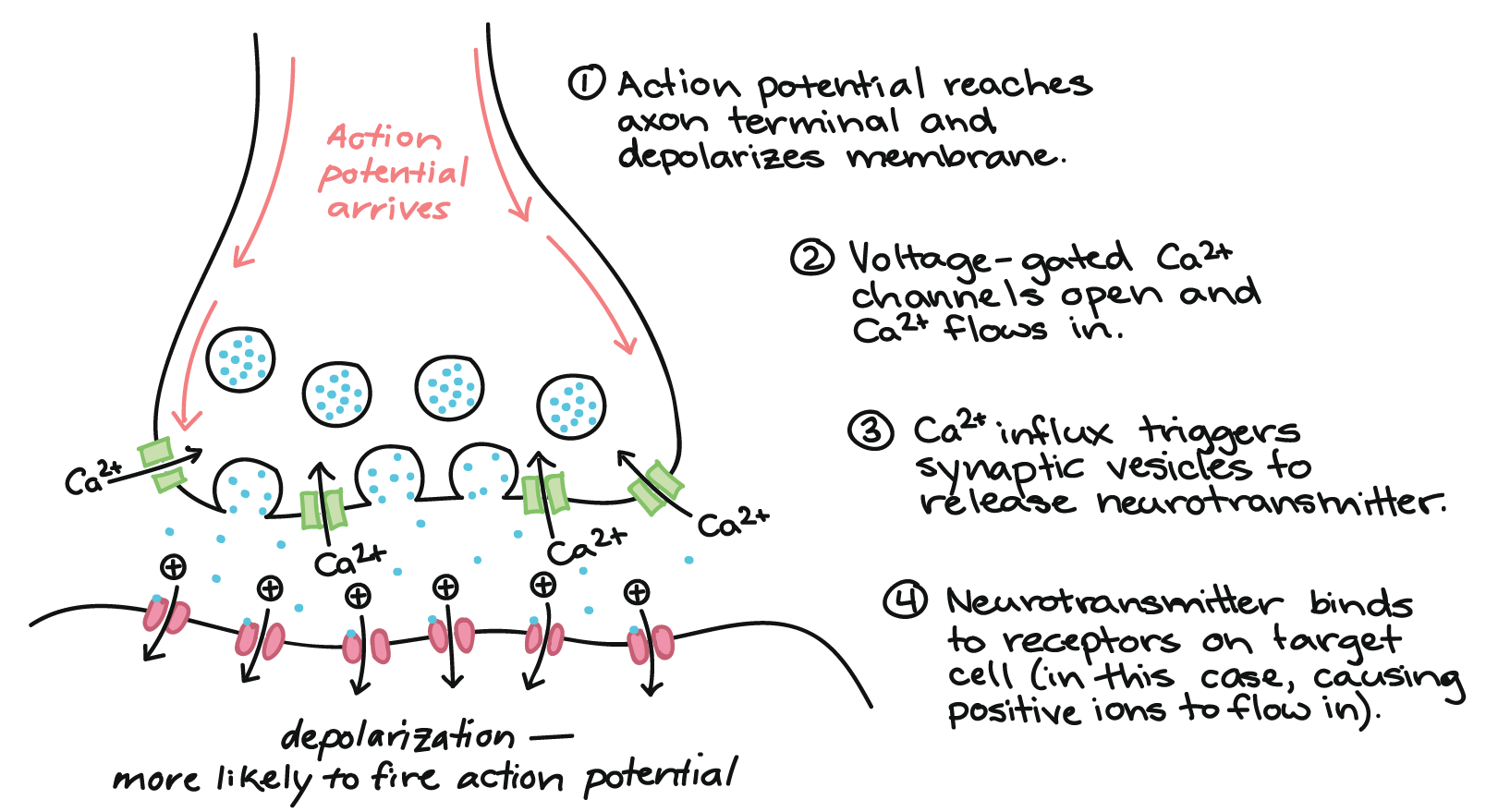
Action potential arrives at the presynaptic bulb / knob.
causes Voltage–gated channels open and calcium ions enter the presynaptic bulb / knob.
Vesicles containing neurotransmitter fuse with the presynaptic bulb / knob membrane, releasing neurotransmitter into the synapse via exocytosis.
Neurotransmitter moves across the synapse and fuses with receptors on the post synaptic membrane.
channels open, leading to depolarisation and generation of a new action potential in the postsynaptic neurone.
If depolarisation exceeds the threshold again at the postsynaptic membrane, then an action potential will be produced and propagated. again
how is continuous stimulation prevented
Acetylcholinesterase is an enzyme that breaks down acetylcholine that is left in the synaptic cleft. This prevents continuous stimulation.
The neurotransmitter is hydrolysed, and products are returned to the presynaptic bulb / knob to be reformed and reused. This process requires ATP.
1
explain the two ways neurotransmitters flow
synapses ensure the unidirectional transmission of impulses.
Spatial summation ; neurotransmitters released from multiple presynaptic neurones act on a single postsynaptic neurone at the same time.
The combined effect of their generator potentials may reach the threshold and trigger an action potential.
Temporal summation ;single presynaptic neurone releases neurotransmitter repeatedlyin rapid succession. The generator potentials build up over time and, if the threshold is reached, an action potential is triggered.
1