mobility and perception
 Call Kai
 Learn
 Practice Test
 Spaced Repetition
 Match
 Flashcards
 Knowt Play
1/64

There's no tags or description
Looks like no tags are added yet.
65 Terms
proproicieption
position of the body
presbyopia
age related loss of focus on close objects (far sighted
myopia
loss of function on distant objects related to eyeshade (nearsighted)
cataracts
opacity of the lens, blocking light to the retina due to age related protein clumping
glaucoma
increased intraocular pressure causing compression of the optic nerve and reduction of peripheral vision
macular degeneration
loss of central vision from deteritioaton of retina, usually correlated with age
Ophthalmoscopy
direct visualization of back of eye
conducive
Alteration of middle ear that prevents sound waves from entering
sensorineural
•Alteration of inner ear, nerves, or brain processing
snellen test
measures visual acuity at a distance
Rosenbaum test
– measures close visual acuity
Tonometry
– measures intraocular pressure
conducive - •Cerumen impaction
build-up of earwax preventing sound conduction
conducive - otitis media
Otitis media – infection of the middle ear structures that causes build-up of fluids, preventing sound conduction
sensorineural - presbycusis
age-related deterioration of ear hair cells and auditory nerves causing loss of high-pitched hearing
•Ototoxicity - sensorinerual
loss of hearing related to medication side effects
Tinnitus – sensorineural
“ringing” in the ears; is a symptom of disease or medication side effects
sensorineural - Meniere’s disease
Meniere’s disease – chronic disease in which there is fluid accumulation in the inner ear causing balance issues, dizziness, and hearing loss
rinne test
⚬Rinne test - tuning fork used to create air conduction (AC) sound and bone conduction (BC) sound (air should seem louder than bone = normal)
weber test
⚬Weber test - tuning fork placed on top of head (sound should be equal in both ears
audiometry
•Audiometry - headphones used to discriminate between different frequencies and volumes
Otoscopy
Otoscopy - direct visualization of the external canal and tympanic membrane
•Paresthesia
•numbness and tingling
neuropathy
•Neuropathy – nerve damage, typically from diabetes, vitamin deficiencies, medications, autoimmune disease
paraplegia
Paraplegia – loss of sensation in lower body
hemiplegia
•Hemiplegia – loss of sensation in half of body (left or right)
quadriplegia
Quadriplegia – loss of sensation from the torso down

primary lesions
atrophy
Wasting away or weakening of a muscle or tissue, usually from lack of use or illness.
contractures
Contractures: Permanent tightening or shortening of a muscle, tendon, or joint that limits movement.
osteoporosis
Osteoporosis: A condition where bones become weak, thin, and more likely to break because bone density decreases.
atelectasis
Atelectasis: Partial or complete collapse of part of a lung, causing the tiny air sacs (alveoli) to not fill properly with air.
secretion congestion
secretion congestion: Build-up of mucus or other secretions in the airways, making it harder to breathe and sometimes causing coughing or abnormal lung sounds.
seborrheic keratosis
(age spots)
⚬Acrochordon
skin tags
lipoma
(adipose “fat” deposit)
nevi
⚬Nevi (freckles/moles)
skin abnormalities
Macule = flat
Papule = small bump
Nodule = deeper lump
Vesicle = small fluid blister
Bulla = big fluid blister
Pustule = pus
Tumor = large solid mass
Wheal = temporary swelling/hive
Plaque = raised, flat area

macule
Macule: A flat, discolored spot on the skin that you can see but cannot feel.
Example: Freckle.
papule
Papule: A small, raised, solid bump on the skin.
Example: Small pimple.
nodule
Nodule: A firm, solid, raised lump that is usually deeper and larger than a papule.
Example: A small lump under the skin.
vesicle
Vesicle: A small, raised blister filled with clear fluid.
Example: Small blister from poison ivy.
bulla
Bulla: A larger, fluid-filled blister.
Think: big vesicle.
pustule
Pustule: A raised skin lesion filled with pus.
Example: Pus-filled pimple.
tumor
Tumor: A solid, raised mass or lump that is larger than a nodule. It may be benign or malignant.
Think: large solid mass.
wheal
Wheal: A raised, swollen area of skin that is usually temporary and often itchy.
Example: Hives or a mosquito bite.
plaque
Plaque: A raised, flat-topped area that is usually wider than it is tall.
Example: Psoriasis patches.
•Primary vs. secondary intention
⚬primary - edges approximated with minimal exposed tissue (i.e. sutures)
⚬secondary - edges left open, more regeneration necessary (i.e. large chronic wound)
undermining
Undermining - a hollow between the skin surface and the wound bed, resulting from death of the underlying tissue
tunneling
Tunneling - a passageway or channel underneath the base of the woundbed that has the potential for infection
pi staging
The ONLY skin wounds that are staged are pressure injuries!!!
No reverse staging as the wound heals
There are SIX total stages according to NPIAP (National Pressure Injury Advisory Panel):
•Stage 1
•Stage 2
•Stage 3
•Stage 4
•Unstageable
Deep tissue injury
blanchable
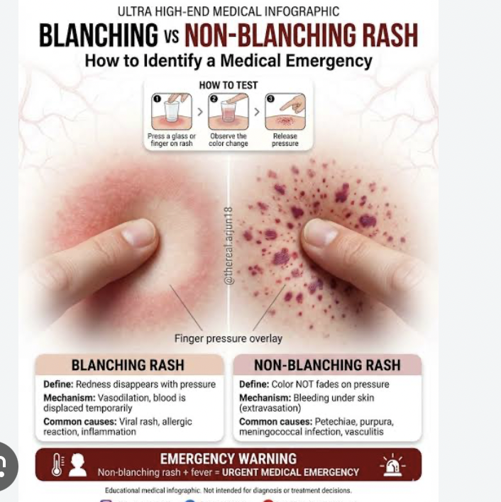
•Blanchable skin turns pale or lighter when pressure is applied, indicating temporary vessel compression.
non blanch able
•Non-blanchable skin remains red/discolored, signaling blood leakage or deep tissue damage
If there is redness over a bony prominence or under a medical device, it is only a stage 1 if it is non-blanching. If it blanches, we document this as “blanchable redness”
stage 1 pressure injury
•Skin is intact (closed wound)

•Non-blanchable area of dark pink or redness over a bony prominence
stage 2 pressure wound
•Partial-thickness with exposed dermis
•Wound bed is viable granulation tissue (pink, red)
•Blisters are considered stage 2, even if intact

stage 3 pressure wound
•Full-thickness with exposed adipose/subcutaneous tissue
•Wound bed is granulation tissue, potentially with slough or eschar
•Depth varies depending on location
•Tunneling or undermining may be present

stage 4
•Deep full-thickness with exposed muscle, bone, tendon, etc.
•Wound bed is granulation tissue, potentially with slough or eschar
•Depth varies depending on location

•Tunneling or undermining may be present
unstagable pressure wound
•Deep full-thickness but extent cannot be determined due to slough or eschar obscuring wound bed
•If slough or eschar are removed, will reveal stage 3 or stage 4

deep tissue injuries
•Non-blanchable maroon or purple discoloration
•Epidermal/dermal separation from blood-filled blister

•Does NOT have slough, eschar, subcutaneous tissue, granulation tissue, muscle, bone or tendon present
phases of wound healing
1.Hemostasis - platelets cause bleeding to stop, temporary clot forms
2.Inflammatory - white blood cells brought to the area, exudate present
3.Proliferative - fibroblasts start to create new granulation tissue
4.Maturation & remodeling - scar tissue forms to create strong, more uniform tissue
homeostasis
Hemostasis - platelets cause bleeding to stop, temporary clot forms
inflammatory
2.Inflammatory - white blood cells brought to the area, exudate present
proliferative
.Proliferative - fibroblasts start to create new granulation tissue
maturation and remodeling
.Maturation & remodeling - scar tissue forms to create strong, more uniform tissue