Embryology and 1st Trimester Anatomy
 Call Kai
 Learn
 Practice Test
 Spaced Repetition
 Match
 Flashcards
 Knowt Play
1/99

There's no tags or description
Looks like no tags are added yet.
100 Terms
Conception occurs approximately __ weeks after the LMP
2
Carnegie staging
Used to categorize the embryo based on age, size and morphology
23 stages or organogenesis that occur in the first 8 weeks of gestation
Embryo develops organs/structures in a specific order that is constant
Gamete
Sperm or ovum
Zygote
Formed by union of sperm and ovum, single cell
Cleavage
Cell division occurs and the single cell forms a cluster
Morula
Cluster of cells formed by zygote division; becomes blastocyst when it enters the UT cavity
Blastocyst
Well organized cluster of cells surrounded by trophoblastic cells
Trophoblastic cells
Produce progesterone and hCG, involved with implantation into chorionic villi then into placenta
Decidua capsularis
Tissue that envelops the blastocyst after implantation
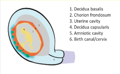
Decidua basalis
Becomes maternal placenta, blastocyst attaches to this layer
Decidua parietalis
Other tissue NOT involved in implantation process
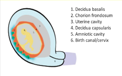
Amnion
Formed from cells inside blastocyst
Chornion
Formed from cells left over from fetal portion of placenta formation
Lack of fusion of chorion and amnion before 16 weeks is associated with
Fetal structural and/or chromosomal abnormalities
Germ layers: Endoderm
Forms respiratory system and GI tract
Germ layers: Mesoderm
Forms circulatory and musculoskeletal systems
Germ layers: Ectoderm
Forms brain, CNS and skin
5 weeks CRL measurement
2-4mm
6 weeks CRL measurement
4-9mm
7-8 weeks CRL measurement
9-16mm
9-10 weeks CRL measurement
23-31mm
11 weeks CRL measurement
41mm
12 weeks CRL measurement
53-67mm
Yolk sac
Performs hematopoiesis and delivers nutrients to fetus before 4 weeks
Produces AFP early in pregnancy
Located within chorion
____________ duct connects yolk sac to fetal pole
Vitelline
Yolk sac can be identified by week __ with TV approach and week _ with a TA approach
5; 7
Yolk sac is usually seen with a GA sac diameter of >__mm TA
13
Yolk sac MUST be seen with a MSD of __mm or greater (TA) _mm or greater (TV)
20; 8
YSD <___ is normal; inner to inner
5.6
Double bleb sign
Amnion sac and yolk sac present = IUP; seen at 5.5 weeks, 2mm CRL
Yolk sac disappears by __ weeks GA
12
In small amount of adults, the yolk sac persists as a __________ in the ileum portion of bowel (Meckel diverticulum)
Diverticulum
Abnormal yolk sac indicates…
Poor outcome of pregnancy
Visualization of the GS separate from the uterine cavity refers to the _______________
Double sac sign
Implantation of GS should occur where?
In UT fundus or mid body
GS is first visualized at __ weeks TV (>2000 units/ml bhCG)
4
>16mm MSD TV should show…
Fetal pole
>25mm MSD TA should demonstrate
Fetal pole
Growth rate of MSD =
1mm/day until 10th week GA
Embryo is usually visible by later in __th week or early _th week suing TV
5; 6
Embryo usually grows __mm/day in first trimester up to 10th week
1
CRL(cm) + 6 =
GA in weeks
CRL(mm) + 42 =
GA in days
LMP date + 7 days + 9 months =
EDD
1st trimester
Week 1 - 13
2nd trimester
Week 14 - 28
3rd trimester
Week 29-delivery
Double decidual sac sign or intradecidual sign due to implantation seen at:
5.5-6 weeks
Tiny CRL and wolk sac seen with TV as early as __ weeks
5.5
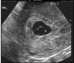
Double bleb sign
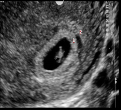
Double decidual sign
6 weeks
Cardiac activity first identified
Normal HR: 100-115bpm
Bean shaped fetal pole
Neural tube is closing
An embryo less than __mm or greater should demonstrate cardiac activity always
7
3 Primary vesicles form at 6 weeks
Prosencephalon: Forebrain, becomes lateral ventricles, cerebrum and thalamus
Mesencephalon: Becomes mid brain and forms aqueduct of sylvius
Rhombencephalon: Hindbrain, forms pons, medulla, cerebellum and 4th ventricle
7-8 weeks
Rhombencephalon can become visible at 7 weeks
Limb buds are visualized
Herniation of midgut into base of cord
Umbilical cord forms
Mineralization of fetal skeleton begins at 8 weeks
9-10 weeks
Heart rate averages 145bpm
Choroid plexus fills lateral ventricles
Organogenesis complete by 10 weeks GA
11 weeks
Cranium and cerebellum are visible
Extremities begin to take shape
Bowel returns to abdominal cavity after cord formation
Fetal intestinal activity begins
NT can be measured (11-14 weeks)
Yolk sac involutes
NT
Magnified image of fetal head in midsagittal plane
Fetal neck must be in neutral position
Amnion must be visualized separately from and NT edges must be clearly demonstrated
Use + calipers measuring widest space
12 weeks
Start of amnion/chorion fusion
Herniation of midgut is considered abnormal after 12 weeks
Skull and femur are adequately mineralized
Can determine fetal gender with TV
Nasal bone evaluation done (need = sign, less echogenic than skin → absent)
CRL
Fetus fills 2/3 of field of view
Midsagittal plane with profile, spine and rump visible
Calipers placed on the outer edge to outer edge
3 measurements taken
Indications for detailed late 1st trimester US 12weeks 0days - 13weeks 6days
Previous child/fetus w/defect or aneuploidy, AMA, Maternal diabetes, positive serum testing, IVF, Abnormal US, teratogen exposure, Maternal BMI over 30kg/m3, Placenta covering internal os under C-section scar
A bladder longer than __mm indicates enlargement and can be related to aneuploidy
7
We should document __ long bones per extremity
3
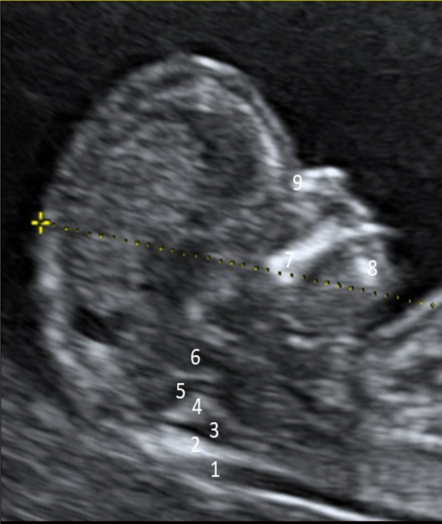
1
NT
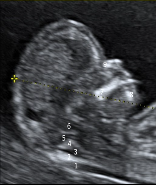
2
Occipital bone
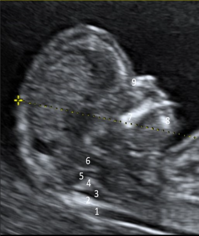
3
CM
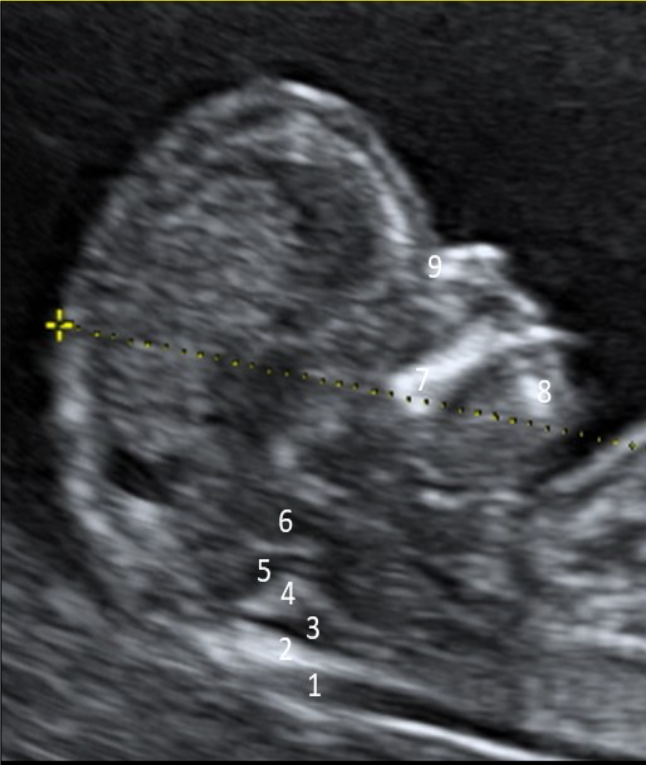
4
CP of 4th ventricle
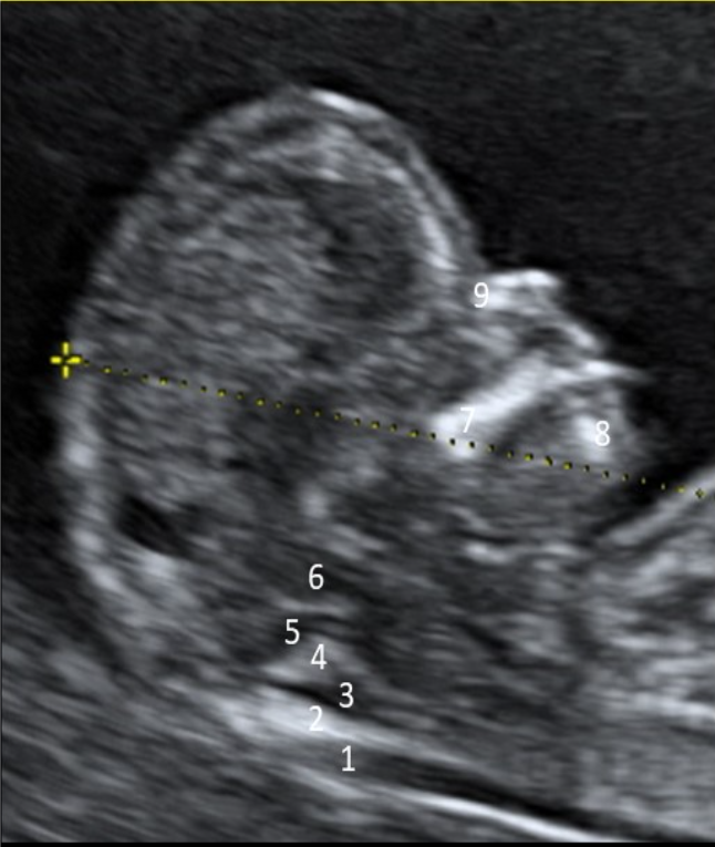
5
4th ventricle
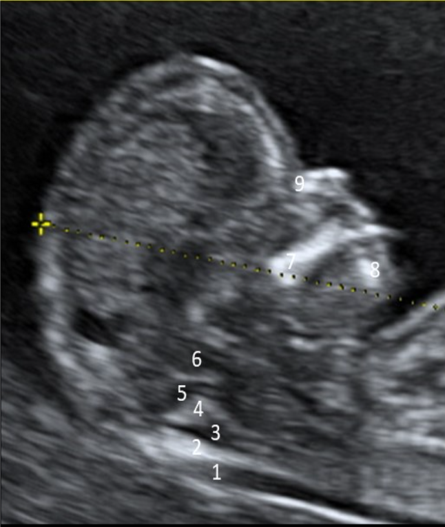
6
Brainstem
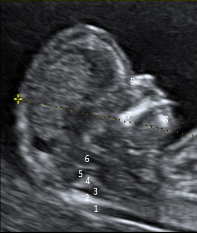
7
Maxilla
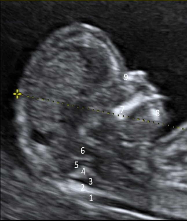
8
Mandible
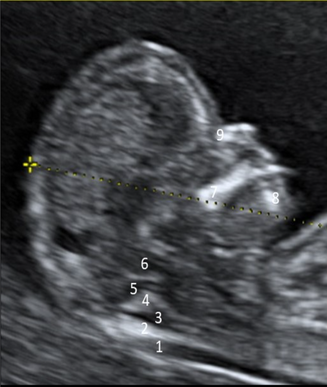
9
Nasal bone
Flow reversal in tricuspid valve is seen in systole and the velocity is typically over __cm/s
60
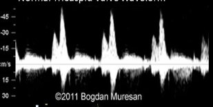
Normal tricuspid valve waveform
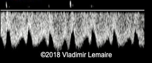
Normal ductus venosus waveform
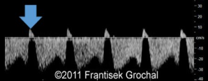
Increased resistance in ductus venosus
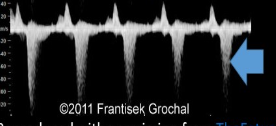
Tricuspid regurgitation
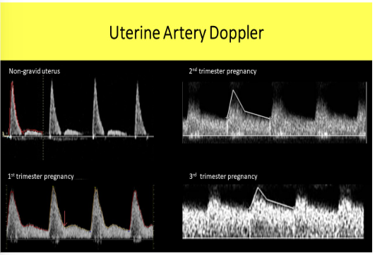
As pregnancy ages the resistance of the uterine artery _______, diastolic flow increases and diastolic notch _________
Decreases; disappears
Conceptual age is the exact date of conception + ________ = Gestational age
2 weeks
The amnion and chorion normally complete fusion no later than week:
16
How is MSD used to calculate gestational age
MSD + 30 = gestational age
MSD normally increases by ____ per day, while the CRL increases by ___per day in the first 10 weeks
1mm, 1mm
What is Naegele rule used for and what is the rule
To estimate the date of delivery
LMP date + 7 days + 9 months = EDD
If M-mode cannot be obtained:
A 2D cine clip of heart motion should be used to document FHT
What should be visible by TV during the 5th week of GA
Double decidual sac sign
Double bleb sign
Decidual reaction
At 6 weeks, a normal embryo with a CRL of _________ will demonstrate a normal HR of __________ that is documented on M-mode
7mm or greater, 100-115bpm
When evaluating a first trimester embryo for cardiac activity, what sound processing parameter should be increased as much as possible?
Frame rate
Organogenesis is normally completed by __ weeks GA
10
All the following can be visualized with TV sonography at the end of 12 weeks GA except:
Herniation of the midgut
What fetal characteristic causes the CRL to be less accurate from 12-14 weeks?
Neck flexion
What fetal biometry is not normally used for dating in the late first tri?
FL
What 3 structures should be visible in transthalmic plane in the late first tri
Thalami, the cerebral peduncles, typically 3rd vent are demonstrated on view used to assess BPD and HC
The 4th ventricle is also called ____________and if compressed it is highly suggestive of:
Intracranial translucency; spina bifida
What 4 structures should be visible in the fetal head in the midsagittal plane during the late first trimester evaluation?
Thalami, midbrain, brain stem, intracranial translucency (4th vent), developing CM
What fetal heart views should be obtained in late first tri eval?
4CH, and 3VT
All the following are documented in the fetal abdomen in the late first trimester except:
Kidneys
Bowel
Liver
Stomach
Bowel
The frontomaxillary facial angle is performed in the late first tri to assess possibility of what 2 anomalies
Trisomy 21 and spina bifida
Regarding US exposure, which of the following can be safely evaluated with spectral Doppler in 1st tri
UT arteries
In first tri, Ductus venosus is best evaluated in what imaging plane?
Right parasagittal
Tricuspid regurgitation documented in the first tri is most commonly associated with:
Trisomy 21