Digestive system - Anatomy ~ SciOly
 Call Kai
 Learn
 Practice Test
 Spaced Repetition
 Match
 Flashcards
 Knowt Play
1/58

There's no tags or description
Looks like no tags are added yet.
59 Terms
four main processes of the digestive system
ingestion
digestion
absorption
elimination
Assimilation
the usage of absorbed molecules
Two organ groups of the digestive system
alimentary canal
esophagus, intestines
accessory organs
salivary glands, pancreas
The mouth
digestion of the food begins
the teeth chew food into smaller pieces (mechanical digestion)
salivary glands secrete saliva into the mouth, breaking down starches into dextrin and maltose (chemical digestion)
Saliva
secreted by salivary glands
form of chemical digestion because it changes the molecular structure of the food
create food boluses
pharynx
shared connection between nasal cavities, mouth, air tract, and esophagus
epiglottis prevents boluses from entering the trachea to the lungs

esophagus/oesophagus
muscle-walled tube that goes from the throat to the lower digestive tract
directly under pharynx
muscular contractions called peristaltic waves propel food down the esophagus

peristalsis
muscular contractions that push food down toward the stomach
helps digestion work against gravity

stomach
breaks down food boluses into a liquid-like mixture by churning the food (mechanical) and secreting gastric acid (chemical) while they wait for the next step of digestion
gastric acid also kills any bacteria that may be in the food
layers from innermost to outermost: mucosa, submucosa, oblique muscle, circular muscle, longitudinal muscle, and serosa
Three main sections: fundus (top part/roof), body (bulk of the stomach), pylorus (leftmost region)

duodenum
the first part of the small intestine (around 25 cm in length)
stationary and fixed behind sheets of connective tissue called peritoneum
glands secrete a thick alkaline fluid that counteracts the acidic nature of the chemicals the food bile has absorbed

gallbladder
secretes pancreatic juice and bile into the duodenum

liver
produces bile which is stored in the gallbladder
~700 mL a day
produced by hepatocytes
breaks fat particles up —> emulsification
divided by the falciform ligament
attached to the underside of the diaphragm by the coronary ligament
4 lobes: right, left, caudate, quadrate
can regenerate

jejunum and ileum
second and third parts of the small intestine
make up the bulk of it
food boluses are digested even further, some nutrients are absorbed through the walls
ileum is the longest portion of the small intestine
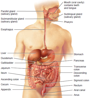
large intestine
shorter than the small intestine, but larger in diameter
water and electrolytes are removed in the large intestine
microbes such as bacteria aid in further digestion
consists of cecum, colon, rectum, and anal cavity
mobile and undergoes several muscular contractions called mass movements (propel colon toward the anus to help move food along with peristalsis)
generally does not absorb nutrients

mastication
chewing
first process of digestion
buccinator muscles
assist mastication
deglutition
swallowing
soft palate elevates to prevent food from entering nasal cavity
epiglottis bends down to prevent food from entering air passageway
teeth
chew and break down food mechanically
a child has 20 deciduous teeth while a adult has 32 permanent teeth
types: incisors, canine, premolars, molars
structure: crown (upper surface, bumps on crown are called cusps), neck (region under crown but above alveolar bone), root (region inside alveolar bone)

salivary glands
produce saliva
types of saliva: serous (enzymatic saliva) and mucous (acts as lubricant)
types of salivary glands: parotid, submandibular, sublingual
Parotid glands
largest
slightly posterior to the cheeks
make 25% of the saliva (rises to 50% when eating)
serous

submandibular glands
below the mandible bone
make up 70% of salivary production
serous

sublingual glands
smallest
make up 5% of saliva
below the floor of the oral cavity
mostly mucus

rugae
if the stomach does not contain much food bolus, it is smaller ad the inner surface has lots of these folds
small intestine
around 6m long
function: digestion of food and absorption of nutrients
duodenum, jejunum, ileum
layers: mucosa, submucosa, muscularis-circular, muscularis-longitudinal, serosa
large surface area with many villi and microvilli
peristaltic and segmental contractions move food

Mucosa cell types
Absorptive cells digest and absorb food
Goblet cells produce mucus
Granular cells protect from bacteria
Endocrine cells produce regulatory hormones
Peristaltic contractions
propel chyme forward
similar to contractions in the esophagus
segmental contractions
mix chyme by squeezing in outward motions
DO NOT help move food through the GI tract
pancreas
secretes enzymes into the duodenum
made up of endocrine (produces hormones) and exocrine tissue (produces digestive enzymes)
secretions travel through the pancreatic duct and join the common bile duct
produces hydrogen carbonate which helps counter the acidic quality of chyme
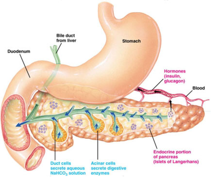
acinar cells
create digestive enzymes in the pancreas
cecum
first part of the large intestine
appendix
small pouch like attachment to the cecum
no real function (vestigial organ)
Colon
ascending, transverse, descending, sigmoid
majority of large intestine
converts chyme to feces
microorganisms help break down components of the feces
stores feces until defecation (feces transported out of digestive tract through anus)
lined with mucus and crypts (have goblet cells to produce mucus)
ascending colon
first part of colon
food travels up through it
transverse colon
second part of colon
food travels left to right
Rectum
straight, muscular tube
thick muscle layers to push food out anal cavity
peritoneum
serous membrane that surrounds the abdominal cavity and organs
inner layer: visceral peritoneum
outer layer: parietal peritoneum
double layer of peritoneum: mesentery
vagus nerve
initiates digestion
ulcers
occur when the mucus in the stomach or the intestines deteriorates, leaving the flesh directly exposed to the acid contained in these organs
causes: bacteria inflames the tissue by living in the mucus, extreme intake of either sour or spicy food with high acid content, the use of NSAIDs (block the ability to make more mucus)
neoplasm
tumor affecting the digestive system
esophageal cancer
includes squamous cell cancer (90-95% of cases worldwide) and adenocarcinoma
symptoms: vomiting blood, regurgitation of food, heartburn, difficulty swallowing, chest pain
tests: barium swallow, chest MRI, endoscopic ultrasound, esophagogastroduodenoscopy, biospy, PET scan
treatment: surgery, chemotherapy, radiation
Stomach cancer
influencing factors: presence of Helicobacter pylori or ulcers caused by this bacteria, surgery preformed on ulcers, smoking, anemia, long-term gastritis, obesity, high salt diet, heavy drinking, genetics, long-term use of NSAIDs
symptoms: indigestions, nausea, heartburn, bloating, loss of appetite, severe stomach pain, blood in stool, weight loss, vomiting, continued bloating and heartburn, yellowing of eyes and skin
most commonly carcinomas, sometimes lymphomas
tests: gastropic examination, CT scans, biopsies (to determine malignancy)
treatment: surgical removal, chemotherapy, radiation, targeted therapy
extremely low survival rate, more common in males
carcinomas
cancer that develops in the epithelial tissue
Colorectal cancers (CRC)
affect lower portion of large intestine
influencing factors: age, race, obesity, diabetes, smoking, sedentary lifestyle, excessive consumption of red or processed meats, excessive alcohol consumption, crohn’s disease, ulcerative colitis, IBS, genetic disorders
symptoms: rectal bleeding, blood in stool, unexplained weight loss, change in bowel movements, weakness or fatigue
testing: fecal occult blood testing, flexible sigmoidoscopy, colonoscopy, multitarget stool DNA screening test
treatment: laparotomy (cut in the abdomen), chemo, NOT RADIATION, palliative care
Lactose intolerance
the digestive system’s inability to break down a sugar called lactose
causes: body’s lack of an enzyme, called lactase
symptoms: pain due to inability to digest sugar

Hepatitis
inflammation of the liver
cause: virus or high alcohol consumption
may cause liver cirrhosis, cancer, death
Hepatitis A
contagious viral disease
very rare but can be spread by contaminated food/water
only humans transmit
symptoms: flu-like symptoms, jaundice
preventable by vaccine, good hygiene, sanitation
clears in 1-2 months with rest and hydration
Hepatitis B
dangerous liver infection
spreads through sexual contact
easily preventable by vaccine
chronic cases may require medication and possibly liver transplant
symptoms: jaundice and dark urine
Hepatitis C
infection of the liver caused by the hepatitis C virus
spread by contaminated blood
can occur by unsterile tattoo needles or dirty needled used by a hospital
symptoms: loss of appetite, jaundice
diagnosed by blood testing for antibodies or viral RNA
no vaccine
can be treated by medication
Hepatitis D
liver disease caused by hepatitis D virus
prevented by sexual absence
symptoms: abdominal pain, nausea, fatigue
few treatments
Hepatitis E
liver disease caused by hepatitis E virus
liver gets inflamed
spread by fecal matter contaminated water
symptoms: jaundice and lack of appetite
goes away in about 4-6 weeks
Appendicitis
appendix becomes infected and inflamed
treatment: removal of appendix
Diverticular disease
general term used to describe the experience of symptoms related to diverticula (small bulges or pockets that develop in the intestine)
considered normal part of aging
influential factors: low fiber and high red meat diets, smoking, obesity, history of constipation, use of NSAIDs, family history
symptoms: abdominal pain, bloating, changes in bowel movements, episodic constipation
treatment: not necessary
diverticulosis
diverticular disease with no noticeable symptoms
diverticulitis
when diverticula become inflamed
Causes: small perforation of one of the diverticula, infection of bacteria inside the bulge in the colon
influential factors: low fiber and high red meat diet, smoking, obesity, NSAIDs, family history
symptoms (much more severe than diverticular disease): severe abdomen pain, fever, diarrhea, rectal bleeding, stricture (section of the colon narrows and prevents stool from passing easily), abscess (pocket of pus sealed off within colon), fistula (diverticulum forms a connection with another organ), peritonitis (inflammation of the membrane tissue covering and surrounding the organs in the abdomen)
diverticulum
an abnormal sac or pouch formed at a weak point in the wall of the alimentary tract
GERD
gastroesophageal reflux disease
contents of the stomach travel back up the esophagus, irritating the lower esophageal area and causing a sensation described as “heartburn”
cause: dysfunctional lower esophageal sphincter (which would typically function to close off the esophagus from the lumen of the stomach)
symptoms: difficulty swallowing, sore throat, regurgitation of food
treatment: antacids, proton pump inhibitors, acetaminophen, avoid problematic foods
crohn’s disease
chronic inflammation of the GI tract
autoimmune disorder
symptoms: chronic pain, fever, diarrhea, constipation, abdominal pain, fatigue, bloody stool, ulcers
testing: colonoscopy, sigmoidoscopy, endoscopy, enteroscopy, MRI or CT scan, x-rays of the stomach, physical examination
ulcerative colitis
inflammation of the large intestine
celiac disease
body has an immune reaction to gluten
symptoms: diarrhea or bloating, flat villi
treatment: avoid gluten