Anatomy of Heart
 Call Kai
 Learn
Learn Practice Test
 Spaced Repetition
 Match
Match Flashcards
 Knowt Play
1/15

There's no tags or description
Looks like no tags are added yet.
Name | Mastery | Learn | Test | Matching | Spaced | Call with Kai | Chat |
|---|
No analytics yet
Send a link to your students to track their progress
16 Terms
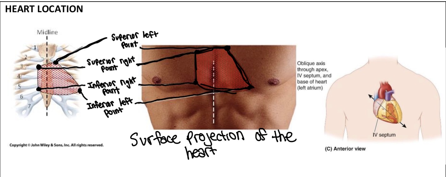
Location of Heart on Surface
Located in the thorax → the heart is inside the thoracic (chest) cavity.
Extending to the 5th intercostal space → the bottom of the heart reaches around the 5th space between the ribs.
To the left of the sternum mainly

Location of Heart in Mediastinum
Mediastinum = the central region of the thoracic cavity (entire space of chest), between vertebrae & sternum.
Contains the heart, great vessels, trachea, esophagus, etc.
Does NOT contain the lungs (the lungs are in the pleural cavities).
Middle portion of the mediastinum = the middle mediastinum, which is occupied mainly by the heart.

Mediastinum Portions
The transverse thoracic plane runs from: Sternal angle anteriorly to T4–T5 intervertebral disc posteriorly
Purpose: to divide the mediastinum into superior & inferior parts.
The inferior mediastinum is divided into 3 parts
From front → back:
Anterior → Middle → Posterior
And the heart is in the middle mediastinum
Surfaces of Heart
Base → the back/posterior part of the heart. Mostly formed by the left atrium, with some right atrium. It’s where the major vessels attach.
Apex → the pointed bottom of the heart. Formed by the left ventricle and points down, forward, and to the left.
Diaphragmatic surface → the bottom surface resting on the diaphragm. Mostly formed by the left ventricle, with some right ventricle.
Sternocostal (anterior) surface → the front surface, facing the sternum/ribs. Mostly the right ventricle.
Pulmonary surfaces → the sides facing the lungs:
Right pulmonary surface → mainly right atrium
Left pulmonary surface → mainly left ventricle

Anterior Heart Features
How each part works step by step:
Superior Vena Cava
Large vein that brings deoxygenated blood from the upper body to the right atrium.
Right Atrium
Receives deoxygenated blood coming from the SVC.
Has an auricle (small pouch) attached to allow it to hold more blood
Right Ventricle
Pumps deoxygenated blood to the pulmonary trunk..
Pulmonary Trunk
Carries deoxygenated blood to the pulmonary arteries then lungs to pick up oxygen.
Left Atrium
Receives oxygenated blood from the lungs.
Has an auricle (small pouch) attached to allow it to hold more blood.
Left Ventricle
Pumps oxygenated blood to the aorta
Has the thickest myocardium because it has to generate high pressure.
Aorta
The body’s largest artery.
Carries oxygenated blood to entire body.
The aorta then gives off three major branches:
Brachiocephalic trunk
→ supplies the right side of the head/neck and right upper limb
Left common carotid artery
→ supplies the left side of the head and neck
Left subclavian artery
→ supplies the left upper limb

Posterior Heart Features
How each part works step by step:
Superior Vena Cava
Large vein that brings deoxygenated blood from the upper body to the right atrium.
Inferior Vena Cava
Brings deoxygenated blood from the lower body to the right atrium.
Right Atrium
Receives deoxygenated blood coming from the SVC & IVC.
Has an auricle (small pouch) attached to allow it to hold more blood
Right Ventricle
Pumps deoxygenated blood to the pulmonary trunk.
Pulmonary Trunk
Carries deoxygenated blood to the pulmonary arteries then lungs to pick up oxygen.
Left Atrium
Receives oxygenated blood from the lungs.
Has an auricle (small pouch) attached to allow it to hold more blood.
Left Ventricle
Pumps oxygenated blood to the aorta
Has the thickest myocardium because it has to generate high pressure.
Aorta
The body’s largest artery.
Carries oxygenated blood to entire body.
The aorta then gives off three major branches:
Brachiocephalic trunk (contains right subclavian artery & right common carotid artery)
→ supplies the right side of the head/neck and right upper limb
Left common carotid artery
→ supplies the left side of the head and neck
Left subclavian artery
→ supplies the left upper limb

Posterior & Anterior Sulcus
Coronary Sulcus
Groove between the surface of right atria and the ventricles for anterior view (left atria and ventricles for posterior view)
contains right & left coronary arteries
What it does: pathway for the coronary arteries and veins that supply/drain the heart muscle.
Anterior/Posterior Interventricular Sulcus
Grooves between the surface of right & left ventricles (depends on anterior or posterior surface)
Contains right & left coronary arteries
What it does: pathway for coronary blood vessels that supply the myocardium.


Pericardium
2 layers of the pericardium:
Fibrous pericardium
Fibrous outer layer that helps anchor heart in place inferiorly to diaphragm
Comes superiorly to surround great vessels
Parietal layer of serous pericardium (wall of body cavity)
2 layers (visceral & parietal)
Parietal layer: sticks directly to the fibrous pericardium
Visceral layer: epicardium
Pericardial Cavity: the space between the parietal & visceral layers of serous pericardium. Has fluids to prevents friction.

Layers of the Heart
Layers of the heart
Pericardium - surrounds/encloses the heart and the roots of great vessels (like the protective sac)
Epicardium (visceral) - directly on heart surface (membrane that protects the muscle heart’s surface to reduce friction)
Myocardium - muscle of the heart (helps contraction and pumping out blood)
Endocardium - linings of chambers of heart (makes inside of heart’s surface smooth to reduce friction)

Coronary Arteries
Brings oxygenated blood TO the heart muscle.
* Right Coronary A. → supplies (to bring) much of the general right side (RV & RA) of the heart
* Right Marginal A. → supplies the right margin/right ventricle
* Left Coronary A. → supplies much of the left side (LV & LA) of the heart
* Left Marginal A. → supplies the left margin/left ventricle
* Circumflex A. → supplies the left margin and ventricles parts of the heart
* Anterior Interventricular A. (Runs in anterior sulcus) → supplies the front of the ventricles
* Posterior Interventricular A. (Runs in posterior sulcus) → supplies the back of the ventricles
Coronary Veins
- Take deoxygenated blood AWAY FROM the heart muscle.
Small cardiac vein → drains (to collect and carry blood away) the right side of the heart
Right marginal vein → drains the right margin
Great cardiac vein → drains much of the anterior heart
Middle cardiac vein → drains the posterior heart
Left marginal vein → drains the left margin
Left Atrium Oblique Vein - drains the left atrium
Left posterior ventricular vein → drains the posterior of the left ventricle
Anterior cardiac veins → drains the anterior right ventricle, directly into the right atrium
Coronary sinus → large collecting vein that receives blood from several cardiac veins and empties into the right atrium


Cusps, Trabeculae Carnae, Chordae Tendinae
Cusps
→ The flaps that form AV valves.
→ Open to let blood move forward and close to stop blood from flowing backward.
Chordae tendineae
→ fibrous cords attached to the cusps to the papillary muscles of the AV valves
→ Holds the valve cusps in place when the ventricle contracts
Trabeculae carneae
→ Irregular muscular elevations projecting on the inside walls of the ventricles.
→ Helps the ventricles contract effectively
Papillary Muscles
A special type of trabeculae carnae attached to the ventricular wall and the chordae tendinae
Pulls on chordae tendineae & prevents AV valve from flipping inside out when ventricles contract

How the Mitral Valves works
Ventricles relax
→ pressure in ventricles is low
→ mitral valve opens
→ blood flows left atrium → left ventricle
2. Ventricles contract
→ pressure in the left ventricle rises
→ blood wants to push back toward the left atrium
3. Mitral/Bicuspid valve closes
→ the cusps come together and seal the opening
→ prevents blood from flowing back into the atrium
4. Papillary muscles contract
→ they pull on the chordae tendineae
→ this helps stop the valve cusps from flipping inside-out/prolapsing into the atrium

Transverse Section of Heart (the
Tricuspid Valve — AV valve
Between the right atrium and right ventricle
Opens: blood moves RA → RV
Closes: prevents blood from going back into the right atrium
Bicuspid/Mitral Valve — AV valve
Between the left atrium and left ventricle
Opens: blood moves LA → LV
Closes: prevents blood from going back into the left atrium
Pulmonary Valve — semilunar valve
Between the right ventricle and pulmonary trunk
Opens: blood moves RV → pulmonary trunk
Closes: prevents blood from going back into the right ventricle
Aortic Valve — semilunar valve
Between the left ventricle and aorta
Opens: blood moves LV → aorta
Closes: prevents blood from going back into the left ventricle

Conducting System of Heart
Atria = rooms that receive blood
Ventricles = rooms that pump blood
Valves = doors that control which direction blood can move
Phase 1: Atrial systole (Both atria are in systole (contracted), while ventricles are in diastole (relaxed))
The blood enters the ventricles (bc they’re relaxed) through the AV valves (between the atrium & ventricles)
ends when the AV valves slam shut
Produces a lub sound
Phase 2: Ventricular systole (Ventricles are in systole (contract), while the atria are in diastole (relaxed))
Blood in the right ventricle exits through the pulmonary artery trunk to the pulmonary arteries due to ventricle pushing blood out
Blood in the left ventricle exits into the aorta
ends as the ventricles complete their pumping job
Phase 3: Atrial and ventricular diastole
Blood starts filling the heart again.
Blood returning to the heart fills the atria and passively into the ventricles
Semilunar valves closes for the dub sound

Heart Nodes
SA node — starts the signal
SA = sinoatrial node
Located in the right atrium
Creates the electrical signal that makes the both atria contract
2. AV node — briefly delays the signal
AV = atrioventricular node
Located between the atria and ventricles (interatrial septum)
Receives the signal from the SA node.
Delays it briefly so the atria have time to finish contracting and empty blood into the ventricles.
3. AV bundle — carries the signal downward
Carries the electrical signal from the AV node into the ventricles.
4. Right & left bundle branches — split the signal
The AV bundle divides into:
Right bundle branch → right ventricle
Left bundle branch → left ventricle
They carry the signal down toward the bottom of the heart.
5. Subendocardial branches — spread the signal
Also called Purkinje fibres.
Spread the electrical signal throughout the ventricular walls.
This causes the ventricles to contract, pushing blood out.
They travel through the interventricular septum (wall between the ventricles).