Biochemistry: Carbohydrates and Lipids
 Call Kai
 Learn
 Practice Test
 Spaced Repetition
 Match
 Flashcards
 Knowt Play
1/75

Earn XP
Description and Tags
Comprehensive vocabulary flashcards covering the biochemical classification, nomenclature, chemical properties, structures, and physiological roles of carbohydrates and lipids.
76 Terms
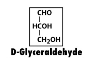
D-Glyceraldehyde Structure
Classification: Aldotriose (3 carbons)
Fischer Projection Configuration:
C1: Aldehyde (−CHO)
C2: −OH on the right
C3: Primary alcohol (−CH2OH)
Significance: The simplest aldose and reference standard for designating D and L stereoisomers.

D-Erythrose Structure
Classification: Aldotetrose (4 carbons)
Fischer Projection Configuration:
C2: Right
C3: Right
Relationship: Epimeric to D-threose at C2.

D-Ribose Structure
Classification: Aldopentose (5 carbons)
Fischer Projection Configuration:
C2: Right
C3: Right
C4: Right (all −OH groups on the right)
Significance: Essential structural component of ribonucleic acid (RNA), ATP, and nucleotide coenzymes.

D-Xylose Structure
Classification: Aldopentose (5 carbons)
Fischer Projection Configuration:
C2: Right
C3: Left
C4: Right
Relationship: The C3 epimer of D-ribose.

D-Glucose Structure
Classification: Aldohexose (6 carbons)
Fischer Projection Configuration:
C2: Right
C3: Left
C4: Right
C5: Right (pattern: Right-Left-Right-Right)
Significance: The primary circulating metabolic monosaccharide in animals.

D-Mannose Structure
Classification: Aldohexose (6 carbons)
Fischer Projection Configuration:
C2: Left
C3: Left
C4: Right
C5: Right (pattern: Left-Left-Right-Right)
Relationship: The C2 epimer of D-glucose.

D-Galactose Structure
Classification: Aldohexose (6 carbons)
Fischer Projection Configuration:
C2: Right
C3: Left
C4: Left
C5: Right (pattern: Right-Left-Left-Right)
Relationship & Role: The C4 epimer of D-glucose; constituent monomer of milk sugar (lactose).

Dihydroxyacetone Structure
Classification: Ketotriose (3 carbons)
Formula & Structure:
CH2OH−C(=O)−CH2OH
Significance: The only simple monosaccharide that lacks a chiral center; interconverted with glyceraldehyde-3-phosphate in glycolysis by triose phosphate isomerase.

D-Erythrulose Structure
Classification: Ketotetrose (4 carbons)
Fischer Projection Configuration:
C1: −CH2OH
C2: Ketone carbonyl (C=O)
C3: −OH on the right
C4: −CH2OH
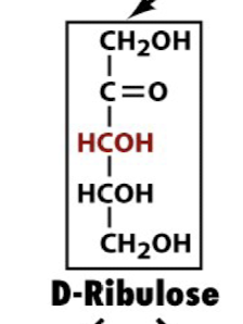
D-Ribulose Structure
Classification: Ketopentose (5 carbons)
Fischer Projection Configuration:
C2: Ketone carbonyl (C=O)
C3: Right
C4: Right
Significance: Keto-isomer of D-ribose; key metabolic intermediate in the pentose phosphate pathway.

D-Xyulose Structure
Classification: Aldopentose (5 carbons)
Fischer Projection Configuration:
C1: −CH 2OH
C2: −OH on the right
C3: −OH on the left
C4: −OH on the right
C5: −CH 2OH
Significance: Important sugar in plant metabolism; variant of D-ribulose.

D-Fructose Structure
Classification: Ketohexose (6 carbons)
Fischer Projection Configuration:
C2: Ketone carbonyl (C=O)
C3: Left
C4: Right
C5: Right
Significance: Fruit sugar; keto-isomer of D-glucose; preferentially cyclizes into a 5-membered furanose ring.
Chair Conformations of β-D-Glucopyranose

Structure: Six-membered pyranose ring in the chair conformation.
Stability Feature: β-D-glucopyranose is the most stable aldohexose because all bulky substituents (the four −OH groups and the −CH2OH group) are in equatorial positions.
α vs. β Anomer: In α-D-glucopyranose, the anomeric C1 −OH is axial (downward), whereas in β-D-glucopyranose it is equatorial (upward).
Triose Phosphate Isomerase (TPI) Mechanism

Reaction: Catalyzes the interconversion of dihydroxyacetone phosphate (DHAP) and glyceraldehyde-3-phosphate (GAP).
Mechanism: General acid-base catalysis via an enediol/enediolate intermediate.
Enzyme Kinetic Property: A catalytically perfect enzyme operating at a diffusion-controlled rate.
Lactose Structure and Linkage

Components: β-D-galactose + β-D-glucose
Glycosidic Linkage: β(1→4) glycosidic bond
Chemical Nature: Reducing sugar (glucose unit contains a free anomeric hemiacetal)
Enzyme: Cleaved by lactase; deficiency causes lactose intolerance.
Sucrose Structure and Linkage

Components: α-D-glucose + β-D-fructose
Glycosidic Linkage: α(1→2) linkage joining both anomeric carbons (C1 of glucose and C2 of fructose)
Chemical Nature: Non-reducing sugar (no free hemiacetal/hemiketal).
Cellulose Structure and Linkage

Polymer: Linear, unbranched homopolysaccharide of up to 15,000 D-glucose units
Glycosidic Linkage: β(1→4)
Conformation: Extended linear ribbons that pack side-by-side into parallel hydrogen-bonded sheets of immense tensile strength; water-insoluble plant structural polymer.
Chitin Structure and Linkage

Repeating Monomer: N-Acetylglucosamine (GlcNAc)
Glycosidic Linkage: β(1→4)
Function & Occurrence: Structural homopolymer forming the exoskeleton of arthropods and cell walls of fungi.
α-Amylose Structure and Conformation

Monomer Unit: D-Glucose
Glycosidic Linkage: Unbranched α(1→4)
Conformation: Left-handed helical coil; isomer of cellulose and linear component of plant starch.
Amylopectin vs. Glycogen Branching Architecture

Linear Chains: α(1→4) D-glucose linkages
Branch Points: α(1→6) glycosidic bonds
Branching Frequency:
Amylopectin: Branch every 24 to 30 residues (plant storage)
Glycogen: Branch every 8 to 12 residues (animal storage; highly branched for rapid mobilization).

Fatty Acids
carboxylic acids with long-chain hydrocarbon side group
Saturated fatty acids pack tightly and form more rigid, organized aggregates
fully reduced and have no double bonds
Unsaturated chains bend and pack in a less ordered way, with greater potential for motion
have double bonds
FA Nomenclature and observations
cis configuration
db are never conjugated
first db occurs between C9 and C10
positions of db are indicated by delta
example: • C18:2(Δ9,12)- Describes the
structure of linoleic acid
Palmitic Acid Structure (16:0)
Formula: CH3(CH2)14COOH
Notation: 16:0
Features: Saturated 16-carbon fatty acid with no double bonds; fully reduced hydrocarbon chain that packs tightly into crystalline aggregates.
mp: 63.1
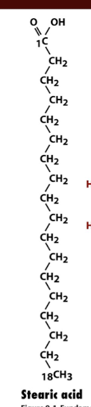
Stearic Acid Structure (18:0)
Formula: CH3(CH2)16COOH
Notation: 18:0
Features: Saturated 18-carbon fatty acid; high melting point (69.6∘C) due to extensive van der Waals contacts.
mp:69.6

Oleic Acid Structure (18:1Δ9)
Notation: 18:1(Δ9)
Structure: 18-carbon monounsaturated fatty acid with a single cis double bond between C9 and C10.
Effect of Double Bond: Causes a 30∘ bend/kink in the hydrocarbon chain, disrupting packing and lowering melting point to 12 degrees C

Linoleic Acid Structure (18:2Δ9,12)
Notation: 18:2(Δ9,12)
Structure: 18-carbon polyunsaturated fatty acid with two cis double bonds separated by a methylene bridge (−CH2−).
Classification: Essential ω-6 fatty acid.
mp: -5
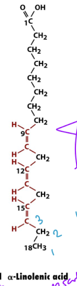
α-Linolenic Acid Structure (18:3Δ9,12,15)
Notation: 18:3(Δ9,12,15)
Structure: 18-carbon polyunsaturated fatty acid with three cis double bonds separated by methylene groups.
Classification: Essential ω-3 fatty acid.
mp: -11
Omega Fatty Acids

Numbering Rule: Numbering begins at the methyl-terminal carbon (ω carbon), opposite to the carboxylic acid carbonyl (C1).
ω-3 Fatty Acid: The first double bond is situated at carbon 3 from the terminal methyl end.
ω-6 Fatty Acid: The first double bond is situated at carbon 6 from the terminal methyl end.
Determinants of Fatty Acid Melting Temperature (T_m)

Chain Length: Melting temperature increases with increasing number of carbons due to greater surface contact and extensive additive van der Waals interactions.
Unsaturation: Introducing cis double bonds causes a rigid bend in the acyl chain, preventing compact crystalline packing and markedly decreasing melting temperature.
Hierarchy Rule: The degree of unsaturation (number of double bonds) takes precedence over chain length in determining melting temperature (e.g., stearic acid 18:0 melts at 69.6∘C, while oleic acid 18:1Δ9 melts at 13.4∘C).

Myristic acid
14:0, saturated, mp=53.9
palmitoleic acid
16:1n-7, unsaturated, mp=-0.5

FA Composition of Three Fat Foods
oil is mostly unsaturated, low mp
butter is saturated and unsaturated, high mp
beef is mostly saturated, highest mp
Triacylglycerol Generic Structure

Backbone: Glycerol (CH2OH−CHOH−CH2OH)
Linkages: Three fatty acid chains (R1,R2,R3) esterified to C1, C2, and C3 of glycerol.
Properties: Nonpolar, hydrophobic energy reservoir stored in adipocytes.
Biological role of triacylglycerols
energy reservoirs which are found in adipocytes
provide long-term energy storage, which is why humans can survive starvation for up to 3 months
provides 6x more metabolic energy than the same amt of glycogen (carbs), which can only supply the energy the body needs for up to a day (short term storage)
Glycerophospholipid Generic Structure

C1: Typically esterified to a saturated C16 or C18 fatty acid chain.
C2: Typically esterified to an unsaturated C16 or C18 fatty acid chain.
C3: Esterified to a phosphate group linked to a variable polar alcohol head group (X).
Function: Major structural amphiphilic building block of lipid bilayers.
Glycerophospholipid Head Groups (X-Groups) to Memorize

−H: Phosphatidic acid
−CH2CH2NH3+: Phosphatidylethanolamine (PE)
−CH2CH2N(CH3)3+: Phosphatidylcholine (PC / lecithin)
−CH2CH(NH3+)COO− : Phosphatidylserine (PS)
−CH2CH(OH)CH2OH : Phosphatidylglycerol
Sphingosine and Ceramide Structures

Sphingosine: 18-carbon amino alcohol containing a trans double bond between C4 and C5, an amino group at C2, and hydroxyl groups at C1 and C3.
Ceramide: Parent unit formed by attaching a fatty acid via an amide bond to the C2 amine of sphingosine.
Sphingolipid Subclasses: Sphingomyelins

Sphingomyelin: Ceramide + phosphocholine or phosphoethanolamine head group at C1 (abundant in myelin sheaths).

Sphingolipid Subclasses: Cerebrosides
Cerebroside: Ceramide + single neutral monosaccharide (glucocerebroside or galactocerebroside).
Sphingolipid Subclasses: Gangliosides
Ganglioside: Ceramide + complex oligosaccharide containing 3 or more sugars, at least one being sialic acid.
receptors for certian pituitary glycoprotein hormones and bacterial protein toxins
specific determinants in cell-cell recognition
play a role in carcinogenesis in cancer
interact with glycan recognition molecules involved in cell adhesion and immune regulation
6% of brain lipids are gangliosides
Tay-Sachs disease
deficiency in GM2 Ganglioside (neurological disorder)
fatal

Steroids
signaling lipids
phosphatidylinositol function regulates cell structure and metabolism
eicosanoids mediate inflammatio, fever, and pain response
pigments/antioxidants
cofactors
derivatives of cyclopentanoperhydrophenanthrene
Cyclopentanoperhydrophenanthrene

Cyclopentanoperhydrophenanthrene: Fused four-ring steroid core (three 6-membered rings A, B, C and one 5-membered ring D) which is part of cholesterol
Cholesterol
three 6-membered rings and one 5-membered ring all fused together
sterol: C3-OH group, important for storage and transport of cholesterol and can condense with a FA to form a sterol ester
found in plasma membrane lipids
increases rigidity due to ring system
Cholesteryl Stearate

Composition: Formed by esterification of cholesterol's C3 hydroxyl group with the 18-carbon saturated stearic acid.
Properties: Highly hydrophobic sterol storage ester associated with lipid transport and atherosclerosis plaque accumulation —> heart attack or stroke
Steroid Hormones: Glucocorticoids
affect carb, protein, and lipid metabolism
all steroid hormones are water insoluble and thus bind proteins for transport
made in adrenal gland
Steroid Hormones: Mineralocorticoids
regulate excretion of salt and water by the kidneys
made in adrenal gland
Steroid Hormones: Androgens and Estrogens
affect sexual development and function
made in the ovaries(estrogen)/testes(androgens)

Addison’s disease
hypoglycemia (low blood glucose)
muscle weakness
sodium/potassium loss
impaired cardiac function
increased stress susceptibility
treatment with glucocorticoids and mineralcorticoids
Shamu has addison’s disease
Cushing’s syndrome
characterized by fatigue, hyperglycemia (high blood sugar), edema(water retention), redistribution of body fat
typically caused by a tumor of the adrenal cortex

Vitamin D
Sterol derivative formed by UV ring disruption between C9 and C10; regulates calcium metabolism.
increases intestinal absorption of dietary calcium
too little —> deformed bones and stunted growth (rickets)
too much —> kidney stones and failure

Vitamin A (retinol)
derived from B-carotene in plant products
oxidized to form retinal which functions as the eye’s photoreceptor
deficiency leads to blindness

Vitamin K
about half of a human’s daily req is supplied by intestinal bacteria
participates in caarboxylation of Glu residues involved in blood clotting
Vitamin E
present as a-tocopherol
hydrophobic molecule in cell membranes that functions as an antioxidant to protect membrane proteins and lipids from oxidative damage
in anti aging and wound healing products

Fat-Soluble Vitamins (D, A, K, E)

Vitamin A (Retinol): Isoprenoid derived from β-carotene that oxidizes to retinal for vision photoreception.
Vitamin K: Essential cofactor for the carboxylation of glutamate in blood clotting proteins.
Vitamin E (α-tocopherol): Lipophilic, membrane-bound antioxidant protecting lipids from oxidative free-radical damage.

Isoprenoids
found in membranes that are soluble in the lipid bilayer
composed from 5C unit with same skeleton as isoprene
Ubiquinone (coenzyme Q) —> 10 isoprenoid units

Phospholipase Cleavage Specificity (A1, A2, C, D)

Phospholipase A1: Cleaves the ester bond at glycerol C1.
Phospholipase A2: Cleaves the ester bond at glycerol C2 (releases arachidonic acid).
Phospholipase C: Cleaves before the phosphate on C3, producing diacylglycerol (DAG) and phosphorylated head group.
Phospholipase D: Cleaves after the phosphate group, releasing head group alcohol (X) and phosphatidic acid.
Enantiomers Definition and Characteristics

Definition: Chiral stereoisomers that are non-superimposable complete mirror images of one another.
Example: D-Glucose and L-Glucose (every single asymmetric chiral center is inverted).
Physical Properties: Share identical chemical properties, melting points, and solubility in achiral environments, but rotate plane-polarized light in equal and opposite directions.
Epimers Definition and Key Distinctions

Definition: Diastereomers that differ in stereochemical configuration at only ONE asymmetric carbon atom.
Key Hexose Examples:
D-Mannose is the C2 epimer of D-glucose.
D-Galactose is the C4 epimer of D-glucose.
Non-Epimer Comparison: D-Mannose and D-Galactose are NOT epimers of each other because they differ at two chiral centers (C2 and C4), making them general diastereomers.
D- and L-Monosaccharide Configuration Convention

Standard: Determined by the orientation of the hydroxyl (−OH) group on the highest-numbered asymmetric chiral carbon (the carbon furthest from the carbonyl) in a Fischer projection.
D-Monosaccharide: The −OH on the reference chiral carbon is oriented to the right.
L-Monosaccharide: The −OH on the reference chiral carbon is oriented to the left.
Total Isomers Formula: A monosaccharide containing n chiral centers has 2n potential stereoisomers.
Anomeric Carbon and Anomers Definition

Anomeric Carbon: The carbonyl carbon (C1 in aldoses, C2 in ketoses) that becomes a new chiral stereocenter during ring cyclization (forming a hemiacetal or hemiketal).
Anomers: Cyclic stereoisomers that differ in configuration exclusively at the anomeric carbon.
Stereochemical Designation:
α Anomer: The anomeric −OH points downward (trans to C6 −CH_2OH in D-pyranoses).
β Anomer: The anomeric −OH points upward (cis to C6 −CH_2OH in D-pyranoses).
Reducing vs. Non-Reducing Sugars

Reducing Sugar: A sugar retaining a free, unlinked anomeric carbon (hemiacetal/hemiketal) capable of ring-opening into a linear chain with a free reactive carbonyl to reduce mild oxidizing agents (e.g., glucose, maltose, lactose).
Non-Reducing Sugar: A sugar in which the anomeric carbons are locked in an acetal/ketal glycosidic bond and cannot undergo ring opening (e.g., sucrose, where both anomeric carbons are joined in an α(1→2) bond).

Aldonic Acids
Aldonic Acid: Produced by the selective oxidation of the carbonyl aldehyde group at C1 to a carboxylic acid (−COOH) (e.g., D-glucose converted to D-gluconic acid).
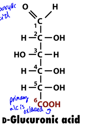
Uronic Acids
Uronic Acid: Produced by the selective oxidation of the terminal primary alcohol group (C6 in hexoses) to a carboxylic acid (−COOH), preserving the C1 aldehyde carbonyl (e.g., D-glucose converted to D-glucuronic acid).

Aldehydes are reduced to form?
alcohols
Sugar alcohols
ribitol, xylitol, glycerol
Homopolysaccharides vs. Heteropolysaccharides
Homopolysaccharide: A complex carbohydrate polymer composed of only a single recurring type of monosaccharide unit (e.g., cellulose, glycogen, amylose, and chitin).
Heteropolysaccharide: A complex carbohydrate polymer assembled from two or more distinct types of monosaccharide building blocks (e.g., peptidoglycan, glycosaminoglycans, and cell-surface ABO antigens).
Biosynthetic Precursors of Glycogen vs. Amylopectin
Amylopectin Synthesis: Plants synthesize amylopectin utilizing ADP-glucose as the activated glucosyl donor substrate.
Glycogen Synthesis: Animals and fungi synthesize glycogen utilizing UDP-glucose as the activated glucosyl donor substrate.
Significance: Tagging glucose with distinct nucleotide diphosphates regulates carbohydrate storage pathways across biological kingdoms.
Enzymes of Starch Breakdown

α-Amylase: Salivary and pancreatic enzyme that cleaves internal α(1→4) glycosidic bonds.
α-Glucosidase: Exoglycosidase that sequentially hydrolyzes one terminal glucose residue at a time.
Debranching Enzyme: Hydrolyzes both residual α(1→4) bonds and branch point α(1→6) bonds to enable complete starch degradation.
Enzymes of Glycogen Breakdown

Glycogen Phosphorylase: Phosphorolytically cleaves α(1→4) glycosidic bonds sequentially inward from the non-reducing ends, releasing glucose-1-phosphate without expending ATP.
Glycogen Debranching Enzyme: Hydrolyzes α(1→6) branch connections to release free glucose and expose remaining linear chains for continued breakdown.
Metabolic Consequences of Carbohydrate-Deficient Diets
Brain Fuel Requirement: The brain cannot utilize unesterified fatty acids directly and relies primarily on circulating glucose for normal metabolic function.
Kidney Stones: Carbohydrate restriction elevates uric acid production, leading to decreased urine solubility and uric acid calculus formation.
Gout: Elevated systemic uric acid crystallizes within joint spaces, precipitating acute inflammatory arthritis.
Caloric Guideline: Carbohydrates should account for approximately 20% of total daily caloric intake in a balanced diet.
Glycosaminoglycans (GAGs) Structure and Function

Structure: Long, unbranched heteropolysaccharides consisting of repeating disaccharide units composed of an amino sugar and an uronic acid.
Physiological Role: Major structural components of connective tissues, extracellular matrix, and synovial fluid providing mechanical joint lubrication.
Hyaluronic Acid: Essential high-molecular-weight GAG that prevents intracranial hemorrhage in premature infants and maintains tissue hydration.
Cartilage Glycan Components: Glucosamine and Chondroitin Sulfate
Constituents: Glucosamine (an amino monosaccharide) and chondroitin sulfate (a sulfated glycosaminoglycan) serve as primary structural building blocks of articular cartilage.
Biological Role: Provide mechanical shock absorption and cushioning between articulating bones in synovial joints.
Therapeutic Synergy: Combined administration (e.g., Dasuquin) stimulates cartilage extracellular matrix synthesis and reduces joint degradation in osteoarthritis.
Proteoglycans and Glycoprotein Linkages (O-Linked vs. N-Linked)

Proteoglycans: Hybrid macromolecules consisting of a core protein covalently bound to one or more glycosaminoglycan chains.
O-Glycosidic Bonds: Oligosaccharides attached via the hydroxyl oxygen of Serine (Ser) or Threonine (Thr) residues.
N-Glycosidic Bonds: Oligosaccharides attached via the amide nitrogen of Asparagine (Asn) or Glutamine (Gln) residues.
ABO Blood Group Antigen Structures

O Antigen (Core): Foundational oligosaccharide chain terminating in an α(1→2)-linked fucose (Fuc) on galactose.
A Antigen: Core modified by addition of an N-acetylgalactosamine (GalNAc) residue via an α(1→3) glycosidic bond.
B Antigen: Core modified by addition of an extra D-galactose (Gal) residue via an α(1→3) glycosidic bond.
ABO Blood Group Transfusion Compatibility
Type O: Expresses only H (O) antigen core; contains anti-A and anti-B antibodies; universal erythrocyte donor, can receive only Type O red cells.
Type A: Expresses A antigen; contains anti-B antibodies; can safely donate to A and AB, and receive from O and A.
Type B: Expresses B antigen; contains anti-A antibodies; can safely donate to B and AB, and receive from O and B.
Type AB: Expresses both A and B antigens; contains neither anti-A nor anti-B antibodies; universal recipient, can donate red cells only to Type AB.