Physiology Exam 3
 Call Kai
 Learn
Learn Practice Test
 Spaced Repetition
 Match
Match Flashcards
 Knowt Play
1/251

There's no tags or description
Looks like no tags are added yet.
Name | Mastery | Learn | Test | Matching | Spaced | Call with Kai | Chat |
|---|
No analytics yet
Send a link to your students to track their progress
252 Terms
What does the cardiovascular system do?
Transport of material: gases, nutrients, waste, communication, degense against pathogens, temperature homeostasis
The heart is a pump in the cardiovascular system,how?
The atria receives blood returning to the heart, the ventricles pump the blood out, the septum divides left and right halves
What are the blood vessels in cardiovascular system?
-veins, arteries, capillaries
-pulmonary and systemic circulation
-portal system joins two capillary beds in series
Where is the heart within the thoracic cavity?
Ventral side; sandwiched between the lungs

What allows for ventricular contraction to squeeze blood upward from the apex of the heart?
The spiral arrangement of ventricular muscle
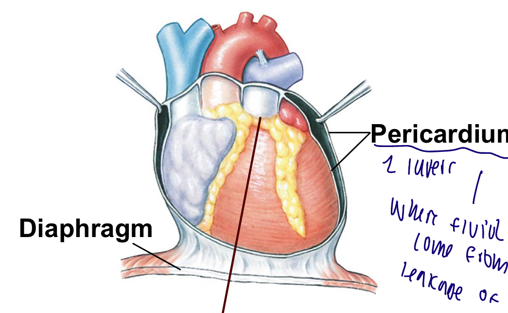
What is the pericardium
It encases a heat with a membranous fluid filled sac
-inner and outer layer to the heart
The internal structure of the heart is
Mostly myocardium, has two heart valves for 1 way flow
Atrioventricular valves: between atria and ventricles, tricuspid valve on right, mitral valve on left
Semilunar valves: between ventricles and arteries, aortic valve, pulmonary valve
Ventricular contraction
Left ventricle contracted, pushing from the wall
The mitral valve on the left will close
Aortic valve will open
Means the papillary muscles and the chordal tendineae tenses up
The atrioventricular valve remains closed to prevent blood backward into atria

Ventricular relaxation
The left ventricle is dilated
The aortic valve is closed
The mitral valve is open
Chordae tendineae and papillary muscles are relaxed
Semilunar valves prevent blood that entered the arteries from flowing back into ventricles

What does cardiac muscle have?
contractile cells and autorhythmic cells/pacemakers
Contractile-pumps blood
Autorhytmic/pacemakers- signal and how nerves react
What does cardiac muscle not have?
organized sarcomeres
Myocardial characteristics
Branched, uninucleate, cells attached to each other by specialized junctions called intercalated disks

Function of desmosomes
transfer force from cell to cell; in intercalated disks
Function of gap junctions
Provide electrical connection to pass rapidly from cell to cell, without this electrical signal it can cause arrhythmia
How much volume do mitochondria occupy in cardiac muscle?
one-third, helps generate energy for pump
Which type of muscle is the slowest to contract and to relax?
Smooth muscle

Where does an action potential in the heart start?
SA node, starts with heart pacemaker cells

Pacemaker potential?
Is the unstable membrane potential in autorhymthic cells
The depolarization is due to Ca channels opening
What does the pacemaker potential look like on a graph?
It depolarizes from -60 to -40 at a constant rate
Gradually becomes less negative until reaches the threshold, thus new triggers action potential

What is important to remember about the pacemaker potential?
It is caused by leakage of potassium out and sodium in (net sodium in); comparable to an hourglass/timer

What happens after the threshold is reached in myocardial autorhythmic cells?
Calcium channels open and let calcium in the cell, further depolarizing the membrane, then calcium channels close at the peak, and potassium channels open and let potassium out, repolarizing the membrane back to -60 and the process starts over once the If channel opens

What effect do parasympathetic nerves have on the heart?
Decreases heart rate through acetylcholine on muscarinic receptors
What effect do sympathetic nerves have on the heart?
Increases heart rate through norepinephrine on beta1-adrenergic receptors
How does parasympathetic stimulation slow the heart rate?
Hyperpolarizing the membrane potential/ rate depolarization decreases, and decreasing permeability, causes K+ increase efflux and Ca decrease influx increasing the time it takes for the potential to reach threshold, slowing heart rate

How does sympathetic stimulation speed up the heart rate?
Depolarizing the membrane potential increases the rate and increasing permeability, sodium increases and calcium influx decreasing the time it takes for the potential to reach threshold, speeding up heart rate

The autonomic control of heart rate
Parasympathetic stimulates hyperpolarizing the membrane potential, slowing depolarization, slowing heart rate
Sympathetic stimulation and epinephrine depolarizes autorhytmic cells, speeding up pacemaker potential and increasing heart rate

What are the phases of the action potential of a cardiac contractile cell?
0. sodium channels open
1. sodium channels close at the peak
2. calcium channels open; fast potassium channels close
3.calcium channels close; slow potassium channels open
4. resting potential

What is the importance of the longer myocardial action potential?
It helps prevent the sustained contraction called tetanus due to calcium, entry, creating the plateau
Why is it important that cardiac muscle don't achieve tetanus?
Cardiac muscles must relax between contractions so the ventricles can fill with blood
How long is the refractory period in cardiac muscle?
Almost as long as the entire muscle twitch; prevents tetanus
EC coupling in cardiac muscle
1. action potential enters from adjacent cell
2. voltage-gated calcium channels open and calcium enters the cell
3. calcium induces calcium release through ryanodine receptor-channels (RyR)
4. local release causes calcium spark
5. summed calcium sparks create a calcium signal
6. calcium ions bind to troponin to initiate contraction
7. relaxation occurs when calcium unbinds from troponin
8. calcium is pumped back into the sarcoplasmic reticulum for storage
9. calcium is exchanged with sodium by the NCX antiporter
10. sodium gradient is maintained by the Na K ATPase

What percentage does the ECF contribute to calcium stores in cardiac muscle?
10 percent
Action potential, excitation-contraction coupling in cardiac muscle
Starts with the heart pacemaker cells
Voltage gated L type ca channels in cardiac muscle cell membrane opens (extra cellular calcium contributes 10%)
Ryanodine receptors open in the SF in excitation contraction coupling in cardiac muscle
Calcium ion is released, calcium binds to troponin to start muscle contractions
The cords bridge cycle as in skeletal muscle
Relaxation in excitation contraction coupling In cardiac muscle
Calcium unbinds troponin
Calcium removed from cytoplasm
Back into the SR with ca ATPase
Out of cell through Na+ Ca2+ exchanger
What is the force generated in cardiac muscle proportional to?
To the number of active crossbridges; determined by how much calcium is bound to troponin, sacromere length affects force of contraction
Where does the right atrium receive blood from?
Superior and inferior vena cava; from the body

Where does the right ventricle pump blood to?
Lungs through pulmonary arteries

Where does the left atrium receive blood from?
Lungs through pulmonary veins
Where does the left ventricle pump blood to?
To the body through the Aorta
What happens if the right side of the heart slows down?
Backup/pressure buildup that causes edema in the body
What happens if the left side of the heart slows down?
Backup/pressure buildup that causes edema in the lungs
Where is the tricuspid valve located?
between right atrium and right ventricle
Where is the bicuspid valve located?
between left atrium and left ventricle
What is important to remember about valves within the cardiovascular system?
To prevent backflow
Sinoatrial SA node for cardiac electrical conduction
Sets the pace of heartbeat at 70 bpm, AV node is 50 bpm and purkinje fibers (24-40 bpm) can act as pacemakers under som,e conditions
Intermodal pathway from SA to atrioventricular AV node
Routes the direction of electrical signals so heart contracts from apex to base
AV node delay is accomplished by slower conduction signals through nodal cells
Purkinje fibers
Transmits electrical signals down the atrioventricular bundle to left and right bundle branches
What are the steps involved in the conducting system of the heart?
1. SA node depolarizes
2. electrical activity goes rapidly to AV node via internodal pathways
3. depolarization spreads more slowly across atria. conduction slows through AV node
4. depolarization moves rapidly through ventricular conducting system to the apex of the heart
5. depolarization wave spreads upward from the apex

What is the P wave associated with?
Atrial depolarization

What is the QRS complex associated with?
Ventricular depolarization

R wave
Atrial repolarization

What is the T wave associated with?
ventricular repolarization

P-R segment? Or P-Q segment?
Conduction through AV nodes and AV bundle

How is heart rate determined?
time between two P waves or two Q waves
The electrical events of the cardiac cycle
-p wave: atrial depolarization
P-Q; conduction through AV node and AV bundle, atria contract
Q wave
R wave: atria, repolarization
QRS; ventricular depolarization
S wave
S-T: ventricles contract
T waves: ventricles repolarization

Normal ECG and abnormal

Diastole
cardiac muscle relaxes
Systole
cardiac muscle contracts
What are the steps of mechanical events of the cardiac cycle?
1. late diastole - both sets of chambers are relaxed and ventricles fill passively, atria are filling with blood from the vein. if the AV valves are open, ventricles are filling
2. atrial systole - atrial contraction forces a small amount of additional blood into ventricles
3. Isovolumic ventricular contraction - first phase of ventricular contraction pushes AV valves closed but does not create enough pressure to open semilunar valves. this means all valves are shut, but atria relax and blood flows in atria
4. ventricular ejection - as ventricular pressure rises and exceeds pressure in the arteries, the semilunar valves open and blood is ejected.
5. Isovolumic ventricular relaxation - as ventricles relax, pressure in ventricles falls, but still higher than atrial pressure. Blood flows back into cusps of semilunar valves and snaps them closed
the AV valve will open again once the ventricular pressure drops below atrial pressure

Where is the first heart sound heard?
following closure of the AV valves; Lub
Where is the second heart sound heard?
following closure of semilunar valve; Dub
Auscultation
Listening to the heart through the chest wall through a stethoscope
What is the pathway of electrical signals of the heart?
SA node, through internodal pathways to the AV node, through the AV bundle, bundle branches, and finally Purkinje fibers
What is the normal max fill volume of a ventricle?
135 mL

What is the normal volume of blood that stays behind in the ventricles?
65 mL

End-systolic volume (ESV)
volume of blood remaining in each ventricle after systole; blood that stays behind after contraction; normally around 65 mL
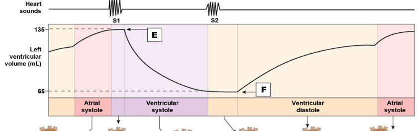
End-diastolic volume (EDV)
volume of blood in each ventricle at the end of diastole; total blood volume; normally about 135 mL

Stroke volume
Amount of blood pumped by one ventricle during a contraction
How do you calculate stroke volume?
EDV-ESV=SV
volume of blood before contraction- volume of blood after contraction= SV
What is the average stroke volume?
70 mL
Cardiac output (CO)
heart rate x stroke volume
What is the average cardiac output?
5 L/min
How does the sympathetic nervous system control cardiac contraction?
catecholamines bind to beta1 receptors to affect voltage-gated calcium channels, where open time increases and theres an increase of calcium entry from ECF ——and phospholamban to create more forceful and faster contractions
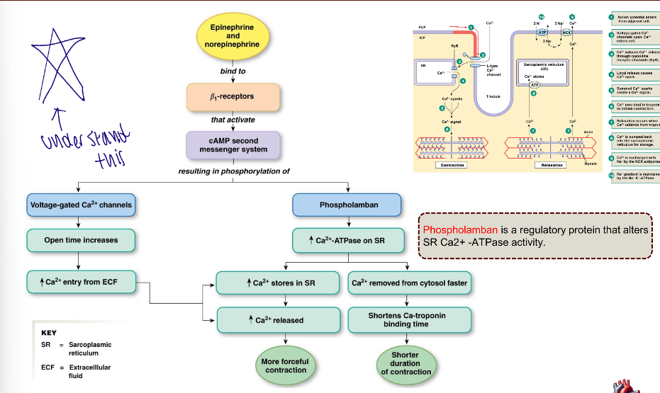
phospholamban
are regulatory protein that increases the Calcium ATPase on SR
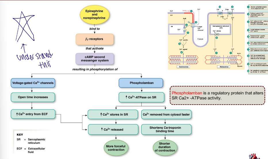
How are more forceful contractions produced in cardiac muscle?
calcium stores are increased in the SR, allowing more calcium to be released, producing a more forceful contraction

How are faster contractions produced in cardiac muscle?
calcium is removed from cytosol faster, shortening calcium-troponin binding time, producing a faster contraction

Inotropic agent
Any chemical that affects contractility
What kind of effects do epinephrine, norepinephrine, and digitalis have on contractility?
positive inotropic effects
What do negative inotropic effects do?
Decrease contractility
What is the force of contraction affected by?
length of muscle fiber and contractility of the heart
What happens when the ventricular wall stretch increases?
Stroke volume increases
Preload
The degree of myocardial stretch before contraction
afterload
force or resistance the heart must overcome to eject blood during systole, like the pressure in the aorta against left ventricle contracts
Frank-Starling Law
stroke volume increases as end-diastolic volume (EDV) increases
What is EDV determined by?
venous return
What is venous return affected by?
skeletal muscle pump, respiratory pump, and sympathetic innervation of veins
What is cardiac output a function of?
Heart rate and stroke volume
What is driving pressure created by?
Ventricles

Where is pressure created and then thereafter transferred to?
Created by contracting muscles and transferred to the blood
What happens to blood pressure if blood vessels dilate?
Blood pressure decreases
What happens to blood pressure if blood vessels constrict?
Blood pressure increases
What does the pressure gradient in blood vessels look like?
High in the Aorta and arteries, medium in capillaries, and low in the veins and Vena cavae

What is the relationship between flow and pressure gradient?
Directly proportional, as pressure increases, so does flow and vice versa
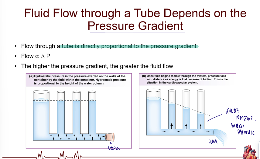
What determines whether or not a fluid flows through a tube?
Whether or not there is a positive pressure gradient; fluid moves from high pressure to low pressure

What is important to remember about flow and pressure gradient?
Flow depends on pressure gradient (delta pressure), not on the absolute pressure
How is flow related to resistance?
flow is inversely proportional to resistance; if resistance increases, flow decreases and vice versa

velocity relation to narrowness
the narrower the vessel, the faster the velocity of flow

How is resistance related to length of the tube? (blood vessel)
directly proportional; resistance increases as length increases
How is resistance related to viscosity? (blood)
directly proportional; resistance increases as viscosity increases