AE 1.5 - Measurements, CAD, Stress Echo, & Wall Segments
 Call Kai
 Learn
Learn Practice Test
 Spaced Repetition
 Match
Match Flashcards
 Knowt Play
1/56

There's no tags or description
Looks like no tags are added yet.
Name | Mastery | Learn | Test | Matching | Spaced | Call with Kai | Chat |
|---|
No analytics yet
Send a link to your students to track their progress
57 Terms
Accurate / proper measurements
1. On-axis image
2. Timing of cycle
- (ie: end-diastole, end-systole, mid-systole)
3. Location
- use landmarks per ASE guidelines
4. Plane
- perpendicular to the structure of interest
Differences between M-mode & 2D measurements
M-mode
- best temporal resolution (best timing)
- 1D information regarding a particular cardiac structure as it relates to time & distance
- the strength of the reflected echo is represented as the brightness of structures appearing on the image display
2D
- main tool for a comprehensive echo study
- 2D image
- the strength of the reflected echo is represented as the brightness of structures appearing on the image display
Simpson's Biplane Method
- take LV & LA dimensions from both 2C & 4C
- use this method to get EF of LV
LV = should be less than 10% difference in lengths between 4C & 2C
LA = should be less than 5mm difference in lengths between 4C & 2C
Why Simpson's Biplane Method is preferred for LV volume calculations
- uses 2D info
- uses info from 2 different views
- allows for more unique volumes/areas along LV length via disks

A4C RV focused view (m-mode)
TAPSE
> 1.7cm
- align m-mode cursor along RV free wall as perpendicular to the lateral tricuspid valve annulus as possible
- distance measured leading edge of annulus to from end-diastole to end-systole

PLAX measurements (diastole)
IVSd
LVIDd
LVPWd
timing
- end-diastole: onset of the QRS, first frame after MV closure or frame in which the cardiac chamber is largest
- taken at MV leaflet tips

PLAX measurements (systole)
LVIDs
timing
- end-systole: frame after AV closure or the time in when cardiac chamber is smallest
- taken at the MV leaflet tips

Zoomed AV measurements
*LVOT
*Aortic annulus
timing
- mid-systole: AV open
- LVOT: should be taken 5-10mm proximal to annulus (before av)

Zoomed aortic root measurements
*Aortic sinuses
*STJ
Ascending aorta
timing
- end-diastole: onset of the QRS or frame before AV opens

High PLAX Asc Aorta measurements
Ascending Aorta
timing
- end-diastole: onset of the QRS or frame before AV opens

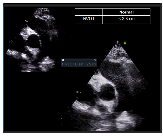
PSAX @ AV RVOT measurements
*RVOT
timing
- mid-systole: PV open
- RVOT: should be taken 5-10mm proximal to annulus
A4C Zoomed LV measurements
LVEDV (4C)
LVESV (4C)
LVEF
- should be less than 10% difference in lengths between 4C & 2C
- trace the LV cavity along the interface of the compacted and noncompacted myocardium of the chamber wall excluding papillary muscles
-- make sure LV length is centered
timing
- ventricular end-diastole: one frame after MV closure or when LV is at its largest
- ventricular end-systole: one frame immediately preceding MV opening or when LV is at its smallest

A4C Zoomed LA measurements
LAVI (4C)
- should be less than 5mm difference in lengths between 4C & 2C
- trace the LA cavity from medial to posterior annulus excluding LAA and pulm veins
-- make sure LA length is centered
timing
- ventricular end-systole: one frame immediately preceding MV opening or when LA is at its largest

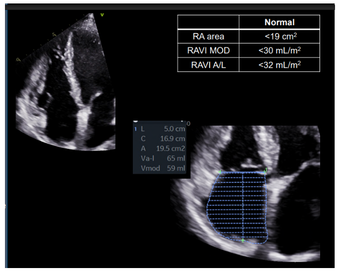
RA focused A4C measurements
RA area/volume
- trace the RA cavity from medial to posterior annulus excluding RAA
- make sure RA length is centered
timing
- ventricular end-systole: one frame immediately preceding TV opening or when RA is at its largest
RV focused A4C measurements
RV dimensions
Basal: basal 1/3rd of RV
Mid: Middle 1/3rd of the RV at the level of LV pap muscles
Longitudinal: mid-plane of tricuspid annulus to the RV apex
timing
- ventricular end-diastole: one frame after TV closure or when RV is at its largest
**Do not measure TV annulus at the base

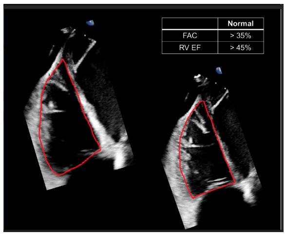
RV focused A4C (FAC) measurements
*RV Fractional Area Change (FAC)
- trace along RV endocardium, including RV trabeculations, papillary muscles & mod band in RV area
timing
- ventricular end-diastole: one frame after TV closure or when RA is at its largest
- ventricular end-systole: one frame immediately preceding MV opening or when LV is at its smallest
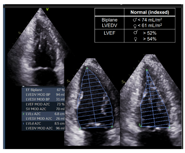
A2C Zoomed LV measurements
LVEDV (2C)- Biplane
LVESV (2C)- Biplane
LVEF- Biplane
- trace the LV cavity along the interface of the compacted and noncompacted myocardium of the chamber wall excluding papillary muscles. Make sure LV length is centered
timing
- ventricular end-diastole: one frame after MV closure or when LV is at its largest
- ventricular end-systole: one frame immediately preceding MV opening or when LV is at its smallest
A2C Zoomed LA measurements
LAVI (2C) - Biplane
- trace the LA cavity from medial to posterior annulus excluding LAA and pulm veins
-- make sure LA length is centered
timing
- ventricular end-systole: one frame immediately preceding MV opening or when LA is at its largest

Subcostal IVC measurements
IVC
measurements must be perpendicular to IVC walls taken inner edge to inner edge
make measurement 1-2 cm from the junction of the IVC into the RA

Subcostal Ao measurements
*Descending Aorta
measurements must be perpendicular to aorta walls taken inner edge to inner edge

SSN measurements
*Aortic Arch
measurements must be perpendicular to aorta walls taken inner edge to inner edge
between Innominate artery and LCCA
make sure not to include any of the branching arteries

Proper imaging views for right -sided measurements
PSAX @ AV level
- RVOT
A4C
- RA focus & area/volume
- RV focus & dimensions & fractional area change
Subcostal
- IVC
- RAP
Proper imaging views for left -sided measurements
PLAX
- IVSd
- LVIDd
- LVPWd
- LVIDs
Zoomed AV
- LVOT
- Aortic Annulus
Zoomed aortic root
- Aortic Sinuses
- STJ
- Ascending Aorta
High PLAX
- Ascending Aorta
A4C Zoomed LV
- LVEDV (4C)
- LVESV (4C)
- LVEF
A4C Zoomed LA
- LAVI (4C)
A2C Zoomed LV
- LVEDV (2C) - Biplane
- LVESV (2C) - Biplane
- LVEF- Biplane
A2C Zoomed LA
- LAVI (2C) - Biplane
Subcostal
- Descending Aorta
SSN
- Aortic Arch
Common measurement errors
1) foreshortening
-- apicals = go low & lateral
-- plax = go high & medial
2) incorrect alignment
-- always stay perpendicular to walls 丄
-- inner vs leading edge
3) poorly defined borders
-- must see walls of structure
Circulation
Pulmonary
RA → TV → RV → PV → Main PA → RPA & LPA → Capillaries
O2 = 75%
Systemic
LA → MV → LV → AV → Aorta → Peripheral arteries → Arterioles
O2 = 95%
Coronary
arteries = to myocardium from Aorta base
veins = away from myocardium & back to RA

Coronary Circulation
fill primarily during ventricular diastole

Major branches of the Left Main Coronary Artery
- left anterior descending coronary artery
- left circumflex coronary artery

LAD
left anterior descending coronary artery
- travels down towards the anterior aspect of the heart
- feeds the anterior, septal, & apical walls
- small branches called perforators supply the IVS
- may supply a portion of the RV apex, moderator band, and the SA node 50% of the time

LCx
- left circumflex coronary artery
- gives rise to obtuse marginal branches

RCA
right main coronary artery
- primary coronary supply to the RV
- also supplies the SA node, AV node, RA, parts of the IVS, IAS, & Inferior LV wall
Posterior descending artery (PDA)
- RCA gives rise to PDA in 85% of patients, this is called “right dominance”
- originates from either the right coronary artery (right dominant), left circumflex artery (left dominant), or both (codominant)

Myocardial regions supplied by each coronary artery
LAD
Anterior wall
Anteroseptal wall
LV apex
LCx
Anterolateral (Lateral) wall
Inferolateral (posterior) wall
RCA
Inferior wall
Inferoseptal wall
RV apex
RV free wall
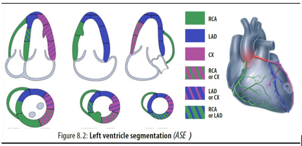
Coronary sinus & venous drainage of the myocardium
Coronary Sinus
- 3 veins drain into coronary sinus: small, middle, & great cardiac vein
- located in the posterior part of the coronary sulcus
- predominantly collects blood from the myocardium & drains into the inferoposterior aspect of the RA
- guarded by a small crescent shaped Thebesian valve
- Anterior Right Ventricular Veins
- Thespian Veins

Basic development of coronary artery disease
-

Angina
Unstable
- characterized by chest pain that occurs suddenly and unexpectedly at rest and/or pain that is more severe or prolonged than prior
Stable
- generally occurs with exercise and is relieved by rest or nitrate medications
Infarction
blood flow completely cut off resulting in tissue death

Myocardial remodeling
- stunning
- myocardial function is still depressed for a variable period of time even when blood flow is restored to area
- induced by acute ischemia
- is temporary
- the changes in the size, shape, structure, and electrical properties of the heart in response to injury, disease, or chronic stress
Ischemic cascade
-

The sequence of events following reduced coronary blood flow
ischemia --> diastolic dysfunction --> systolic dysfunction --> EKG changes --> chest pain
perfusion abnormalities --> wall motion abnormalities --> EKG changes --> angina, MI, death
Echocardiographic appearance of infarcted myocardium
1. Myocardium becomes akinetic
- happens within hours, surrounding tissues may be “stunned”
2. Myocardium thins after infarction
3. Acoustic impedance of myocardium changes as scarring and healing occurs
- becomes more echogenic (very bright)
True aneurysm
- thin, dyskinetic
- wider neck than pseudoaneurysm
- bulge distorts the normal contour of the LV cavity
- contains all layers of myocardium (NOT a rupture)
- apical, inferior, & posterior areas are most common
- low risk of rupture

Pseudoaneurysm
- narrow neck
- free wall of LV ruptures and a hemopericardium is contained
- walls = thrombus & pericardium
- higher risk of free rupture of myocardium – survival is rare
- most common location is basal inferior segment – NOT APICAL

Post-MI Pericarditis
- Dressler's syndrome
- occurs 1-8 weeks after MI
- inflammation of pericardium
- will hear friction rub murmur
Common indications for stress echocardiography
- Evaluation of Symptoms
-- chest pain/angina
-- SOB
-- fatigue
- baseline EKG symptoms
- known CAD
- risk stratify before non-cardiac surgery, post-MI, after intervention (PTCA, CABG)
- evaluation Valve disease
- HCM gradients (hocm)
Contraindications (should not do) for Stress Echo
MI in last 48 hours
Unstable Angina
Uncontrolled HTN
Uncontrolled arrhythmias
Severe AS
Decompensated CHF
Mobile LV thrombus
Acute aortic dissection
Basic exercise stress echo protocol
baseline
- resting images of LV (6)
4C, 2C, 3C, PLAX, Pap muscles, Apex
post-exercise
- continuous capture
- has 60sec to get the 6 images again
Importance of obtaining post-exercise images quickly
- sonographer has 60 seconds from the time the treadmill stops to obtain peak/post exercise images AND with the HR be above target HR
- false negative test from a delay in obtaining images
17-segment model (bullseye)
pay attention to septal insertion landmark
outer --> inner:
- basal --> mid --> apical --> apex
anterior v inferior
septal v lateral

17-segment model
pay attention to septal insertion landmark

Wall segments (chamber views)
4C -- 2C -- 3C
IS AL -- I A -- IL AS
"i saw a lion -- in africa -- it loved angry sheep"

Basal segments
basal anterior
basal anteroseptal
basal inferoseptal
basal inferior
basal inferolateral
basal anterolateral

Mid segments
- has papillary muscles
mid anterior
mid anteroseptal
mid inferoseptal
mid inferior
mid inferolateral
mid anterolateral

Apical segments
apical anterior
apical septal
apical inferior
apical lateral
LISA

Apex segments
apical cap/apex

Relationship between coronary artery territories & wall motion abnormalities
-

Wall motion
hyperkinetic = -1 = more vigorous than normal
normokinetic = 1 = normal wall motion
hypokinetic = 2 = reduced wall motion
severely hypokinetic = 2.5 = severely reduced wall motion
akinetic = 3 = no/little wall motion
dyskinetic = 4 = abnormal motion
aneurysmal = 5 = aneurysmal motion/bulging
