BIO 211 LAB QUIZ 2 PRACTICE
 Call Kai
 Learn
 Practice Test
 Spaced Repetition
 Match
 Flashcards
 Knowt Play
1/142

There's no tags or description
Looks like no tags are added yet.
143 Terms

What part of the heart is this?
Apex

What region of the heart is this?
Base
The heart is enclosed within a tough, double layered, sac called the:
Pericardium

Identify layer of heart
Endocardium

Identify layer of heart
Myocardium

Identify layer of heart
Epicardium
What layer of the heart walls lines the chamber?
Endocardium
What layer of the heart walls is the heart muscle itself?
Myocardium
What layer of the heart walls surrounds the heart?
Epicardium

Identify the chamber.
Right atrium

Identify the chamber.
Left atrium

Identify the chamber.
Right ventricle

Identify the chamber.
Left ventricle
Define: “internal ridges of myocardium in right atrium and both auricles”
pectinate muscle

Identify this structure on the model:
Pectinate muscle

Identify structure on heart: (red arrow)
Pectinate muscle

Identify the feature
Fossa ovalis

Identify the feature
Fossa ovalis

Identify the feature
Coronary sinus opening

Identify the structure: (flap)
Right auricle

Identify the structure: (flap)
Left auricle

Identify the structure
Ligamentum arteriosum

Identify this structure:
Interventricular septum
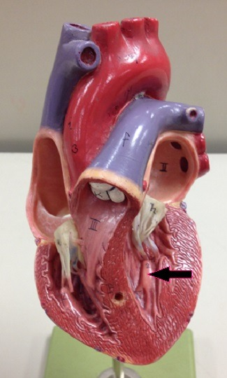
Identify this structure
Papillary muscle

Identify those ridges:
Trabeculae carneae

Identify the structure:
Chordae tendineae

Identify feature, orange arrow
Moderator band

Identify the groove:
Anterior interventricular sulcus
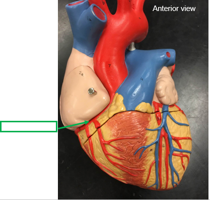
Identify the groove:
Coronary sulcus
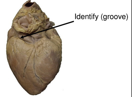
Identify groove:
Coronary sulcus

Idenitfy
posterior interventricular sulcus

Identify
posterior interventricular sulcus
What valves are under the Atrioventricular portion?
Bicuspid valve and Tricuspid valve
What valves are under the Semilunar portion?
Pulmonary and aortic semilunar valve

Identify the valve
Bicuspid valve
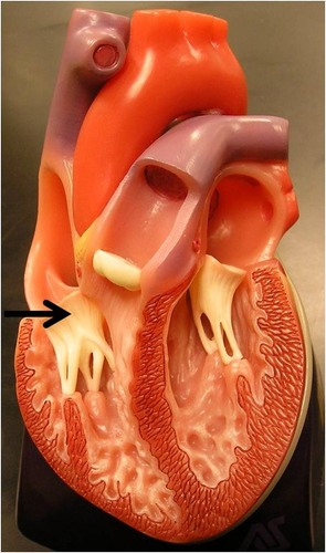
Identify the valve
Tricuspid valve

identify the valve
Pulmonary valve

Identify
Aortic semilunar valve

Identify artery
Right coronary artery

Identify artery
Right coronary artery

Identify artery
Left coronary artery

Identify artery
Left coronary artery
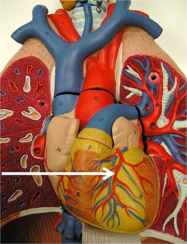
Identify artery
Anterior interventricular artery

Identify artery
Circumflex artery

Identify the artery
Posterior interventricular artery

Identify the vein
Great cardiac vein
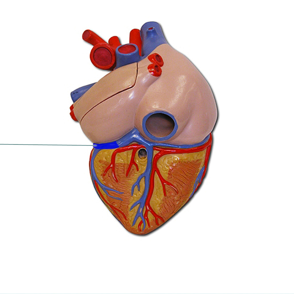
Identify the vein (coronary sulcus)
Great cardiac vein

Identify the vein
Middle cardiac vein

Identify
Coronary sinus
The middle cardiac vein and great cardiac vein empty into
coronary sinus
The coronary sinus empties into
Right atrium

Identify the vessel
Ascending aorta

Identify the vessel feature
Aortic arch

Identify
Intraventricular septum

Identify the vessel
Pulmonary trunk

Identify the vessel
Superior vena cava
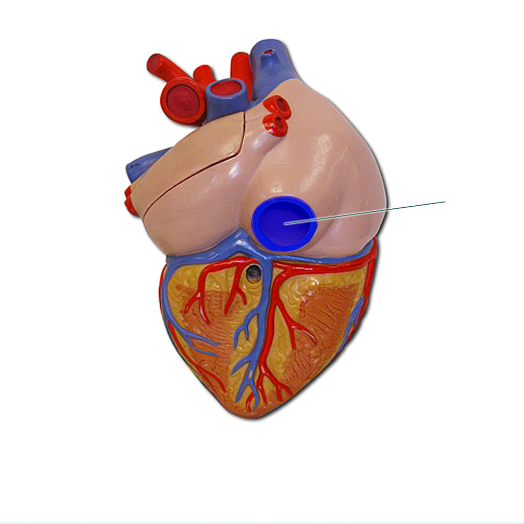
Identify the vessel
Inferior vena cava

Identify
Coronary sinus opening

Blood flow begins with
Superior/Inferior vena cava and coronary sinus
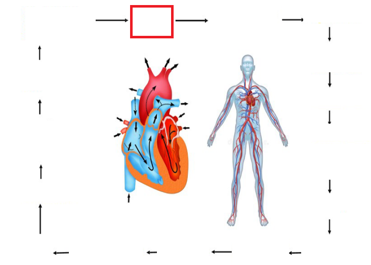
2nd stage in blood flow
Right atrium

3rd stage in blood flow
Tricuspid valve

4th stage in blood flow
Fourth ventricle
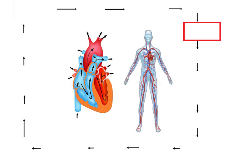
5th stage in blood flow
Pulmonary valve
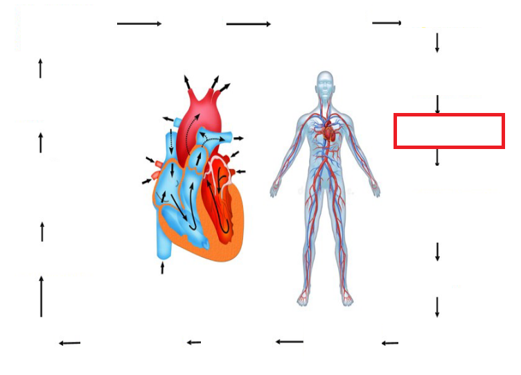
6th stage in blood flow
Pulmonary trunk
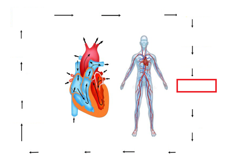
7th stage in blood flow
Pulmonary arteries
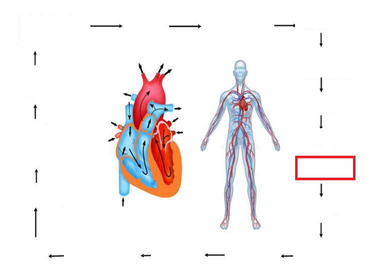
8th stage in blood flow
Gas exchange in lungs

9th stage in blood flow
Pulmonary veins

10th stage in blood flow
Left atrium

11th stage in blood flow
Left AV valve

12th stage in blood flow
Left Ventricle

13th Stage in blood flow
Aortic semilunar valve

14th stage in blood flow
Aorta

15th stage in blood flow
systemic arteries

16th stage in blood flow
gas and nutrient exchange in peripheral tissue

Identify the feature on the model
Intercalated disc
Listening to heart sounds is defined as
auscultation
machine used to measure electrocardiography
electrocardiograph
a print out of the heart’s electrical activity is
electrocardiogram

What part of the wave is this?
P wave

What part of the wave is this?
QRS wave

What part of the wave is this?
T wave
sound of closure of the atrioventricular valves (systole)
Lubb (S1)
Used to listen for “sounds of Korotkoff” (sounds from returning blood) when taking
blood pressure
Stethoscope
Stethoscope checks for
initial and ending korotkoff
Ways to raise BP:
Standing up, exercise, smoking, cold weather
Systolic
Ventricular contraction
Diastolic
Ventricular relaxation

What is the name of this? (fetal)
ductus arteriosus

What is the name of this? (fetal)
Foramen ovale

Identify(fetus)
ductus venosus

Identify (adult)
ligamentum venosus

Identify (fetus)
Umbilical vein

Identify (fetus)
Umbilical arteries

Identify (adult)
umbilical ligaments

Identify (adult)
Ligamentum teres
Ways to decrease BP
Sleep, resting
sound of the closure of the semilunar valves (diastole)
Dubb (S2)
Name the 5 structures involved in the Cardiac Conduction system in order.
SA node → AV node → Bundle of his → R/L Bundle of Branches → Purkinje fibers
Which structure is the heart's natural pacemaker?
SA node
Which structure briefly delays the electrical impulse?
AV node