ANPS Exam 1
 Call Kai
 Learn
Learn Practice Test
 Spaced Repetition
 Match
Match Flashcards
 Knowt Play
1/175

There's no tags or description
Looks like no tags are added yet.
Name | Mastery | Learn | Test | Matching | Spaced | Call with Kai | Chat |
|---|
No analytics yet
Send a link to your students to track their progress
176 Terms
Life’s Heirarchy:
-Atoms are organized into molecules
-Molecules are organized into cells
-Cells are organized into tissues
-Tissues are organized into organs
-Organs are organized into organ systems
-Organ systems make up the entire organism
Elements:
-Element: pure substance made of only one type of atom
-Elements and atoms are the smallest building blocks of the human body
-CHNO are the most abundant elements in the human body
-An atom is the smallest unit that retains all chemical properties of an element
-what determines the chemical properties of an element?
subatomic structure
Subatomic Structure:
Subatomic particles include:
1. Neutrons – no charge, found in nucleus
2. Protons – positive charge, found in nucleus
3. Electrons – negative charge, found in outer shell
-Atomic number = number of protons
-Mass number = number of protons and neutrons
-Atoms interact through the electrons in their outer electron shell (valance shell)
Stability:
Atoms are more stable with their outer shell filled with electrons:
• First shell holds 2 electrons
• Second shell holds 8 electrons
• Third shell holds 8 electrons
Bonding and octet rule:
-Atoms interact through the electrons in their outer electron shell
-The “octet rule” states that atoms tend to interact in such a way that they end up having 8 electrons in their outer shell (hydrogen and helium are exceptions).
Ions:
-Ions are atoms that have gained or lost electrons and become electrically charged as a result
-Ionic bonds form between 2 ions to neutralize charges
-Positively charged ions are called cations (ex. Na+): CAT paws
-Negatively charged ions are called anions (ex. Cl-): An- Anti negative
-Ions carry charge and allow for the electrical signaling to occur
-Cations: sodium, potassium, magnesium, hydrogen, carbonate
-Anions: Chloride, bicarbonate, phosphate
Common Ions in the human body:
-Sodium: Na+
-Potassium Ion: K+
-Calcium: Ca2+
-Magnesium Ion: Mg2+
-Hydrogen Ion: H+
-Chloride ion: Cl-
-Bicarbonate: HCO3
-Phosphate ion: PO3/4-

Ionic bonds:
-a type of chemical connection formed through the electrostatic attraction between oppositely charged ions created by the transfer of electrons between atoms
-In an ionic bond, one atom has given up one or more electrons, leaving it with a positive charge, while the other
atom has taken on one or more electrons, leaving it with a negative charge
-Strong bonds formed by the attraction between cations and anions: in order to neutralize charges
-Ions are atoms that have gained or lost electrons and become electrically charged as a result
-Ions are the building blocks/intermediate between ionic bonds
Bonds:
-Chemistry of the human body involves chemical bonding to create ionic, inorganic, and organic molecules
-Bonds tie one atom to another to create bigger chemical structures in the body
-ionic bonds, covalent bonds, and hydrogen bonds (polar electrostatic attractions)
Covalent Bonds:
-Stronger than Ionic because of sharing electrons vs transferring
-Non polar covalent: electrons shared equally, no partial or full charges
-Polar covalent: electrons shared unequally because of electronegativity differences: Ex. H2O, partial negative at O b/c slightly more electronegative
-The formation and destruction of covalent bonds often requires proteins called enzymes
Covalent bonds involving carbon and hydrogen make up the bulk of many organic molecules:
• Covalent bonds involving carbon and hydrogen can be polar or nonpolar, depending on the bonding partner.
• When hydrogen and carbon form a covalent bond with each other, this bond is nonpolar .
• When hydrogen or carbon form a covalent bond with more “electron hungry” atoms like oxygen and nitrogen, the resulting bond is polar
***The partial positive charge on hydrogen can interact with negative charges on other molecules or anions through hydrogen bonding

Hydrogen Bonds: type of electrostatic attraction
-Hydrogen frequently forms covalent bonds with other elements; b/c of hydrogen’s subatomic structure, it’s not very “electron hungry” and may have unequal sharing of electrons in covalent bonds (polar covalent bonds): EASY TO CREATE ELECTRONEGATIVITY DIFFERENCES WITH HYDROGEN
-Hydrogen bonds: The attraction between the partially positive (δ+) hydrogen atom of a polar molecule may be attracted to negatively charged atoms of other molecules
-Hydrogen bonds create weak forces that affect the structure and properties of compounds such as water and proteins.
-similar to an ionic bond, but weaker because the hydrogen atom involved is not fully + charged.
Water molecules interacting: surface tension
-Individual water molecules are attracted to one another by hydrogen bonds between the oxygen atom (slight negative charge) of one molecule and a hydrogen atom (slight positive charge) of another water molecule
-Each water molecule is held together by polar covalent bonds
-One water molecule is attracted to another by HYDROGEN BONDS
-H-bonds hold water drops together: Hydrogen bonds between water molecules give water surface tension, which is why water forms drops and why water striders can walk on water
Properties of water:
-Solute: substance being dissolved (Ex. salt)
-Solvent: Liquid (Ex. H2O)
-Solubility: Most molecules in the body dissolve in water (especially polar molecules)
-Reactivity: Water is involved in many of the chemical reactions in the body Dehydration synthesis forms bond; hydrolysis breaks bond
-High heat capacity: Water can absorb and retain a great deal of heat, so it takes a lot of energy to change temp
-Cushioning: Water surrounds certain body organs, and can protect against physical trauma (Ex. CSF protecting brain)
Water molecules and solutions:
-The polar water molecule is attracted to any substance with charged atoms, such as ions, or molecules with polar covalent bonds
-Solubility comes from electrostatic forces
-Polar and charged substances readily interact with water and thus are called hydrophilic - this includes most substances in the body (ions, proteins, carbohydrates, nucleic acids): polar molecules
-Nonpolar substances do not readily interact with water and thus are called hydrophobic (most lipid molecules)
Water as a solvent:
-The polar water molecule acts as a solvent because it will interact with and dissolve any substance (solute) with charged atoms
-Water will dissolve polar and charged (hydrophilic) substances that readily interact with water - these substance includes most substances in the body (ions, proteins, carbohydrates, nucleic acids).
-Universal solvent
PH:
-pH : the measure of the concentration of hydrogen ions (H+) in a solution
-MOST BASIC (pH 14): Low H+ concentration
-MOST ACIDIC (pH 0) High H+ concentration
-Solutions such as bodily fluids can be classified by their pH as: neutral (pH = 7), acidic (more H+ pH < 7), basic (less H+ pH > 7)
-PH important for homeostasis: Maintaining this normal range of pH is essential for physiological function
-Each digit jump in pH represents 10-fold change in hydrogen ion concentration: from PH of 5 to PH 8 is 1000x less concentrated
-The pH of body fluids like blood and extracellular fluid are normally maintained over a very small range (7.35-7.45) which is slightly basic, but very close to neutral.
Bonds:
tie one atom to another to create bigger chemical
structures in the body
Organic compounds : the building blocks of the human cell
-Most biological macromolecules contain carbon, hydrogen and oxygen (nitrogen in some)
Biological macromolecules are organic compounds that fall into one of four major classes:
1. Lipids: Fatty acids
2. Carbohydrates: Monosaccharides
3. Proteins: Amino Acids
4. Nucleic acids: Nucleotides
Carbon:
-Each of the 4 biomolecules contains carbon as a major component
- Organic compounds always contain carbon
- What is so special about carbon? It’s stucture makes it so the valence shell need 4 more electrons for stability, meaning it can form single, double, and triple bonds with itself and other atoms.
-Carbon has 6 protons, 6 neurons, and 6 electrons.
Based on this information, predict how carbon will
interact with other atoms through chemical bonds
- The structure of the carbon atom gives it unique bonding properties
- Carbon can share its 4 outer electrons in many different ways; allowing for covalent bonding with 2,3, or 4 other atoms
- Carbon may form single bonds, double bonds, triple bonds
**The pattern of bonds formed by carbon influences the shape of the
molecule; thus, carbon-based molecules can have wide range of sizes and shapes
Lipids:
Hydrophobic (generally): many non-polar covalent bonds
Five classes:
1. Fatty acids
-building blocks for triglycerides and phospholipids
2. Triglycerides: body fat, adipose tissue
-energy storage, insulation, protection
3. Phospholipids: primary component of cell membranes
4. Steroids: include cholesterol and sex hormones
5. Eicosanoids: signaling molecules
Fatty acids and triglycerides:
Fatty acids:
- Long chains of mostly carbon and hydrogen
- Fatty acids are non-polar, the lack of charges makes
these molecules largely unattractive to water -hydrophobic
Triglycerides: three fatty acids attached by dehydration synthesis to one molecule of glycerol
Phospholipids:
-Phospholipids are an important class of lipids that make up the cell
membrane that surrounds all cells
-Phospholipids are categorized as amphipathic because they are polar in one part of the molecule and non-polar in another
-In water, will spontaneously rearrange into bilayer
Steroids:
• Cholesterol is a key steroid molecule
• Steroids are hydrophobic
• Hormones derived from cholesterol include sex hormones (“sex steroids”), and hormones regulating metabolism (“corticosteroids”)
• Also important in lipid digestion (found in bile): emulsification
Biomolecules : Carbohydrates
-Typically have an –H and an –OH group attached to carbon giving these molecules their name: “hydrated carbon”
-The “sugar” group; an important energy source that can be used to generate ATP
-Formed from building blocks called monosaccharides which are assembled into larger molecules (disaccharides & polysaccharides)
***Hydrophilic due to the presence of polar covalent bonds
-Glycogen is used to store glucose: Glycogen is a polymer formed by many glucose molecules linked together

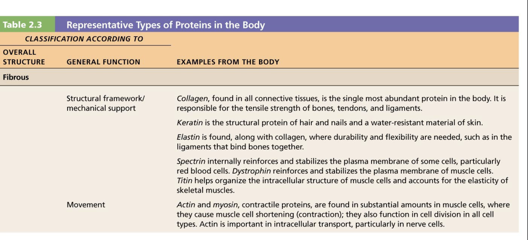
Biomolecules : Proteins
-Proteins serve a wide variety of vital functions in the body
-Proteins are made of building blocks called amino acids
-There are 20 different amino acids in the body but all share a common core structure
Every amino acids contains:
- a central carbon atom
- an amino group
- a carboxylic group
- a radical (R) group that differs between amino acids
-Variable R groups give different properties to the amino acids
-Some are hydrophilic, Some are hydrophobic, Some positively charged, Some negatively charged
-When a long chain of amino acids is formed, the atoms interact with those of neighboring amino acids and the environment around the protein

Amino acids:cont.
-Amino acids are linked end-to-end with peptide bonds to form linear
strands; strands may be short (peptides or oligopeptides) or long (proteins)
-Dehydration synthesis forms peptide bond; hydrolysis breaks peptide bond
Protein structure:
-The primary structure of a protein refers to the linear
sequence of its amino acids
-The secondary structure of a protein refers to the helix or sheet
that forms when its amino acids start to interact with one
another by formation of hydrogen bonds with nearby atoms
-The tertiary structure of a protein refers to the final 3D shape of the
protein after folding occurs due to additional hydrogen and covalent
bonds as well as hydrophobic interactions within the protein: For many proteins, this is the finished, functional form
-Some proteins in the body are formed from multiple smaller
proteins that bind together. In this case the individual proteins are
referred to as subunits of the final protein, and we refer to the
assembled protein as having quaternary structure

Protein shape is sensitive to environment
-The interactions between amino acids that create the finished protein are critically sensitive to pH and temperature. Proteins pushed outside their optimal temperature and pH range become temporarily or permanently denatured and will cease to function.
-The ability of the body to maintain a stable temperature and pH allows cell reactions to proceed under optimal conditions

Biomolecules : Nucleic Acids
Nucleic acids provide the genetic instructions for making proteins.
They store and transfer information at the molecular level within the
cell
-Includes :
DNA (deoxyribonucleic acid)
RNA (ribonucleic acid)
-Related molecules:
ATP (adenosine triphosphate) : energy molecule
GTP (guanosine triphosphate) : cell signaling
NAD+ and FAD : role in mitochondrial ATP production
-Nucleic Acids are made of building blocks called nucleotides
-Each nucleotide contains:
- a phosphate group (PO4)
- a sugar group (ribose or deoxyribose)
- a nitrogen containing base
-Nucleic Acids are made of building blocks called nucleotides
-2 types of nitrogen bases:
Purines are double-ringed molecules (adenine, guanine)
Pyrimidines have a single ring (cytosine, thymine, uracil)
RNA and DNA:
RNA is built by linking RNA nucleotides together into a single strand
-Each RNA nucleotide contains:
- a ribose sugar
- a phosphate group
- and one of these 4 bases : Adenine, Guanine, Cytosine, Uracil (A, G, C, U)
-A strand is formed by linking the sugar group of one nucleotide to the phosphate group of another nucleotide
-The strand has a phosphate-sugar “backbone” with bases pointing away from the backbone
-DNA is built from DNA nucleotides assembled into a double stranded helix
-Each DNA nucleotide contains
- a deoxyribose sugar
- a phosphate group
- and one of these bases : Adenine, Guanine, Cytosine, Thymine (A, G, C, T)
-Each strand has a phosphate-sugar “backbone” with bases pointing away from the backbone
-The two strands of DNA are parallel to one another, with the bases pointing toward the other strand
-The two strands are held together with hydrogen bonds
-The DNA strands are coiled into a double helix
-The two parallel DNA strands are held together by hydrogen bonds formed between complementary bases on opposite strands.
*Adenine pairs with Thymine (A-T)
*Guanine pairs with Cytosine (G-C)
Adenosine Triphosphate (ATP)
-ATP is a high energy compound which stores cellular energy in high energy bonds
-Made by adding a phosphate group to adenosine diphosphate (ADP) in a process referred to as phosphorylation
-Removal of terminal phosphate group converts ATP to ADP, releasing energy
-ATP is the energy form driving most cellular activity
-Many cell reaction are powered by ATP: **be able to know reactions requiring ATP hydrolysis
All components of the cell derive from the basic chemical substances
water, ions, organic and inorganic molecules
The cell is the smallest living unit that can perform all vital physiological functions
-Cells can grow, reproduce, obtain nutrition, and interact with their environment.
-Eggs (ova) and sperm are classified as “sex cells”; all other cells in the body are considered “somatic cells”.
-Every cell has an outer boundary called the cell membrane or plasma membrane
-Inside the cell we find organelles and intracellular fluid
-Cell components are built with proteins, carbohydrates, lipids and nucleic acids.
-Cells are surrounded by extracellular fluid, which is often called interstitial fluid
**Human Cells are Diverse in Shape and Function; structure= function
The structure of a “typical” human cell
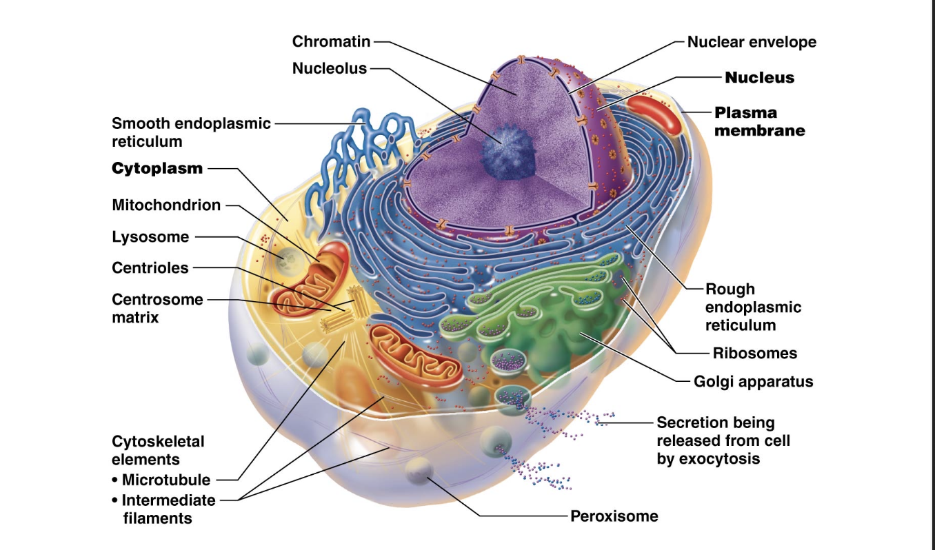
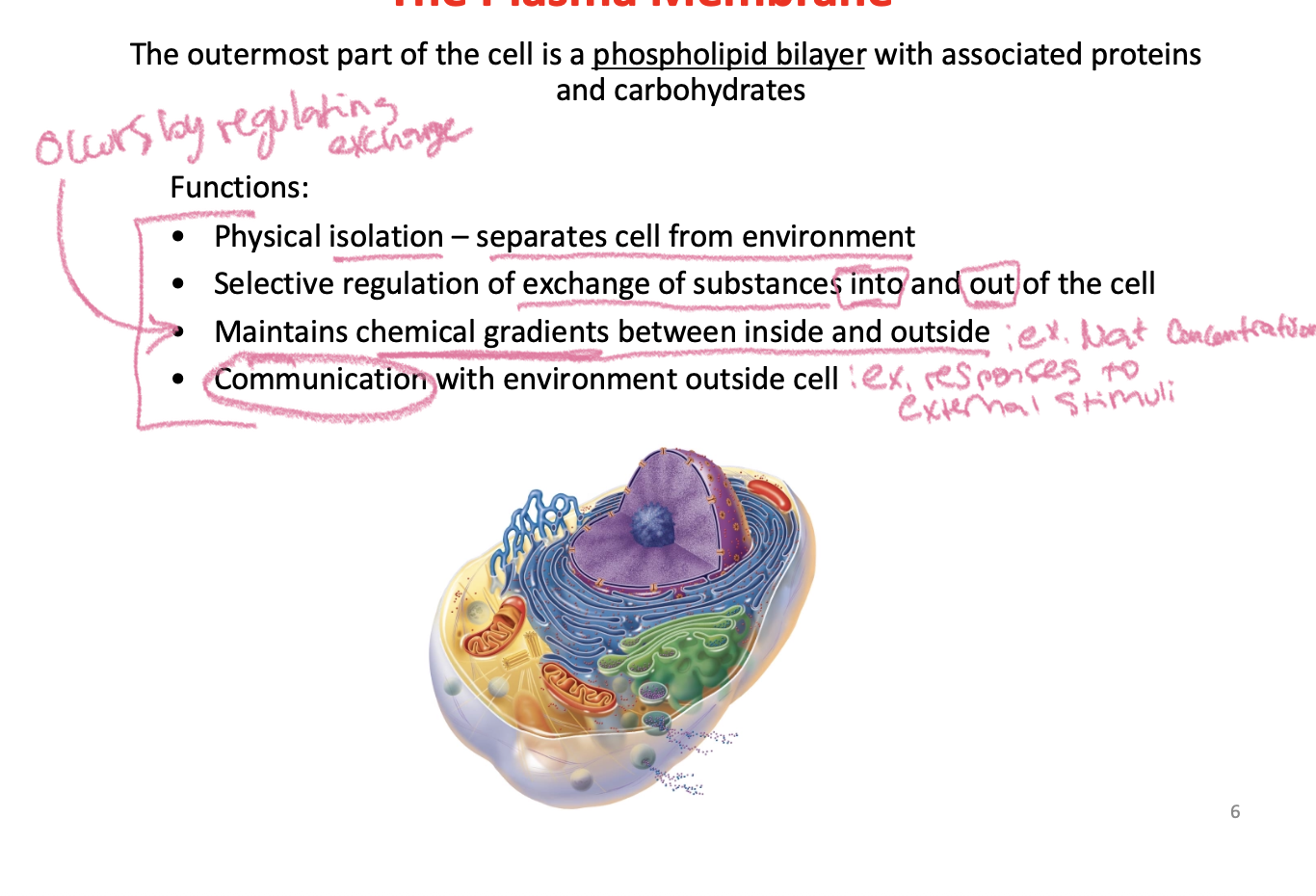
The Plasma Membrane:
-Phospholipid molecules give the cell membrane its structure
-Ampipathic: a molecule with both polar and nonpolar components
Phospholipid molecules in water assemble themselves into a bilayer:

What can and can’t pass through the cell membrane with/without proteins:

Cholesterol is an important lipid component of the plasma membrane

Membrane Proteins give the cell its unique function

Protein Structure Review:

Membrane proteins: Transport Proteins

Receptor Proteins:

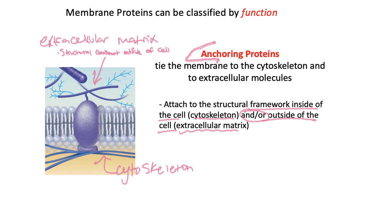
Anchoring Proteins
Cell – Cell Junction Proteins

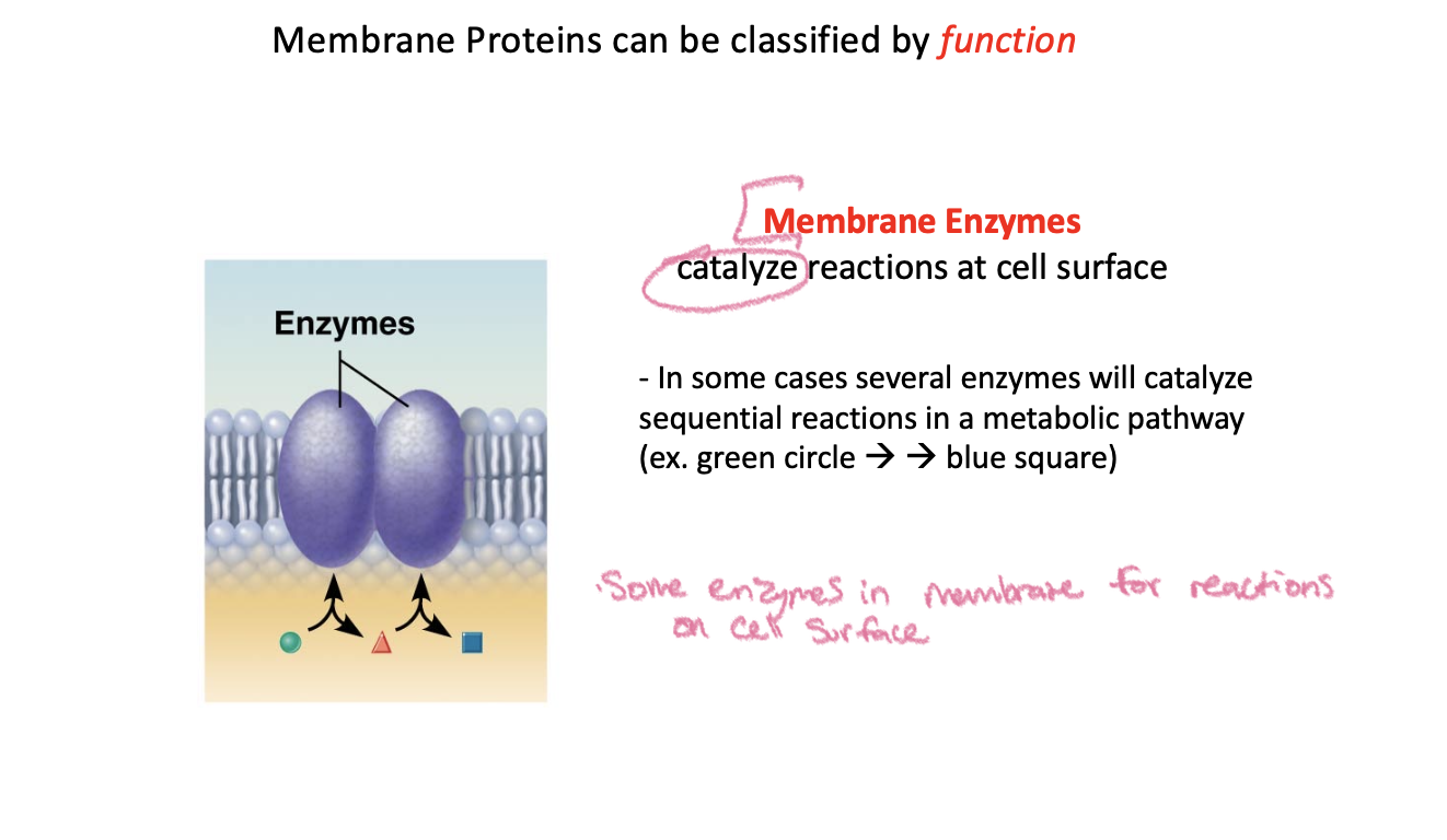
Membrane Enzymes
Recognition Proteins

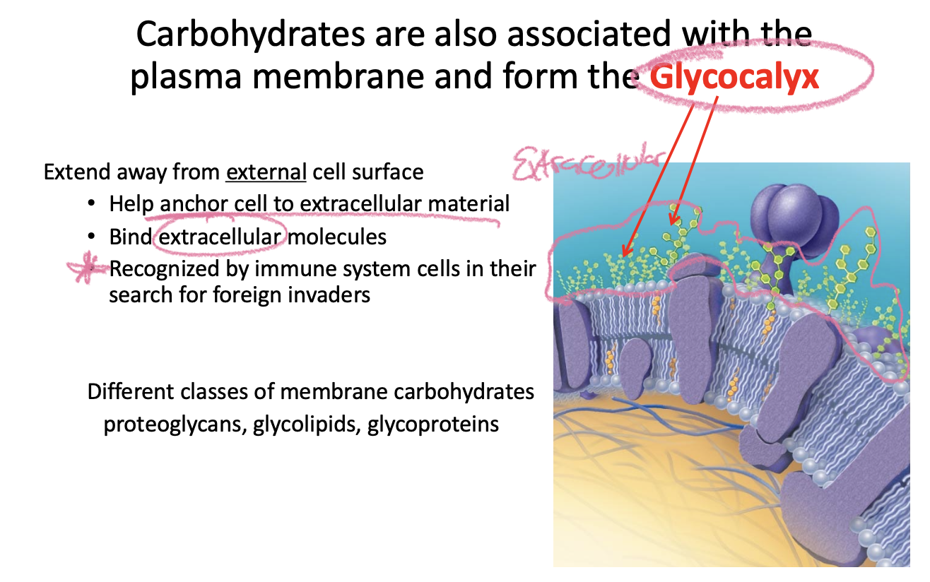
Carbohydrates are also associated with the plasma membrane and form the Glycocalyx:
The Fluid-Mosaic Membrane Model

An example demonstrating fluidity of plasma membrane:

Regulating the movement of materials across the plasma membrane is a key role of the plasma membrane:

Membrane transport processes fall into two groups:

Passive transport

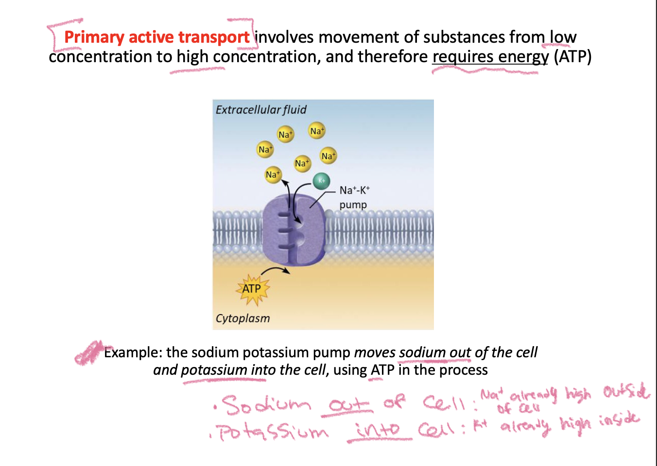
Primary active transport
Secondary active transport

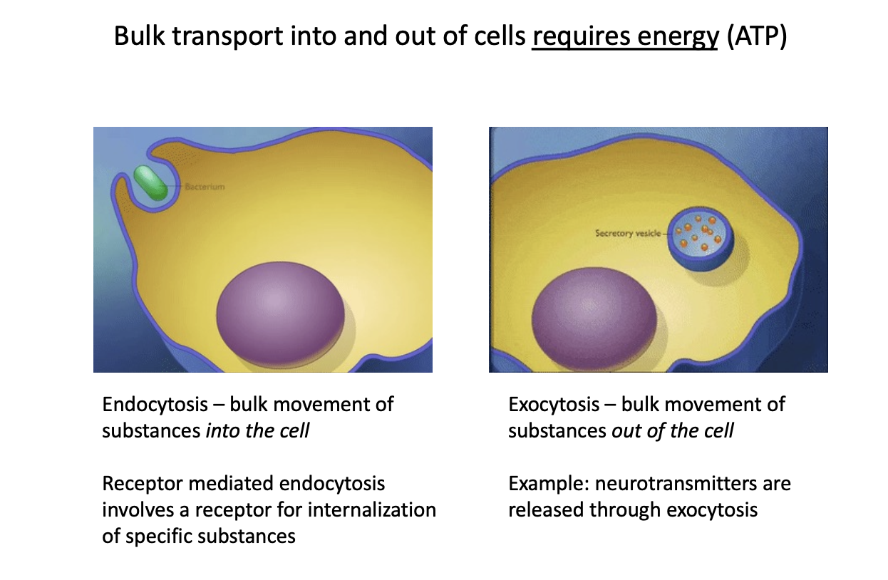
Bulk transport: into and out of cells requires energy (ATP
aquaporins:
-Water can move freely across the plasma membrane in almost all cells, due to the presence of aquaporins
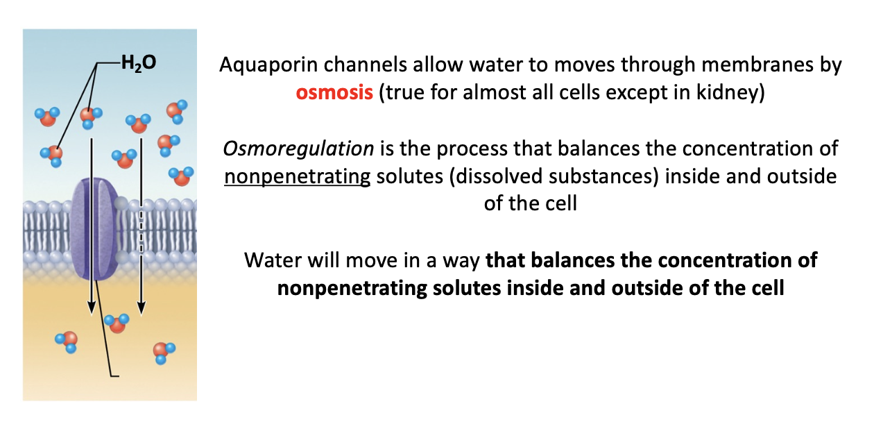
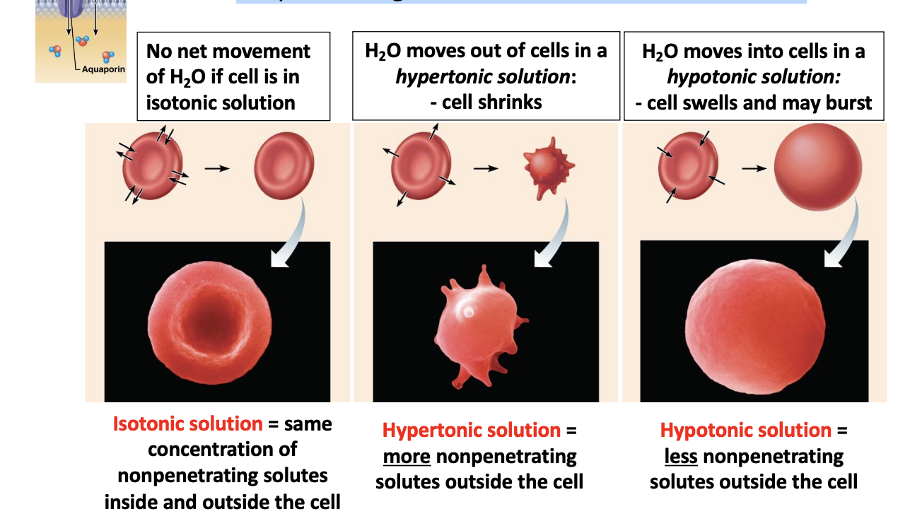
Osmosis:
-influenced by the concentration of nonpenetrating solutes: water will move in a way that balances the concentration of nonpenetrating solutes inside and outside of the cell
Final Notes on the Plasma Membrane: *don’t need to memorize
-Phospholipid molecules are small, and outnumber proteins 50:1 in a plasma membrane
-About 50% of a typical cell plasma membrane is protein some cells as little as 25% membranes of organelles can be as much as 75% protein
~ 1700 different proteins in a plasma membrane – so many variations within each class (e.g. hundreds of different transport proteins) *text images typically show one or two to illustrate a process
-Proteins may not be uniformly distributed within membrane
Contents of the interior of the cell

“Plasma membrane” vs. “membrane”
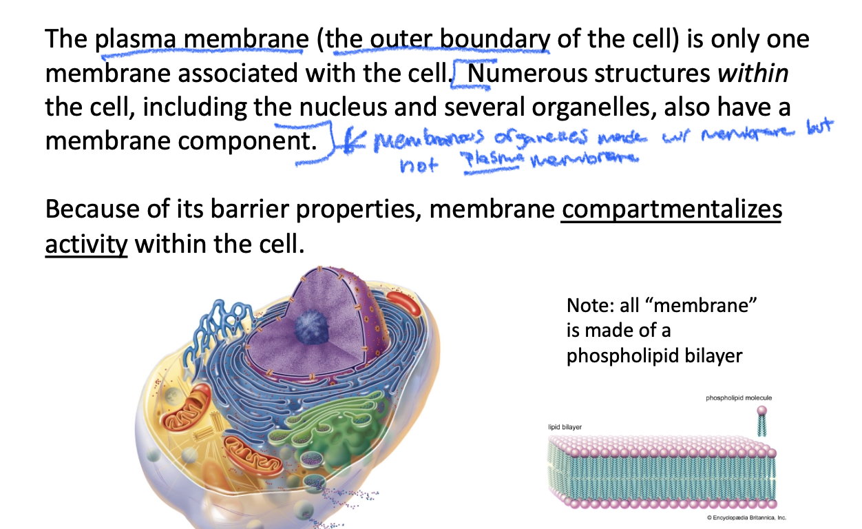
Organelles with Membrane

Double membrane:
-Membrane surrounded by another membrane
-Nucleus, mitochondira
The Nucleus:
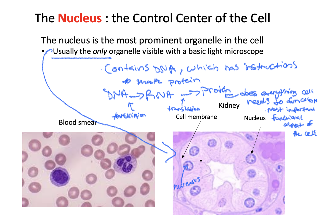
Contents of the nucleus:
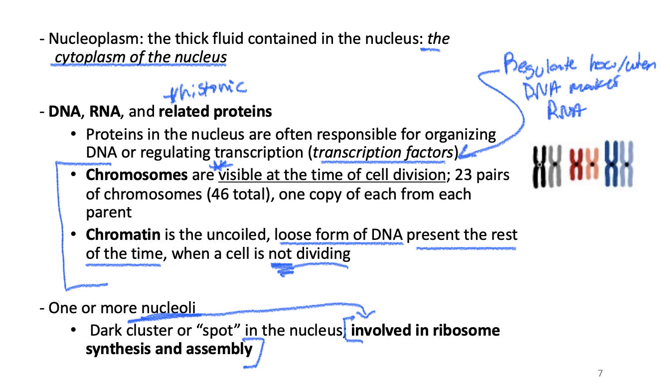
DNA: visible chromosomes vs. chromatin
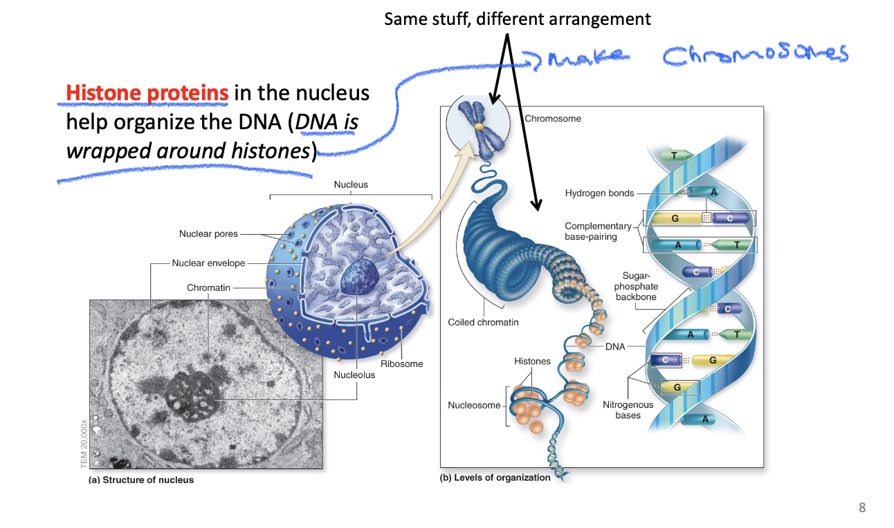
Proteins are built from instructions in the DNA

Protein Synthesis : the Big Picture

3 different kinds of RNA
-The genetic instructions for making a protein are contained in the sequence of DNA and RNA nucleotides
-Specific triplets of RNA nucleotides (codons) will encode a particular amino acid.
-Ex. AUG à methionine (Met); ACG à threonine (Thr) ….

Three cytoplasmic organelles involved in protein synthesis and processing: Ribosome, Endoplasmic Reticulum, Golgi apparatus

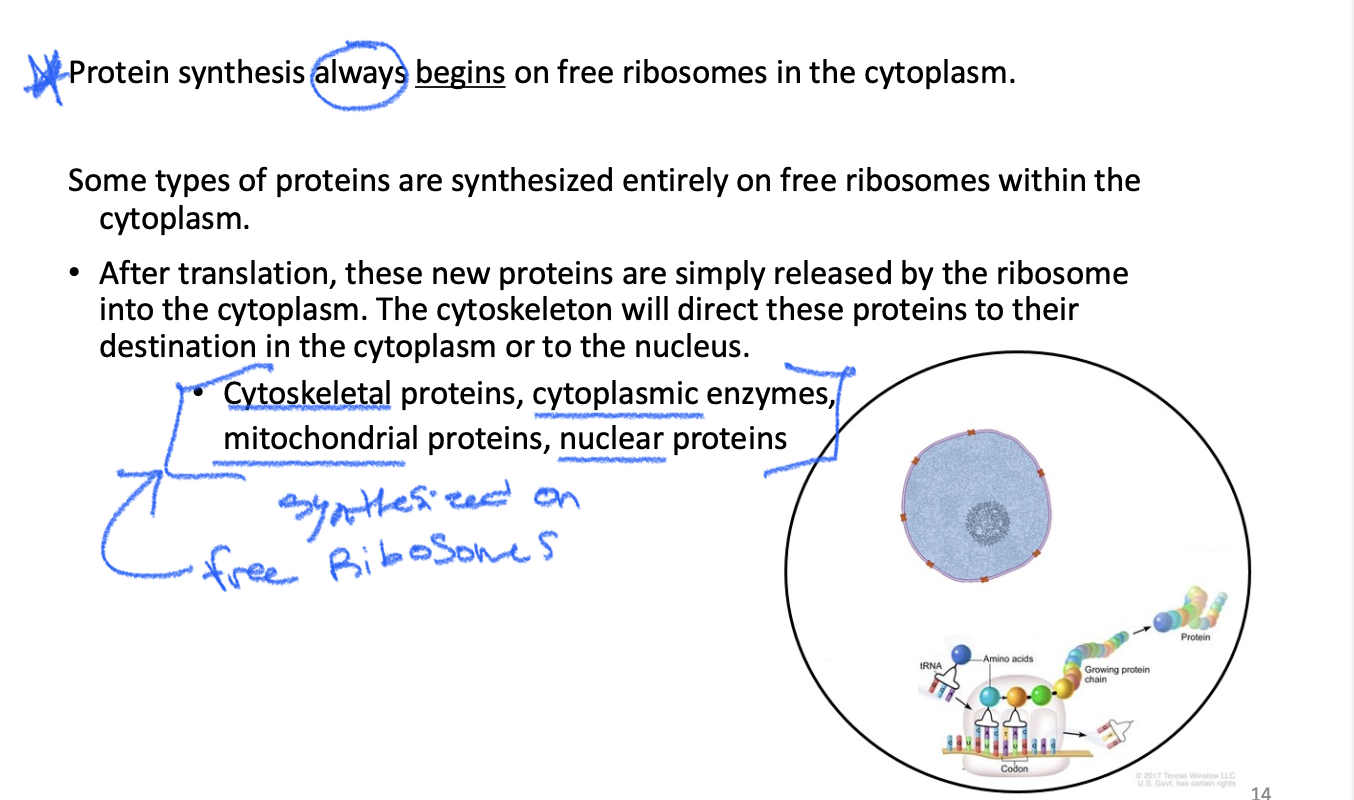
The Fate of Newly Synthesized Proteins
These proteins are not entering the ER/Golgi system.
They are made completely on free ribosomes and then go to places like:
Cytoplasm
Nucleus
Mitochondria
Path:
Free ribosome → protein → destination
Protein does NOT have an ER signal → ribosome stays free → protein is made in cytoplasm → it does NOT enter ER/Golgi
Some proteins must be synthesized at the rough endoplasmic reticulum.
These proteins DO enter the ER and travel through the endomembrane system.
Their final destination can be inside OR outside the cell:
Lysosome → inside the cell
Cell membrane → part of the cell
Outside the cell → secreted by exocytosis
Path:
Free ribosome → rough ER → vesicle → Golgi → destination
Protein has an ER signal → ribosome is directed to rough ER → protein enters ER → Golgi pathway

Mitochondrion

The Cytoskeleton

The “typical” human cell
-All have same components but look a little different depending on their location and function
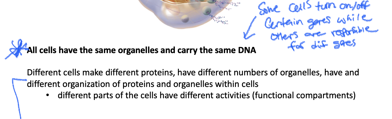
Cells
-Smallest unit of life that can preform all vital physiological functions
-Cells can grow, reproduce, obtain nutrition, and interact with the environment
-Eggs/sperm; sex cells, meiosis
-Body cells: Somatic cells, mitosis
*cells grow and reproduce during the cell cycle
Somatic cells vs. Gametes:
-Somatic cells: comprise our organs and tissues, 23 PAIRS (46 total) of chromosomes- diploid 2n, one copy of each chromosome from mom and one copy from dad
*Divide by mitosis: 2 identical diploid cells formed wax with 23 pairs of chromosomes
-Gametes: Comprise sex cells, 23 SINGLE chromosomes- haploid 1n, one copy of each chromosome
*Divide by meiosis: Asymmetrical division, 4 NON-identical haploid cells form in order to have genetic variation in offspring
-Cell division is essential for: growth and development, and to replace dead/dying cells- development + maintenance
mitosis:
-Equal division of a event cells into two identical daughter cells
-Through mitosis: single zygote (fertilized egg) can develop into the 35 trillion+ cells in human body
-Cell division doesn’t stop after we reach full size: rate of division = rate of cell loss (maintenance): if more cells are dying without new ones being born → diseases
Stem cells:
Early embryonic cells: pluripotent, meaning they can form every kind of cell in the human body
Differentiation: process by which stem cells become specialized cells via communication with other cells
Adult stem cells: Most organs have partially differentiated stem cells that can become any type of cell in that specific zone. EX. bone marrow, lining of intestine, muscle
Fully differentiated adult cells: Divide throughout life and produce cells of the same type, & can increase rate of division with certain signals (not just stem cells used for cell replacement): EX. liver cells, endothelial cells lining blood vessels, fibroblasts in skin
* experimenting with stem cells in a clinical setting, bone marrow transplantation is the only one currently in use
The cell cycle:
-The stages of a activity from a cells formation until its division
-2 Identical daughter cells produced in mitotic division: genetically identical
-Cells spend most time in interphase (G1, S, G2), growing and preforming daily functions (like protein synthesis making diff proteins to determine a cells function)
-Mitotic phase: prophase, metaphase, anaphase, and telophase; sequence of events that divide the nucleus
* interphase and cytokinesis are part of the cell cycle, but mitosis is the division of the nucleus
Two different DNA configurations:
-Chromatin: how DNA is found most of the time in lose dispersed form, exposed for process of protein synthesis
-Chromosomes: During mitosis DNA becomes tightly packed into condensed chromosomes; allows for division of nuclear (genetic) material
G0 phase
-What happens when cells exit the cell cycle
-Most adult cells stop dividing; these cells are in G0 phase & live out their lives doing day-to-day maintenance activities
Interphase: G1 phase
-Normal activities: synthesis of proteins and RNA for maintenance activities
-Preparation: Synthesis of proteins needed for S phase, duplication of organelles, cell grows in size
*length of G1 phase varies: hours, days, weeks, longer
-G1= growth, doing regular cellular processes then setting up for division
Interphase: S-phase
-DNA replication: DNA polymerase synthesizes new strand via semi-conservative replication and complementary base pairing (new strand is hard old DNA/ new)
-Sister chromatids: Duplicated copies of original 46 chromosomes, during S-phase there are 92 total chromosomes
-Duplication of centrioles: form mitotic spindle
-Length of S-phase: about 6 hrs
Interphase:
G1= Cell Growth: The cell increases in physical size and recovers from the previous division.
Protein and Organelle Synthesis: It creates extra enzymes, structural proteins, and organelles (like ribosomes and mitochondria) needed for the upcoming DNA replication.
Metabolic Activity: The cell carries out its everyday specialized jobs within the organism.
The G1 Checkpoint: A critical control point checks if the cell is big enough, has enough energy, and has undamaged DNA before allowing it to move forward into the S (synthesis) phase.
S= DNA Replication: The cell makes an exact copy of its nuclear DNA. Every chromosome is duplicated.
Sister Chromatid Formation: Each chromosome goes from a single strand to two identical strands called sister chromatids, which remain joined at a region called the centromere.
Centrosome Duplication: The centrosome (the cell's microtubule-organizing center) duplicates. These pair up and will later help pull the chromosomes apart during mitosis.
Histone Production: The cell rapidly produces histone proteins, which act as spools to tightly wrap and organize the newly synthesized DNA.
G2:
-Protein synthesis in preparation for mitosis, continued growth
-Length of G2: about 4 hours
-DNA replication checkpoint
Mitosis: Prophase:
-Chromatin condenses into visible chromosomes, each with 2 sister chromatids joined at a centromere
-Centriole pairs move to opposite ends of the cell: Anchor microtubule network that assemble mitotic spindles between centrioles
-Nuclear membrane dissolves in late prophase, spindle fibers attached to chromatids
Bio:
Chromosomes shorten, thicken, and become visible
Each chromosome has been duplicated
Duplicated chromosomes are sister chromatids
Sister chromatids are attached at the centromere
Nuclear membrane breaks down
Mitosis: Metaphase
-Sister chromatids align at middle of cell at the “metaphase plate”
-Metaphase checkpoint so make sure chromosomes are lined up at center of cell and attach to spindle fibers
Nuclear membrane is broken down
Duplicated chromosomes line up at the center of the cell
Spindle fibers control chromosome movement
Sister chromatids attach to opposite spindle poles
Mitosis: Anaphase
-Microtubules (mitotic spindles) pull sister chromatids to opposite ends of cell: centromeres split
-Results in equal separation of genetic material
Mitosis: telophase
-Microtubule spindle disassociates
-Nuclear membranes reform around each group of chromatids to form 2 new nuclei
-Chromosomes → chromatin: disperse back into chromatin arrangement
Sister chromatids reach opposite ends
Each side has a diploid set of chromosomes
Nuclear membranes reform
Chromosomes decondense
Cytokinesis divides the cytoplasm
Cell cycle: cytokinesis
-Division of the cytoplasm
-An event that is part of telophase and overlaps with the end of mitosis (timing overlaps, but separate process)
-Actin network pulls membrane into cleavage furrow at midline: Actin like a ring that squeezes, results in furrow
-Furrow grows and eventually separates cytoplasm into 2 separate cells
Cell cycle checkpoints:
-Regulation: monitors the progress of events, gets dysregulated in cancer
-G1 checkpoint: passes this checkpoint if cell size is adequate enough to divide into 2 cells, nutrient availability is sufficient, & growth factors (signals from other cells) are present
-G2 checkpoint: passes if cell size is adequate, and chromosome replication is successfully completed/accurate, if damaged DNA, cycle pauses for repair
-Metaphse checkpoint: pass if all chromosomes are attached to mitotic spindle (occurs in late prophase, early metaphase)
Cell cycle regulation: genes
-Protooncogenes: genes whose normal protein products stimulate the cell cycle if cell environment is correct: tells division to continue if checkpoints are passed
*Mutated versions: Oncogenes- she strive cell cycle, regardless of checkpoints, leading to abnormal growth (cancers)
-Tumor suppressor genes: Normal function to repress cell division by maintaining checkpoints: create checkpoints to slow division
*Mutated versions: fail to repress cycle, leading to abnormal growth
*either over, or under stimulation leads to unregulated growth
Apoptosis
-Programmed cel death
-normal cells follow control signals, and their rate of cell division/level of differentiation is kept under control
-Types of stress that can lead to apoptosis: DNA damage, viral infection, growth factor deprivation
**Cancer cells do not undergo apoptosis and continue to divide abnormally
Carcinogens:
-Substances that cause cancer
Induce DNA damage → mutations- Ex. tobacco smoke, UV radiation
Stimulate self perforation: hormones, viral infection
-Terms:
-Carcinoma: cancer of epithelial cell origin (90%)
-Leukemias/lymphomas: cancer of blood lines (8%)
-Sarcomas: solid tumors of other connective tissues (rare)
Cancer cells ignore control signals:
-a tumor results from abnormal proliferation of a cell
-benign: remains confined to its original location, does not invade surrounding tissue
-malignant: invades adjacent tissue and spreads to other parts of the body, called metastasis
Tissues:
-Collections of specialized cells and cell products organized to preform one or more select functions
-Histology: microscopic study of tissues
-All somatic cells in the body are classified into 1 of these 4 tissue types…. Epithelial, Nervous, Connective, and muscular
Why do we study tissue types?
-each tissue type has unique characteristics determined by the properties of cells: cells determine characteristics of tissues
*Ex. muscle cells contract, while bone cells provide provide support
-Different cell types have different composition of proteins, carbohydrates, lipids, and nucleic acids, so different cell types stain differently and have different functions: all made of same stuff, but composed differently – Ex. molecular make up of orange dictates function (protection)
-Organ: body structure built with at least two tissues that carries out a specific function
* organ is built with layers; usually one tissue predominates in each layer. Understanding tissue properties helps us understand organ function
Histology:
-what we see is dependent on how the tissue is sectioned and stained – most of our histology work will use drawings of organs