NB Exam 2: Gyri, Sulci & Fissures (lateral, medial, coronal)
 Call Kai
 Learn
 Practice Test
 Spaced Repetition
 Match
 Flashcards
 Knowt Play
1/44

There's no tags or description
Looks like no tags are added yet.
45 Terms

Lateral view: name the gyrus at #1 (card 1)
Superior frontal gyrus • Function: Frontal lobe: reasoning, planning, motor movement; part of the prefrontal cortex (BA 9, 8, 6 region) • Damage: Prefrontal damage: depressive profile (apathy, abulia, perseveration) or manic profile (lability, distractibility, confabulation)


Lateral view: name the gyrus at #1 (card 2)
Middle frontal gyrus • Function: Prefrontal cortex (dorsolateral, BA 9/46): executive control; frontal eye fields (BA 8) sit here: eye movements • Damage: Prefrontal profiles (depressive/manic); frontal eye field damage: eyes deviate toward the side of the injury


Lateral view: name the gyrus at #1 (card 3)
Inferior frontal gyrus • Function: Broca's area (BA 44 pars opercularis + BA 45 pars triangularis), the 'third frontal convolution': speech-organ blueprint, syntax, planning verbal responses • Damage: Broca's aphasia


Lateral view: name the gyrus at #1 (card 4)
Precentral gyrus • Function: Primary motor cortex, BA 4 (motor strip): sends motor plans to muscles via the pyramidal pathway; motor homunculus • Damage: Loss of voluntary movement (plegia/paresis); damage to its inferior part, behind Broca's: apraxia of speech


Lateral view: name the gyrus at #1 (card 5)
Postcentral gyrus • Function: Primary sensory cortex, BA 3, 1, 2 (sensory strip): touch, vibration, proprioception, stereognosis; sensory homunculus • Damage: Loss of touch, vibration, proprioception and stereognosis (no named disorder on your slides)


Lateral view: name the gyrus at #1 (card 6)
Superior parietal gyrus (lobule) • Function: Parietal lobe; somatosensory association area BA 5, 7: uses sensory feedback to refine movement, incl. fine speech movements • Damage: Not specified on your slides


Lateral view: name the gyrus at #1 (card 7)
Inferior parietal gyrus (lobule) • Function: Made of the supramarginal (BA 40) + angular (BA 39) gyri: sounding out words, reading, math • Damage: Phonological dyslexia (40); alexia, acalculia (39); R side: hemispatial neglect


Lateral view: name the gyrus at #1 (card 8)
Supramarginal gyrus • Function: BA 40: helps us sound out words; stores auditory representations of phonemes (phonology) • Damage: Phonological dyslexia: trouble reading new words/nonwords, decoding, writing single letters


Lateral view: name the gyrus at #1 (card 9)
Angular gyrus • Function: BA 39, where the temporal lobe meets the parietal: reading and math; metaphors, sense of embodiment. Right side: visuospatial processing • Damage: Alexia, acalculia, out-of-body experiences; right angular damage: hemispatial neglect (ignores the left side)


Lateral view: name the gyrus at #1 (card 10)
Parieto-occipital gyrus • Function: Border region between the parietal and occipital lobes (labeled on the textbook figure; no function on your slides) • Damage: Not covered on your slides


Lateral view: name the gyrus at #1 (card 11)
Superior occipital gyrus • Function: Occipital lobe: vision (visual association cortex, BA 18/19 region) • Damage: Visual cortex damage: Anton syndrome or visual agnosia (stroke, TBI)


Lateral view: name the gyrus at #1 (card 12)
Lateral occipital gyrus • Function: Occipital lobe: vision (visual association cortex) • Damage: Visual cortex damage: Anton syndrome or visual agnosia


Lateral view: name the gyrus at #1 (card 13)
Inferior occipital gyrus • Function: Occipital lobe: vision • Damage: Visual cortex damage: Anton syndrome or visual agnosia


Lateral view: name the gyrus at #1 (card 14)
Superior temporal gyrus • Function: Holds primary auditory cortex BA 41/42 (Heschl's gyrus: receives sound, tonotopic) and Wernicke's area BA 22 (attaches meaning) • Damage: Wernicke's aphasia: fluent jargon, poor comprehension of others' and own speech


Lateral view: name the gyrus at #1 (card 15)
Middle temporal gyrus • Function: Inferior temporal area (BA 21): auditory and language processing, reading facial emotions • Damage: May play a role in hallucinations (overactive memory functions)


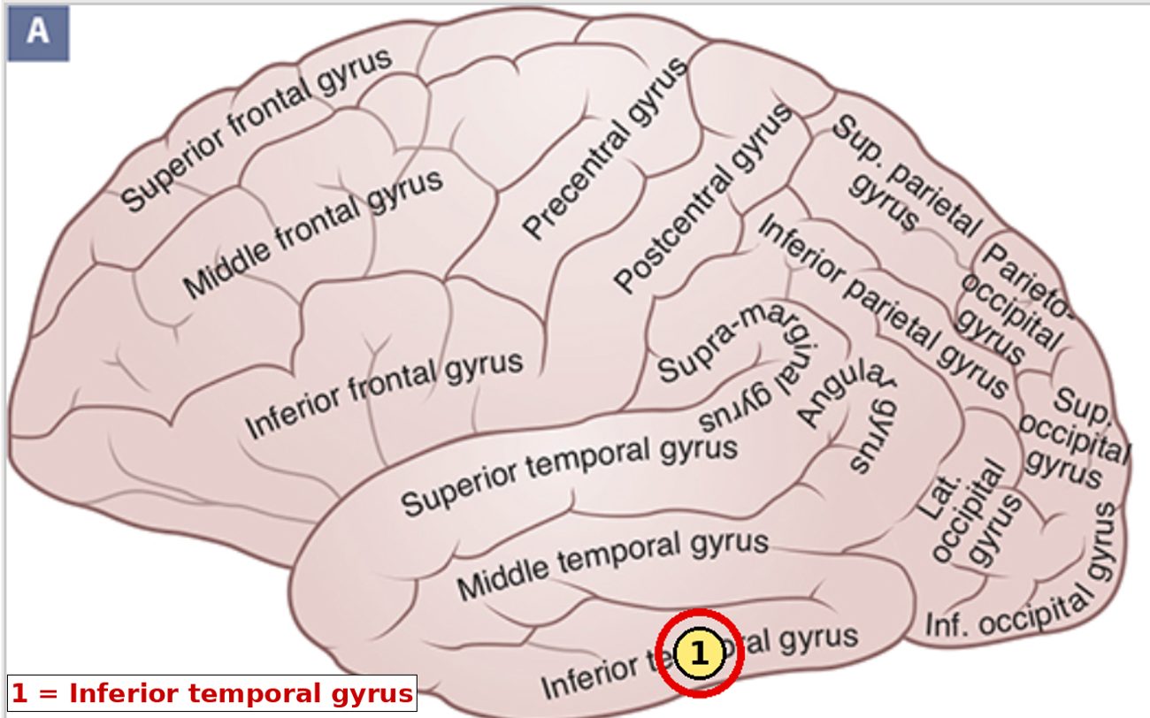
Lateral view: name the gyrus at #1 (card 16)
Inferior temporal gyrus • Function: Inferior temporal area (BA 20): auditory and language processing, reading facial emotions • Damage: May play a role in hallucinations
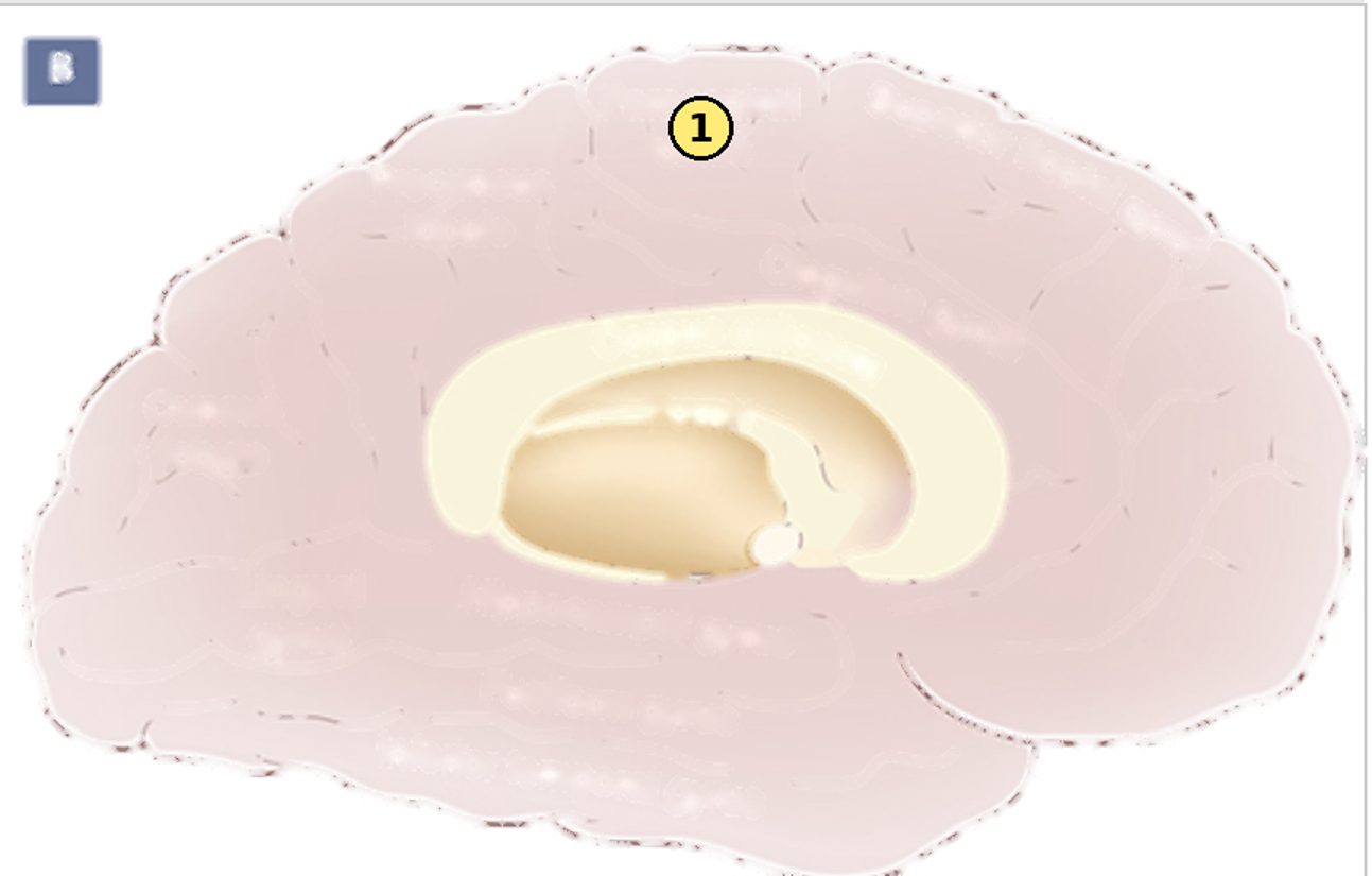
Medial view: name the gyrus or lobule at #1 (card 17)
Paracentral lobule • Function: Top of the medial surface where the motor strip (BA 4) and sensory strip (BA 3,1,2) wrap over (from the Brodmann map) • Damage: Not specified on your slides


Medial view: name the gyrus at #1 (card 18)
Superior frontal gyrus (medial) • Function: Medial frontal lobe: premotor (6), frontal eye fields (8), prefrontal (9) • Damage: Prefrontal depressive/manic profiles


Medial view: name the gyrus or lobule at #1 (card 19)
Precuneus lobule • Function: Medial parietal lobe, in front of the parieto-occipital sulcus (listed on your slide; no function given) • Damage: Not covered on your slides


Medial view: name the gyrus at #1 (card 20)
Cingulate gyrus • Function: Limbic system + cognition (BA 23-26, 30-33). Anterior: cognitive control, error/conflict detection; posterior: autobiographical memory, emotion. ACC filters, PCC focuses • Damage: Not specified on your slides


Medial view: name the structure at #1 (card 21)
Corpus callosum • Function: Main interhemispheric connection: band of white matter fibers (axons) joining the right and left hemispheres • Damage: Agenesis of the corpus callosum (failed ventral induction, Ch 3)


Medial view: name the structure at #1 (card 22)
Fornix • Function: Limbic structure under the corpus callosum (listed on your medial-view slide; no function given) • Damage: Not covered on your slides


Medial view: name the gyrus or lobule at #1 (card 23)
Cuneus lobule • Function: Medial occipital lobe, above the calcarine sulcus: vision • Damage: Visual cortex damage: Anton syndrome or visual agnosia


Medial view: name the gyrus at #1 (card 24)
Lingual gyrus • Function: Medial occipital, below the calcarine sulcus: vision • Damage: Visual cortex damage: Anton syndrome or visual agnosia


Medial view: name the gyrus at #1 (card 25)
(Para)hippocampal gyrus • Function: Memory (BA 27, 28, 34, 35, 36): hippocampus = declarative memory; entorhinal cortex (28, 34) = relay between cortex and hippocampus • Damage: Declarative memory problems (facts, events)


Medial view: name the gyrus at #1 (card 26)
Fusiform gyrus • Function: BA 37 (occipitotemporal gyrus): remembering and naming seen objects; visual lexicon • Damage: Anomia (word finding), lexical agraphia


Medial view: name the gyrus at #1 (card 27)
Inferior temporal gyrus (medial view) • Function: Inferior temporal area (BA 20): auditory/language processing, facial emotions • Damage: May play a role in hallucinations


Lateral view: name the sulcus at #1 (card 28)
Superior frontal sulcus • Function: Boundary between the superior and middle frontal gyri • Damage: Sulci are boundaries: damage here affects the gyri on either side (prefrontal)


Lateral view: name the sulcus at #1 (card 29)
Precentral sulcus • Function: Just in front of the precentral gyrus; separates premotor (BA 6) from the motor strip (BA 4) • Damage: Affects neighboring premotor/motor cortex: apraxias, motor loss


Lateral view: name the fissure at #1 (card 30)
Central fissure (sulcus) • Function: Separates the frontal from the parietal lobe; motor strip (4) in front, sensory strip (3,1,2) behind • Damage: Neighbors: motor loss (in front), sensory loss (behind)


Lateral view: name the sulcus at #1 (card 31)
Postcentral sulcus • Function: Behind the postcentral gyrus; separates the sensory strip from the parietal association cortex (5, 7) • Damage: Neighbors: sensory loss


Lateral view: name the sulcus at #1 (card 32)
Inferior frontal sulcus • Function: Boundary between the middle and inferior frontal gyri (Broca's is just below it) • Damage: Neighbor below: Broca's aphasia


Lateral view: name the sulcus at #1 (card 33)
Parieto-occipital sulcus (lateral) • Function: Separates the parietal from the occipital lobe • Damage: Neighbors: parietal association / vision


Lateral view: name the fissure at #1 (card 34)
Lateral fissure (Sylvian) • Function: Separates the temporal lobe from the frontal and parietal lobes; the insula lies deep inside it; auditory cortex (41/42) sits on its lower bank • Damage: Neighbors: Broca's (front), Wernicke's/auditory (below)


Lateral view: name the sulcus at #1 (card 35)
Lateral occipital sulcus • Function: Occipital lobe landmark (the textbook figure labels this spot 'Lat. occipital gyrus'; your deck lists the lateral occipital sulcus) • Damage: Neighbors: vision


Lateral view: name the sulcus at #1 (card 36)
Superior temporal sulcus • Function: Boundary between the superior and middle temporal gyri (below 41/42 and 22) • Damage: Neighbor above: Wernicke's aphasia


Lateral view: name the sulcus at #1 (card 37)
Middle temporal sulcus • Function: Boundary between the middle and inferior temporal gyri • Damage: Neighbors: inferior temporal area (20/21)


Medial view: name the sulcus at #1 (card 38)
Parieto-occipital sulcus (medial) • Function: Separates the precuneus (parietal) from the cuneus (occipital) • Damage: Neighbors: parietal / vision


Medial view: name the sulcus at #1 (card 39)
Cingulate sulcus • Function: Runs just above the cingulate gyrus • Damage: Neighbor: cingulate cortex


Medial view: name the sulcus at #1 (card 40)
Calcarine sulcus • Function: Medial occipital lobe: primary visual cortex (BA 17) lies along it; cuneus above, lingual gyrus below • Damage: Visual cortex damage: Anton syndrome or visual agnosia


Medial view: name the sulcus at #1 (card 41)
Collateral sulcus • Function: Inferior medial surface; borders the parahippocampal gyrus • Damage: Neighbor: memory (parahippocampal)


Medial view: name the sulcus at #1 (card 42)
Inferior temporal sulcus • Function: Inferior surface; borders the inferior temporal gyrus • Damage: Neighbor: inferior temporal area


Coronal view: name the fissure at #1 (card 43)
Longitudinal fissure • Function: Separates the right and left hemispheres (sagittal); the dura folds into it • Damage: Midline landmark; agenesis of the corpus callosum sits beneath it


Coronal view: name the fissure at #1 (card 44)
Lateral fissure (Sylvian) • Function: Separates the temporal lobe (below) from the frontal/parietal lobes (above); insula deep inside • Damage: Neighbors: Broca's, auditory cortex, Wernicke's


Coronal view: name the structure at #1 (card 45)
Corpus callosum
• Function: White matter band joining the right and left hemispheres, roofing the lateral ventricles
• Damage: Agenesis of the corpus callosum (ventral induction)
