Vestibular System and Olfactory Pathways: Anatomy, Reflexes, and Disorders
 Call Kai
 Learn
 Practice Test
 Spaced Repetition
 Match
 Flashcards
 Knowt Play
1/47

There's no tags or description
Looks like no tags are added yet.
48 Terms
what is the function of the vestibular system
- keeps us upright/balanced by drving posture adjustments that stop us from falling
- keeps haze stable during head movement
**function declines with age (why fall risk ↑ )
- if system misfires --> vertigo and motion sickness
what structures makes up the vestibular apparatus? descrip the materila and flid in the apparatus
1. 3 semicircular cannals
- ant.
-post.
- lateral
2. otolith organs
- utricle
-saccile
structure:
- a inner membranous labrynth filled with endolymph fluid
- surrounded by bony lanbrinth containing perilymph fluid
*notes this is continuous with the cochlea and uses the same kind of hair cells
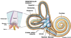
what is the structure of the semicircular canals and what does it contain
Three canals positions perpendicular from eachotther in three planes → any rotation can be detected
- ampulla/ampullaris = swelling at base of each canal contaiing hair recepttors crista ampullaris
- cupula = gelatinous structure sittin gafter the hair cells
- endolymph fluid = durign rotational movement fluid moves and pushes the cupula bending te hair cells

function of semicircular canals
detect rotational (angular) acceleration of the head. eg. spinning

hair cells structure
Each vestibular hair cell has
- one tall kinocilium and
- rows of shorter stereocilia
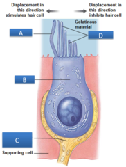
hair cell bending direction
toward kinocillium --> depolarizationm --> ↑ firing rate
away from kinocillium --> repolarization --> ↓ firing rate
*ussually hair cells firing stealdiy at rest and go up or down with bending

Horizontal Canal Push-Pull Mechanism (Right vs. Left)
turning right speeds up the right horizontal canal's nerve and slows the left (a push-pull pair)

what happens after the firing rate of the hair cells ↑ or ↓
1. Vestibular nerve (CN VIII) carries the signal to the vestibular nuclei in the brainstem.
2. The brain compares firing rates from both sides (push-pull mechanism).
3. This helps determine the direction and speed of head rotation.
4. The brain coordinates responses to maintain balance, posture and stable vision (vestibulo-ocular reflex).
what happens in canals when you start spinning
- endolymph lags
- sends strong rotational signal via hair cells
what happens in canals when you keep spinning at steady speed (how long it take)
- endolymph fluid catches up
- cupula straightens up and signal fades
(takes ab 20 s)
what happens in canals when you stop spinning - why does it make you dizzy
- the fluid doesnt stop instantly instead keeps going
- makes you feel like your spinninng other way
leads to dizziness or nysrtagmus (where eyes start beating to compensate for spinnning thats not happening
what structure do the ottolith organs, saccule and utricle have
1. macula = patch of hair cells
- roughly vertical in utricle
- vertical in saccule
2. otolithic membran
- Gelatinous layer sitting on top of the hair cells.
- Contains otoliths
3. otoliths/otoconia
- Small calcium carbonate crystals embedded in the otolithic membrane.
- crystals are heavier than the fluid, so gravity and acceleration drag the membrane and bend the hair cells

what is the function of the otolith organs? how is signals dif from canals
sense both
linear acceleration (a car pulling away)
gravity (tilting the head)
- signal doesnt fade like canal (rotational acelleration

how do otolith organs tell bw tilting head back and accelerating fowards
look the same to the otoliths
the brain sorts it out using the canals and vision

Fun fact: fish otoliths
Fish have the same otolith organs, but each holds one solid stone (an otolith) instead of our many tiny otoconia
- Each species has its own shape
- Growth rings record age, like a tree
what is the vestibular nerve
the part of cranial nerve VIII (vestibulocochlear nerve) responsible for carrying balance and head-movement information from the inner ear to the brain.

vestibular/scarpas ganglion
where the cell boding of the sensory neurons are that receive infor from hair cells in vestibular apparatus
--> sends sigals toward brainstem
Where does the vestibular nerve send information?
1. Hair cells in the vestibular apparatus detect movement.
2. Signals travel through the vestibular nerve (CN VIII).
3. Information reaches the ...
- vestibular nuclei in the medulla and pons
- some go directly to cerebellum (flocculonodular lobe * the only primary sensory fibres that reach the cerebellum directly
4. The brain processes the information to coordinate:
- Balance and equilibrium
- Posture
- Eye movements (vestibulo-ocular reflex)
**look at pic
Where the vestibular nuclei project
1. Eye-muscle nuclei (III, IV, VI)
through the MLF - the vestibulo-ocular reflex
2. Vestibulospinal tracts:
- lateral: same side, whole cord - antigravity extensors for balance
Eg. muscle in the trunk
- medial: both sides, neck - keeps the head steady with gaze
3. Cerebellum, the opposite vestibular nuclei (push-pull),
4. the thalamus → cortex for the conscious sense of motion
(smallest portion)

The vestibulo-ocular reflex (VOR)
reflex that keeps your eyes focused on a target while your head is moving.
One of the fastest reflexes in the body (~10 ms) - the same abducens/MLF circuit as Lecture 12
what is the pathway for the VOR for horizontal head movemnts (eg. head turns right)
Head turns RIGHT → Eyes move LEFT.
Head turns right → right horizontal canal excited → signal to right vestibular nuclei which sends signal to
→
1. cross to the left abducens nucleus → contract left lateral rectus muscle
2. via the MLF → contract the right medial rectus
causes Both eyes turn left, exactly as far as the head turned right - gaze stays put

what is the pathway for the VOR for horizontal head movemnts (eg. head turns down)
Same logic, different canals and muscles
Head nods down → anterior canals excited → vestibular nuclei → MLF travels to → oculomotor nuclei → superior rectus and inferior oblique contract → eyes move up
*look at notes diagram
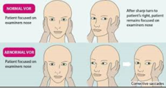
why do chicken heads not move when the body is moves
Birds steady the whole head with vestibular and visual reflexes acting on the neck; we mostly steady the eyes with the VOR
what are 3 vestibular disorders
1. BPPV = crystals in the wrong place.
2. Vestibular neuritis = one vestibular nerve is affected.
3. Motion sickness = sensory mismatch between vision and vestibular input.
what is bppv
benign paroxysmal positional vertigo
- most common cause of vertigo
- when there is a build up of otoconia, some break ff and drift into a canal (ussually posterior)
- these can push onto the cupula and
- when person lies down crystals sink around canal, draggin the fluid which bend cupula --> causes seconds of spinning and nystagmus
how to diagnose BPPV
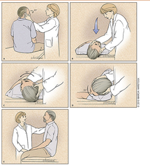
Dix-hallpike test
- lie back with right ear down to get crystals moving
- Patient's head is turned 45° to the right, then quickly laid backward with the head extended and right ear down.
Dislodged crystals move within the posterior canal, triggering:
Upward-beating nystagmus
Torsional nystagmus (twisting toward the affected right ear)
When sitting back up, the nystagmus may reverse direction.
Frenzel lenses:
used in the dix haplike test to help:
1. see the pts eyes well lit
2. to stop the pt from fixing their gaze as it blurs their environment givign them no single point to focus on --> so it doesnt dampen the nystagmus
how is dppv treated
epley maneuver
1. Start in the Dix-Hallpike position: right ear down, head back
2. Turn the head 90° to the left
3. Roll onto the left side, nose toward the floor
4. Sit up: the crystals drop back into the utricle, where they do no harm
*Each position is held until the spinning stops (about 30 s). Schematic for a right posterior canal;
circle = head, pointer = nose.
Vestibular neuritis:
sudden loss/damage of one vestibular nerve → days of vertigo and nystagmus
Motion sickness:
the vestibular and visual signals disagree - e.g., reading in a moving car
Why do we care about smell?
alot of flavour is just smell
- odors go from nose to back f mouth and reaches the cortex without first relaying to the thalamus
**its a direct line to emotion and memory
loss of smell is
- an early sign of parkinsons dieseaser
- covid 19
olfactory in other animals (how many receptors, how many receptor types in humans)
- smell matters more to other animals
- humans vs dogs = 6mill vs 300mill olfactory receptors neurons (*note not type as humans have ab 400 types)
what is the olfactory system
sensory system responsible for detecting and processing smells (olfaction).
structures:
1. Olfactory epithelium
2. Olfactory nerve (CN I)
3. Olfactory bulb
4. Olfactory tract
5. Olfactory cortex and limbic structures
what is the olfactory epithelium
patch of tissue located at the very top of the nasal cavity called the nasal mucosa that contains the sensory neurons responsible for detecting smell.
- the only neurons directly exposed to the outside world
also contains:
- trigeminal (CN V) endings for pain eg. sting of ammonia, cool of menthol to tell u to avoid a smell

structure of olfactory epithelium
- bipolar neurons
- specialized epithelium
- consists of basal cells, supporting cells, and olfactory receptor cells
- lies just below the mucus layer
- see picture in study guide
Olfactory receptor types (howmany)
- humans have 400 types vs 1000 in mice
- each olfactory receptor neuron expresses only one receptor type
why does covid 19 make u lose smell
- virus infects support cells in olfactory epithelium
- **NOT the acc receptor neurons bc the virus lacks its entry receptor (ACE2)
- most ppl thie epithelium heals (96% within 6 months)
- 1 in 20 have lasting loss
parosmia
- while recovering from covid 19 some find smells distorted
- eg. coffee smells rotten
- smell training can help
what is the olfactory bulb
brain structure located at the bottom of the frontal lobe, just above the nasal cavity. It is the first major processing station for smell information.
strcutures of the olfactory bulb
- cribiform plate: area of the ethmoid bone taht the receptor axons of the CN1 pss through to get to the bulb
- mitral cells: carry the signal from the globeruli along the olfactory tract
- olfactory golmeruli: all neurons with the same recemptor convers on a couple of glomeruli ( a map of odour features)

is smell side dependent
NO
- olfactory bulb projects mostly to the same side (ipsilateral) of the brain.
- Unlike many other sensory systems, smell does not rely heavily on crossing to the opposite side.
- eople generally cannot tell which nostril a pure odour went into
**only odours that also trigger the trigeminal nerve can be placed
how are right and left olfactory systems connected
- communicate through the anterior commissure.
This allows information from both sides of the nose to be shared between the brain hemispheres.

how is the olfacotry system connected to the limbic system
Olfactory tract goes to→
- piriform (primary olfactory) cortex (to preceive smells)
- amygdala (link smell to emotion)
- entorhinal cortex (connect smell to memorY_
how is the olfacotry system connected to the thalamus
there is no thalamic relay
Most sensory systems → Thalamus → Cortex.
Olfaction → Primary olfactory cortex directly (bypasses initial thalamic relay).
*there may be a small portion that go through a side route through thalamus to orbitofrontal cortex (fo conscious identification of odours)
Take-home points fill in blank
Canals sense ____ (__adapt); otoliths sense ___ and ___ (__ adaption)
Canals sense rotation (and adapt); otoliths sense tilt and linear acceleration (no adaption)
Hair cells signal direction: toward the kinocilium = ____ firing, away = _____
Hair cells signal direction: toward the kinocilium = more firing, away = less
The VOR uses the _____, _____ and _______ to hold gaze steady; vestibulospinal tracts ______
The VOR uses the vestibular nuclei, MLF and eye-muscle nuclei to hold gaze steady; vestibulospinal tracts hold us upright
Smell: _____ receptor type per neuron, glomeruli in the bulb, and a ____ route to the limbic system
Smell: one receptor type per neuron, glomeruli in the bulb, and a direct route to the limbic system