Chapter 15 - Mutation and DNA Repair
 Call Kai
 Learn
 Practice Test
 Spaced Repetition
 Match
 Flashcards
 Knowt Play
1/91

There's no tags or description
Looks like no tags are added yet.
92 Terms
Learning goals: Mutations
Differentiate de novo mutations from inherited mutations
de novo mutation means a new change in the DNA sequence
Inherited means the change was transmitted from a parent
Understand and be able to describe when and where mutations occur
Be able to describe the key experiment (Luria-Delbruck fluctuation test) that demonstrated the natural occurrence of mutations
Be able to classify different types of mutations (where: cell type, chromosome type, sequence effects)
Be able to describe how mutations arise:
Chemistry of the nucleic acids
Biochemistry of the machinery
Effects due to “outside agents” damaging DNA
Types of de novo mutations
De novo Mutations can be spontaneous or induced
Spontaneous mutations happen naturally and randomly and are linked to normal biological or chemical processes
Rates of spontaneous mutations vary greatly Different loci, and different organisms (Table 15.2)
Induced mutations are the result of an external agent, either natural or artificial. These agents are called mutagens
Rates of spontaneous mutations vary
Varies by up to 105-fold; what could account for this?
Types of mutations (loss vs gain of function)
Size and context of the gene
Repair mechanisms
Number of divisions in germline
Essentiality of genes

Why do spontaneous mutations occur?
An observation poses a rhetorical problem: When a population of bacteria are exposed to an infectious virus like bacteriophage T1, bacteria that are resistant to that virus are observed.
Q: How did these T1 resistant mutants arise?
Two hypothesis:
1. (correct) Mutations occur spontaneously without reason. Advantageous mutations can be selected for in subsequent generations, but their occurrence is not caused by selective or evolutionary pressure.
2. (previously popular but incorrect) Mutations occur in response to a new selective or evolutionary pressure.

Many mutations occur spontaneously
The Luria-Delbrück fluctuation experiment demonstrated that mutations are not adaptive but are pre-existing and spontaneous
Salvador Luria and Max Delbrück relaxing
They received a Nobel prize in 1969 for this work
Are spontaneous mutations pre-existing? The Luria-Delbrück Fluctuation Test
Question: Are mutations induced equally in each of these small tubes, or are they only selected for in some tubes where the resistant bacteria were already present (in unobservably small numbers)

Are mutations induced or pre-existing?
If Mutation Induced By Selection:
Number of mutants should be a function of the number of cells before selection began
If mutations occurred spontaneously:
Mutations occurred during culture before selection. Some plates should have many more colonies if this happened early during the culture period. Therefore, there should be a lot of variability in the number of cells!
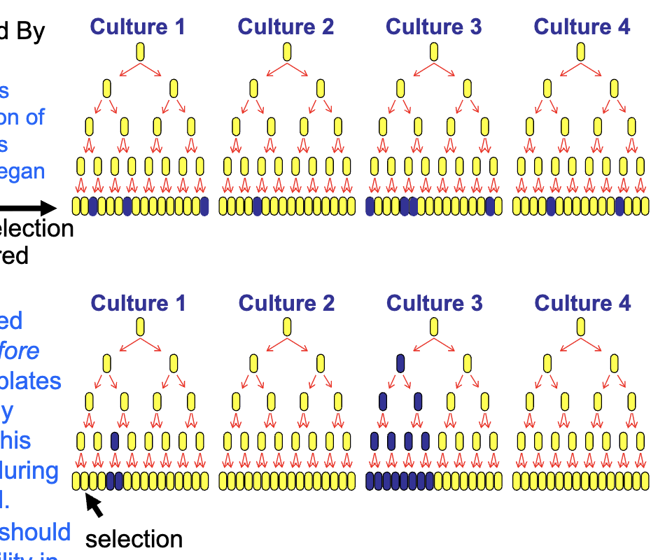
Are spontaneous mutations pre-existing? The Luria-Delbrück Fluctuation Test 2
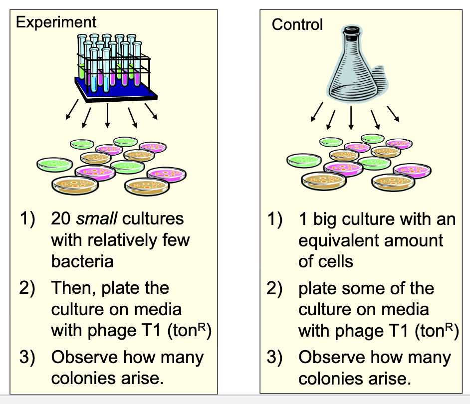
Samples from bulk culture have low variance (due only to sampling errors)
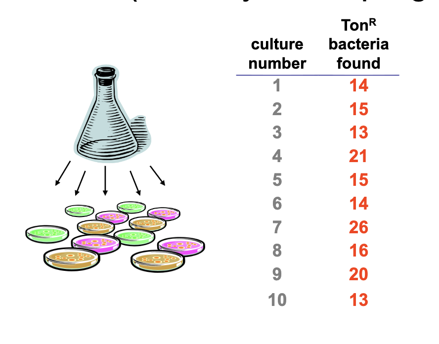
Individual cultures have a very high variance (due to jackpots)
Mutations occur spontaneously without reason (and already existed in these tubes before selection).
Advantageous mutations were selected for in subsequent generations, but their occurrence was not caused by selective pressure.
Many ways to classify mutations
Descriptions will often use several descriptors.
Where & when
Cell type (germ-line, somatic)
Chromosome type (autosome, X, or Y)
Phenotypic effect(s)
Neutral, lethal, conditional, etc.
Type of molecular change
Location in gene (promoter, ORF, etc.)
Kind of change: point versus extended
Substitution mutation vs. insertion/deletion
Transition vs. transversion (types of substitutions)
Mutations have many causes
Spontaneous mutations
Chemistry of the nucleic acids
Tautomeric shifts
Depurination (apurinic sites)
Deamination
Mistakes by DNA polymerase (biochemistry)
Insertion of an incorrect base (without correction)
Slippage of repeated sequences
Induced mutations
Due to the actions of outside agents
UV light
Ionizing radiation
Mutagens: acridine dyes, base analogs, alkylating agents, aflatoxin, etc.
More learning goals
Mutation
Be able to define mutation and describe it using correct terminology (i.e., classify by type, phenotypic effects and location) Classification
Be able to describe and interpret the experiments that test the concept mutations occur randomly and not in response to environmental conditions Luria-Delbrück Experiment
Understand the mechanisms by which mutations arise: spontaneous (due to intrinsic chemistry or biochemistry acting on the molecule) vs. induced (outside agents) Sources of Mutation
Be able to describe the mechanism leading to the lesion
Be able to describe the lesion itself
DNA Repair
Be able to describe the repair mechanisms associated with fixing each kind of lesion
Be able to describe what happens when the DNA repair machinery is mutated (non-functional)
Big, big picture
Mutations are any change to the DNA sequence
They are the source of genetic variation, can be acted upon by natural selection. They are also sources of genetic damage leading to cell death, disease, etc.
Mutations occur spontaneously and can be induced
Mutations have different effects depending on where and when they occur, and what the exact change is
Organisms rely on DNA repair mechanisms to counteract mutation
Repair machinery is overlapping and is employed differently in G1, S (during replication) and G2 (sister chromatids) phases
When the repair machinery itself is broken (due to mutation), then there even more propensity for mutation leading to increased risk for cancers, disease, aneuploidy
Many ways to classify
NOTE: mutations are explained by multiple descriptors
Where & when:
Cell type (germ-line, somatic)
Chromosome type (autosome, X or Y)
Phenotypic effect
Neutral, lethal, conditional, etc.
Type of molecular change
Where relative to a gene (promoter, in the ORF, etc.)
What kind of change:
Substitution: transition or transversion (types of substitutions)
Insertion or deletion (indel): how large?
What cell types do mutations occur in?
Somatic mutations occur in any cell type except germ cells and are not heritable
These mutations might lead to cancer
Germ-line mutations occur in gametes and are inherited
These are generally not evident in the parental generation
These are now part of the offspring’s genome
What chromosomes do mutations occur in?
Autosomal mutations occur within genes located on the autosomes
X-linked or Y-linked mutations occur within genes located on the X or Y chromosomes
Consequences of mutations
The consequences of mutations differ depending upon where (cell type, chromosome) and when (early in development or after maturation) the mutation occurs:
Somatic mutations (diploid organism):
When an autosomal recessive mutation occurs, it is unlikely to result in a detectable phenotype unless X-linked in males.
Dominant mutations tend to affect phenotype.
Germ-line mutations:
Inherited dominant autosomal mutations will be expressed phenotypically in the first generation.
X-linked recessive mutations arising in the gametes of a homogametic female may be expressed in hemizygous male offspring.
Classification of mutations
The classification can be made based on the molecular event.
Insertion or deletion of one or more nucleotide (aka, indels which is short hand for insertion/deletion)
Substitution (replacement) of a nucleotide with another
If a pyrimidine replaces a pyrimidine or a purine replaces a purine, a transition mutation has occurred.
If a purine and a pyrimidine are interchanged, a transversion mutation has occurred.
Transitions are more common than transversions.
The consequences depend on where the change is relative to neighboring genes.
Frameshift mutations
Frameshift mutations result from insertions or deletions (indels) of a base pair in a coding region (ORF).
Indels affect the translation of the gene into protein.
Indels can also affect transcription by disrupting promoters and regulatory regions

Point mutations
Point mutations are base substitutions in which one base pair is altered.
They can occur outside of a coding region:
Probably will not have an effect, unless they affect transcription of a gene or chromatin structure.
They can occur within a coding region:
Missense mutations change a codon resulting in an altered amino acid within a protein-coding portion of a gene.
A nonsense mutation changes a codon into a stop codon and results in premature termination of translation.
A silent mutation alters a codon but does not result in a change in the amino acid at that position of the protein.
Could alter (add, remove) a splice junction.
Classification of mutations by effects on phenotypes
Neutral mutations, the vast majority of all mutations, occur in the large portions of the genome that do not contain genes and therefore they do not affect RNA or protein production.
Mutations can be classified according to their phenotypic effects as:
loss-of-function (i.e., recessive)
gain-of-function (i.e., dominant)
morphological
nutritional (biochemical)
behavioral
regulatory
Lethal mutations interrupt an essential process and result in death.
The expression of conditional mutations depends on the environment in which the organism finds itself.
A good example is a temperature-sensitive mutation.
Mutations occur randomly
A common misconception is that mutations occur in response to a new condition.
The Luria-Delbrück fluctuation test demonstrated that mutations are not adaptive but they occur spontaneously.
Theory behind the Luria-Delbruck fluctuation test
If mutations are induced and occur in response to a challenge, then the rate of mutation is a function of the number of cells.
Prediction: a constant number not affected by the culturing conditions

Theory behind the Luria-Delbruck fluctuation test 2
If mutations occur spontaneously and are independent of the challenge, then the rate of mutation depends on time and the number of progeny with it.
Prediction: fluctuation in numbers in the different cultures
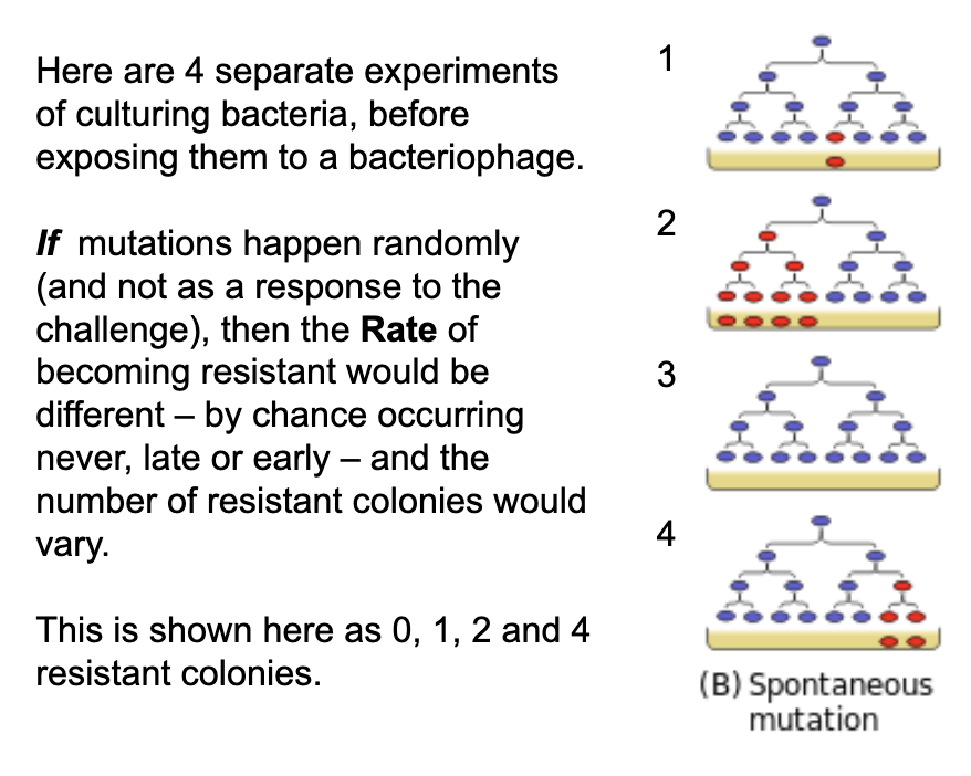
Theory behind the Luria-Delbruck fluctuation test 3
If mutations occur spontaneously and are independent of the challenge (resistance to infection by the phage T1), then the rate of mutation depends on time and the number of progeny with it
Predict: fluctuation in numbers in the different cultures

Theory behind the Luria-Delbruck fluctuation test 4
If mutations are induced and occur in response to a challenge (resistance to infection by the phage T1), then the rate of mutation is a function of the number of cells
Predict: a constant number not affected by the culturing conditions

Experimental set-up

Experimental set-up 2
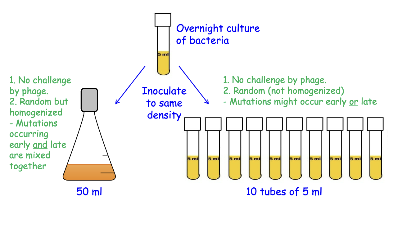
Experimental set-up 3
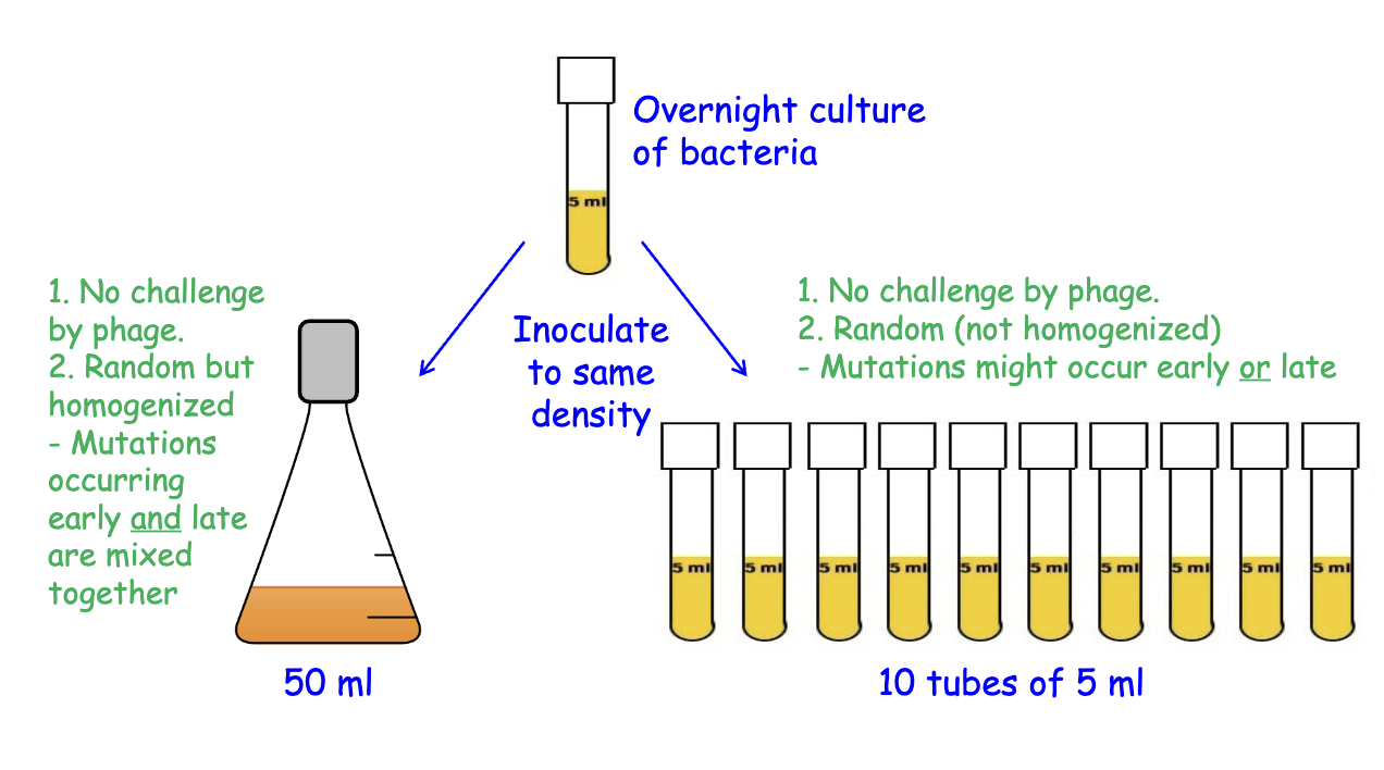
Experimental set-up 4

The Luria-Delburck Experiment Demonstrating that Mutations are Spontaneous

Mutations
A mutation is an alteration in DNA sequence

Sources of mutations
Spontaneous:
Due to the chemistry of the nucleic acids
Tautomeric shifts
Depurination (apurinic sites)
Deamination
Note: the problems occur during replication
Tautomeric shifts
Definition:
The spontaneous isomerization of a nitrogen base to an alternative hydrogen-bonding form, possibly resulting in a mutation. Reversible shifts of proton position in a molecule.
Bases in nucleic acids shift between keto and enol forms or between amino and imino forms.
Tautomeric shifts in nucleotides can result in mutations due to anomalous base pairing
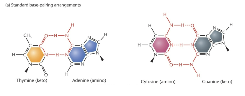
Tautomeric shifts

Tautomeric shifts

Shifts during replication
Tautomers represent different isomers of each nucleotide.
Nucleotides exist in an equilibrium between preferred and less- preferred tautomers.
keto →←enol amino →← imino
However, a less-preferred isomer can form or be incorporated into DNA during replication; that rare form will be stabilized by H-bonds.
But, it would likely revert back to the preferred form at the next replication.

What are the most common causes of spontaneous mutation
DNA base damage by depurination and deamination are the most common causes
Depurination
Apurinic sites
The spontaneous loss of a purine base due to hydrolysis of the glycosidic bond.
The glycosidic bond joins the sugar to the base.
This process is also called “depurination”.

Depurination
Apurinic sites are problems during replication

Deamination
Hydrolysis of the amine from adenine (to form the base hypoxanthine) and cytosine (to form uracil).
Hypoxanthine pairs with C and uracil pairs with G
This causes transition mutations: an AT base pair → GC, and the CG base pair becomes TA

Deamination of meC
A primary source of mutation is when 5-methyl cytosine is deaminated.
5meC is a common modification of C in euk.
5meC becomes T when it deaminates.
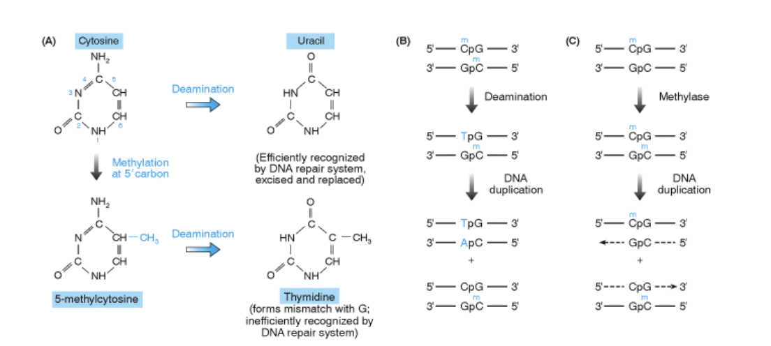
DNA damage
Spontaneous
Deamination
Spontaneous hydrolysis

Sources of mutations
Spontaneous
Due to the chemistry of the nucleic acids
Tautomeric shifts
Depurination (apurinic sites)
Deamination
Due to mistakes by DNA polymerase (biochemistry)
Insertion of an incorrect base (with no correction)
Insertion of a base analog instead of a “regular” nucleotide
Slippage of a repeated sequence
Polymerase Errors
Insertion of an incorrect base
DNA polymerase occasionally inserts incorrect nucleotides, generally due to mis-pairing.
In this case, the insertion is not corrected via the proof- reading activity of DNA polymerase.
These types of errors predominantly lead to point mutations (substitutions).
DNA synthesis: error rate of 1 per 1000-10,000 bases (10-3-10-4)
→ Proofreading during synthesis: error is 1 per 1-10 million (10-6-10-7)
Our haploid genome is ~3 billion bp; this would be 600-6000 errors per cell cycle
→ Post-replication repair, decreases this error rate even more to about 1 per 1-10 billion (10-9-10-10):
this is now ~6-60 mutations per cell cycle
Incorporation of analogs
Base analogs can substitute for purines and pyrimidines during nucleic acid replication.
These base analogs are mutagenic if they form base pairs with a different nucleotide more often than the normal nucleotide.
E.g., 5-BrU undergoes the tautomeric shift more frequently than T; thus it can base pair more easily with G

Slippage during replication can lead to small insertions or deletions
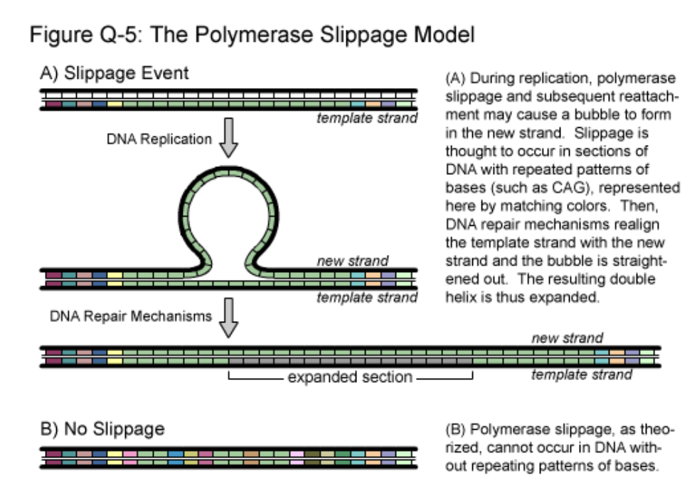
A model describing polymerase slippage during replication

Trinucleotide repeat sequences are responsible for human disorders
In several cases, a correlation exists between the number of repeats and the age of manifestation of mutant phenotypes.
The greater the number of repeats, the earlier disease onset occurs. In affected individuals, the number of repeats may increase in each subsequent generation, a phenomenon known as genetic anticipation.
NOTE: note the summary table but don’t memorize

Sources of mutations
Due to the chemistry of the nucleic acids
Tautomeric shifts
Depurination (apurinic sites)
Deamination
Due to mistakes by DNA polymerase (biochemistry)
Insertion of an incorrect base (with no correction)
Insertion of a base analog instead of a “regular” nucleotide
Slippage of a repeated sequence
DNA damage
Replication errors

Induced mutations
Due to the actions of outside agents
Alkylation
UV light
Ionizing radiation and oxidation
Acridine Dyes (intercalation)
Transposable Elements
DNA Damage
Damage of DNA causes lesions that are chemically altered bases or damage to the sugar- phosphodiester backbone.
Lesions exist in the short-term and evoke DNA repair mechanisms to fix the lesion.
Lesions are repaired and the process of repair can convert the lesion into a mutation (recall that mutations are any changes to the DNA sequence).
Chemicals or treatments that induce DNA lesions cause mutations and cancer, and are therefore referred to as mutagens or carcinogens.
Alkylating agents
Alkylating agents donate an alkyl group to amino or keto groups in nucleotides to alter base-pairing affinity
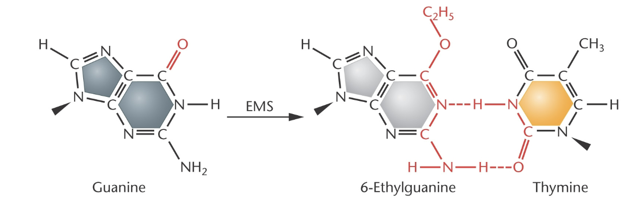
Alkylating agents 2
Alkylating agents donate an alkyl group to amino or keto groups in nucleotides to alter base-pairing affinity, block DNA replication, prevent transcription

Mustard gas is bi-functional
It will crosslink DNA (within and between duplexes);
Thus, replication, recombination, and transcription will be affected

Mustard Gas
Mustard gas (Yperite) was first used by the German Army in September 1917. It was one of the most lethal of all the poisonous chemicals used during the war.
It was almost odorless and took twelve hours to take effect. Yperite was so powerful that only small amounts had to be added to high explosive shells to be effective. Once in the soil, mustard gas remained active for weeks.
It was used by former Iraqi President Saddam Hussein against the Kurds in 1987-1988.
ISIS used mustard gas against Syrians in 2015-2016.
Biological effects:
The skin of victims of mustard gas blister, the eyes become sore, and they vomit.
Mustard gas causes internal and external bleeding and attack the bronchial tubes, stripping off the mucous membrane. This is extremely painful and, in WWI, most soldiers had to be strapped to their beds because of the pain.
It usually took a person four or five weeks to die of mustard gas poisoning.
One nurse, Vera Brittain, wrote: "I wish those people who talk about going on with this war whatever it costs could see the soldiers suffering from mustard gas poisoning. Great mustard-colored blisters, blind eyes, all sticky and stuck together, always fighting for breath, with voices a mere whisper, saying that their throats are closing and they know they will choke."
Other agents also alkylate DNA
The major guanine adduct formed by benzo(a)pyrene, a common polycyclic hydrocarbon found in cigarette smoke and other combustion residues.
The major guanine adduct of Aflatoxin B1, produced by a mold (Aspergillus) that grow on peanuts and grain crops.
A 2004 outbreak killed 120 people (of 317 affected) in Kenya
Children are particularly sensitive: stunted growth, liver damage, etc.
Dog food was recalled in 2014

UV radiation
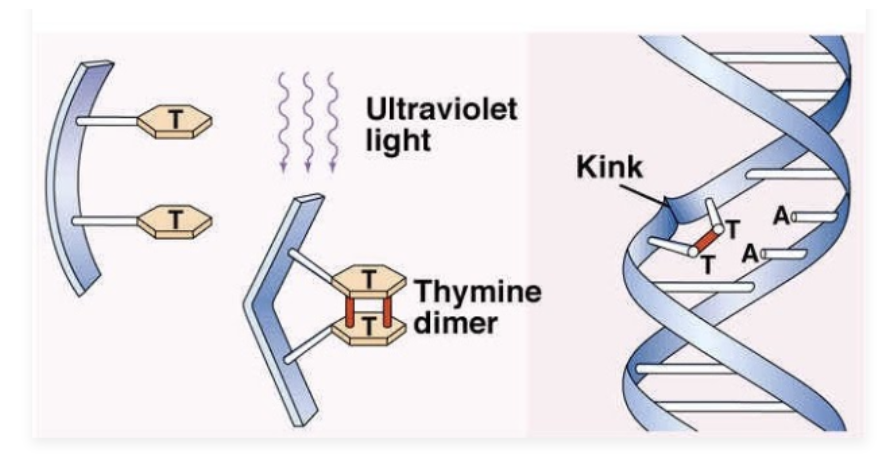
UV radiation creates pyrimidine dimers that distort the DNA conformation in such a way that errors tend to be introduced during DNA replication.

Pyrimidine dimer
Also called a cyclobutane ring
The sugar-phosphate backbone is kinked and the adenine bases are unpaired
Ionizing radiation
Ionizing radiation causes damage to DNA, directly and indirectly through reactive oxygen species.
DNA damage is broken covalent bonds.
Strand breakage (double- and single-strand breaks)
Damage to bases

Ionizing radiation
Ionizing radiation in the form of X rays, gamma rays, and cosmic rays are mutagenic.
The rate of formation of mutations is directly proportional to the dosage of X-rays (Muller experiment)

Oxidation
Oxidative damage can be caused by both high- energy radiation as well as reactive oxidants formed by normal cellular metabolism.
Reactive oxidants are superoxides (O2-), hydroxyl radicals (-OH) and hydrogen peroxide (H2O2)
They react with bases to modify DNA (examples shown to the right, there are hundreds of modifications), to break the glycosidic bond, and to make single-strand breaks in the chain.

Acridine dyes
Acridine dyes cause frameshift mutations by intercalating between purines and pyrimidines.
Ethidium bromide, used as a fluorescent agent to detect DNA in the lab, is an acridine dye.

Intercalation of ethidium bromide in DNA
Acridine dyes such as proflavin and ethidium bromide have a flat, planar structure, conjugated double bonds, and are of the same dimension as a base pair.
They can intercalate between adjacent base pairs and distort the DNA helix, leading to insertion or deletion (indel) mutations.

Doxorubicin
Doxorubicin is an anticancer drug derived from the Streptomyces bacterium. It is a chemotherapeutic agent used to treat solid tumors and blood cancers.
bone sarcomas, and cancers of the breast, ovary, bladder, and thyroid; lymphoblastic leukemia, acute myeloblastic leukemia, Hodgkin lymphoma, and small cell lung cancer.
The primary mechanism of action is its ability to intercalate within DNA base pairs, causing breakage of DNA strands and inhibition of both DNA and RNA synthesis.
DNA damage full diagram

Mico- and nanoplastics are genotoxic
Microplastics can lead to chromosome structural changes (e.g. inversions, deletions, et al.) by causing DSB, aneuploidy by disrupting chromosome segregation, and can damage DNA leading to replication and transcription errors.

DNA damage repaired
IMPORTANTTT STUDYYYY
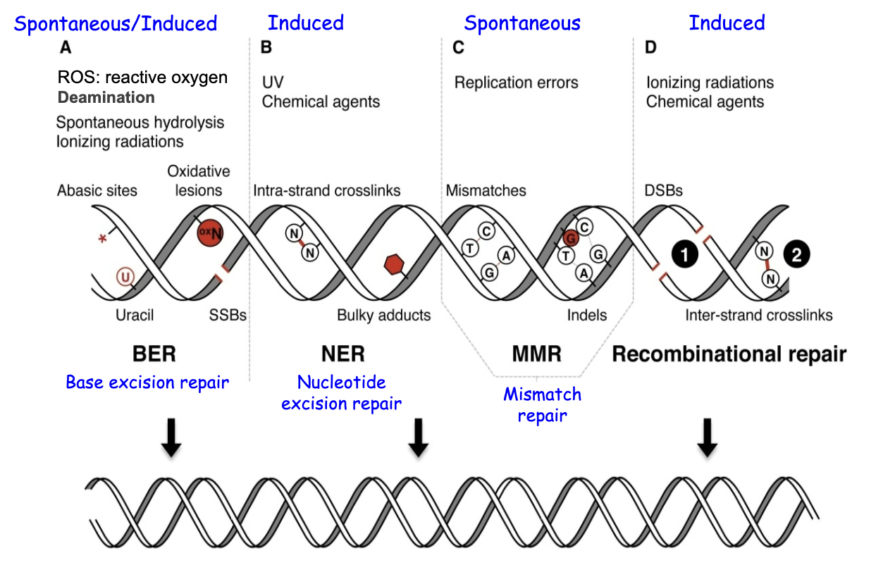
Repairing DNA
Living systems have evolved a variety of elaborate repair systems that counteract both spontaneous and induced DNA damage.
These repair systems are essential to the maintenance of the genome.
Defective repair systems lead to increased mutations and cancer.
Repair mechanisms
Reversing bonds (lyases); excision repair; mismatch repair; repair during DNA synthesis by special polymerases; homology repair; non-homologous end-joining.
Some mechanisms only work at particular phases of the cell cycle – e.g, during S-phase or G2.
Base excision repair (BER)
For U in DNA and apurinic sites.
Base excision repair (BER).
Removal of the uracil base first to make an apurinic site.
Involves recognition of the erroneous base by DNA glycosylase
Cleave the DNA backbone by an endonuclease to remove the deoxyribose sugar.
Synthesize DNA to fill-in the one missing nucleotide

Photoreactivation repair (PRE)
Photoreactivation repair removes pyrimidine dimers caused by UV light.
The process depends on the activity of a protein called the photoreactivation enzyme (PRE), found in bacteria, plants and some eukaryotes, but it was lost during evolution of placental animals.
PRE is a DNA photolyase.
Energy to break the covalent bonds comes from light in the blue wavelengths (300-600 nm). The enzyme has the ability to capture that energy and then to split the bonds.

Nucleotide excision repair (post-replication)
Nucleotide excision repair (NER) repairs bulky lesions.
Bulky lesions are alkylations, pyrimidine dimers, and other modifications to the DNA.
Excision of a large segment of the DNA (catalyzed by the uvr genes in E. coli).
Individuals with xeroderma pigmentosum have lost the ability to undergo nucleotide excision repair.

Xeroderma pigmentosum: disease caused by NER deficiency
Gene products perform different steps in the pathway to DNA repair

DNA Repair Systems
Post-replication:
Mismatch repair corrects errors that remain after proofreading.
There is a bias to correcting the newly made DNA (as opposed to the original DNA strand).
The original DNA strand is recognized based on DNA methylation of the parental strand.
Newly replicated DNA is hemi-methylated, with the newly synthesized DNA as lacking methylation.

Mismatch repair
Corrects mistakes that remain after proofreading
Old strands are methylated but new ones are not yet
This allows a repair bias: remove the newly synthesized DNA and fill in
Removal: cut with an endonuclease, degrade with an exonuclease, and synthesize with DNA polymerase.

SOS Repair and Translesion Synthesis (repair during replication)
Cells encode special DNA polymerases that can synthesize through DNA lesions.
SOS repair in bacteria and translesion synthesis in eukaryotes are enzymes that respond to DNA polymerases that are stalled at a site of lesions or gaps in the DNA sequence (see next slide, from previous lecture).
DNA synthesis becomes error-prone.
The fidelity of copying a DNA template is reduced allowing DNA synthesis to continue by incorporating random nucleotides.
This mechanism uses special DNA polymerases
Although SOS repair is itself mutagenic, it may allow cells to survive DNA damage that might otherwise kill them.
E. coli DNA polymerases - focus on repair

Translesion Synthesis

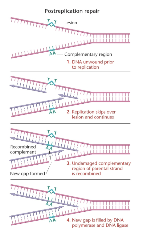
Recombination-based (post-replication repair)
Post-replication repair occurs when DNA replication skips over a lesion and requires homologous recombination to correct the damage.
This involves unwinding of the DNA duplex and strand invasion (recall the discussion of the Holliday structure).
The non-damaged parental strand is copied, unwound and used to correct lesions and gaps on both sister chromatids.
Video link (newly added on 11/7)
Non-homologous end joining

DNA damage repaired

Cancers
Cancer cells contain genetic detects affecting genomic stability and DNA repair.
A lack of genome stability is seen as aneuploidy.
Some cancers that are heritable have mutations in the repair machinery:
BRCA1 and BRCA2 are linked to breast, ovarian and prostate cancers
p53 to Li-Fraumeni syndrome
Genes encoding the nucleotide excision repair machinery lead to xeroderma pigmentosum.
BRCA1 and DNA Repair

DSB Repair Video
slide 49, 11/6
Sources of mutations again
Spontaneous
Due to the chemistry of the nucleic acids
Tautomeric shifts
Depurination (apurinic sites)
Deamination
Due to mistakes by DNA polymerase (biochemistry)
Insertion of an incorrect base (with no correction)
Insertion of a base analog instead of a “regular” nucleotide
Slippage of a repeated sequence
Induced
Due to the actions of outside agents
Alkylation
UV light
Ionizing radiation, oxidation
Acridine Dyes (intercalation)
Transposable Elements
Transposable elements
In general:
Discovered by Barbara McClintock in maize
“Jumping genes”, mobile DNA elements
When they move, they can disrupt genes to cause new mutations.
Found in all domains of life (prokaryotes and eukaryotes)
~45% of the human genome is composed of transposable elements
Geneticists use them to cause mutations and then can screen for phenotypes, and can see where the transposon inserted itself.

Two types (one has an RNA intermediate)

Hermes transposition
Researchers use transposon mutagenesis to discover genes involved in processes/pathways of interest.
Hermes is an insect, class II transposon that has been adapted for use in fungi, including C. glabrata.
Hermes was modified to have a selectable gene (e.g. NATR)
Transposition is induced in very large numbers, and selected for on nourseothricin plates
The loci that have received the Hermes transposon will be found by DNA sequencing
Genes that can be mutated will have transposition events; essential genes cannot be disrupted.
Some of the genes in the inositol pyrophosphate pathway are apparently essential (and not the focus of the Genetics lab this semester)
Hermes transposition
Generally the transposon is transformed into cells on a plasmid

Transposition in C. glabrata
Example
