OB/PEDS - EXAM 2
 Call Kai
 Learn
 Practice Test
 Spaced Repetition
 Match
 Flashcards
 Knowt Play
1/269

There's no tags or description
Looks like no tags are added yet.
270 Terms
Pediatric Hematologic Considerations
Production of blood cells begins at 8 weeks in utero
Fetus receives iron through the placenta
Erythropoietin is derived primarily from the liver in the fetus
Kidneys are the primary source after birth
Iron-Deficiency Anemia
Iron deficiency is most common nutritional disorder in the world
Adolescents and children go through rapid periods of growth and require more iron (at risk)
Peak prevalence in children 12-24 months (off formula) and then adolescence
Disproportionately impacts children and pregnant women
Occurs when the body doesn’t have enough iron to produce hemoglobin
Iron-Deficiency Anemia - Etiology
Impaired absorption
Increased requirement
Runners
Chronic blood loss
GI bleed
Dietary lack
Vegan - need beans, greens (have iron)
Adequate dietary intake of iron required for body to make enough hemoglobin
Iron-Deficiency Anemia - Risk Factors
Prematurity
Immature gut and kidneys (poor absorption)
Exposure to lead
Old homes have lead based paint (children ingest) or parents in the workforce
Exclusive breast-feeding beyond 4 months old without iron supplementation
AAP recommends 1st 6 months for breastfeeding
Cow’s milk without adequate iron intake
Fill up and not eat or microhemorrages in gut = slow blood loss
Switch at 12 months to cows milk
Low socioeconomic status
Poor access, poverty
Use of medications that decrease iron absorption
PPIs, H2 blockers, antacids, thyroid, antibiotics
Iron-Deficiency Anemia - Diagnostic Criteria
For child 6 months to < 5 years:
Ferritin (blood test) < 15 micrograms/L
AND
Hemoglobin < 11 g/dL (for children 0.5 to 5 years)
REMEMBER THIS ONE
For children 5 to < 12 years:
Ferritin < 15 micrograms/L
AND
Hemoglobin < 11.5 g/dL (for children 5-12 years)
Iron-Deficiency Anemia - Clinical Manifestations
Mild cases: often no symptoms
Reduced growth in prolonged cases (decreased iron and hemoglobin)
Common symptoms (d/t poor circulation of O2): pallor, fatigue, SOB, weakness, headache, dizziness, irritability, + murmur (blood is thinner)
Other possible symptoms: pica (eat odd things; ice, dry wall (child), rocks (child)), unsteady gait, muscle weakness, difficulty feeding
Iron-Deficiency Anemia - Nursing Assessment
Obtain thorough history
Ask about availability of iron-rich foods
Chronic: special diets (vegetarian)
Inspect mucous membranes and skin for pallor (pull lip/eyelid in AA)
Assess nails for spooning (chronic)
Auscultate for murmur
Assess spleen for enlargement (splenomegaly)
Patient education on what foods to eat
Iron-Deficiency Anemia - Nursing Interventions
Goal - determine and treat the underlying cause
Breastfeeding mothers should increase their daily intake of iron (like red meat)
> 4 months need iron supplementation if just exclusively breastfeeding
Formula-fed babies should have iron-fortified formula
Limit cow’s milk intake to < 24 ounces/day for children > 1 year of age
Decreases risk of microscopic GI bleeding and increases appetite for other foods
Cows milk = decreases absorption of iron from foods in GI tract
Avoid NSAIDS when possible
Can cause GI bleeding, irritation, and iron loss
Iron-Deficiency Anemia - Caregiver Education
Give iron 1 hour before meals to maximize absorption
With syringe behind teeth or straw (d/t staining) in older children
Don’t give with milk
Citrus increases absorption
Stools may be black in color
Constipation may occur - increase fiber and water intake
Stool softener → miralax or apple juice
Common Sources of Iron
Red meats (iron from this source is easiest for body to absorb)
Tuna
Eggs
Enriched grains
Dried beans
Leafy green vegetables
Iron-fortified cereals
Dried fruits
Picture: heme iron (blood source) vs. non-heme iron

Sickle Cell Disease
Inherited hemoglobinopathy (autosomal recessive)
Both carriers ~25% chance of offspring with disease
Vaso-occlusive (temporary blockage of vessels - pain) phenomena and hemolytic anemia are the clinical hallmarks
3 most common types:
HbSS (homozygous) - symptoms are most severe (“so severe”)
HbSC - defect causing a structural variant in B-globin gene; causes deficiency and structural variations but milder features
HbS beta thalassemia - have a decreased production of normal hemoglobin
Thalassemia minor (B-thalassemia trait) - mild microcytic anemia (no treatment needed)
Thalassemia intermedia - child needs blood transfusions
Thalassemia major - ongoing medical attention, blood transfusions, and iron removal (chelation therapy)

Sickle Cell Disease - Diagnosis
Newborn screening in all 50 states
Doesn’t distinguish between sickle cell disease and sickle cell trait
Electrophoresis confirms diagnosis of sickle cell disease

Mechanism of Vaso-Occlusion
Normal RBC
Round shape
Soft/flexible to allow laminar flow of blood
Average lifespan 120 days
Sickled RBC
Crescent shape
Sticky/rigid, inhibits laminar blood flow
Average lifespan 20 days

What happens in vaso-occlusion?
Stress or traumatic event triggers sickling of cells
Physical (dehydration) + increased emotional state
Blood becomes more viscous as sickled cells clump together
Normal blood flow to tissues in that area is blocked (can be anywhere)
Local tissue hypoxia followed by ischemia and possible infarction
Pain due to decreased circulation in the area
Increased metabolic need from pain causes tachycardia and tachypnea which leads to further sickling
Morphine (serious pain meds) and hydrate with fluids
Sickle Cell Disease - Nursing Assessment
Skin - pallor, breakdown, lesions (where blockages are), jaundice (d/t breakdown of RBCs)
Oral mucosa - pallor, dry/cracked
Auscultate chest - murmur, adventitious or decreased breath sounds (acute chest = blockage in lungs)
Palpation of extremities and abdomen - warmth/tenderness/ROM joints, abdominal tenderness, organomegaly
Vital signs - elevated temperature, HR, RR, and hypotension (d/t decreased perfusion and dehydration)

Sickle Cell Disease - Dactylitis
Severe swelling and tenderness that affects fingers and toes (“sausage fingers”)

Sickle Cell Disease - Acute Chest Syndrome
Severe lung complication of SCD
Creates a pneumonia like illness
One of leading causes of morbidity, hospitalizations, and death in children and adults
Picture - dark = air

Sickle Cell Disease - Nursing Care
Prevention
Vaso-occlusive crises
Infection
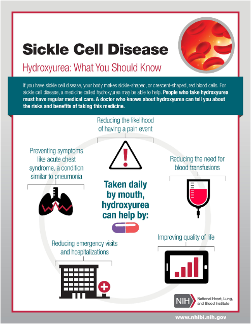
Hydroxyurea
Preventive med
Increasing amount of fetal hemoglobin and makes RBCs increase, rounder, and more flexible
Done by increasing certain type of hemoglobin (F) or fetal hemoglobin because it is found in newborns
Penicillin
Preventive med
Decreased risk of infection (which would trigger a crisis)
Managing Sequelae
Pain management
Treat underlying condition
Increase fluid requirements
Supplemental O2 PRN
No ice packs (may use heat; ice causes vasoconstriction while heat on affected joints unoccludes and vasodilates)
Education + Support
S/S of vaso-occlusive events
Health maintenance w/ PCP and specialist
Med administration
Genetic testing
Hemophilia
Inherited, blood borne disorder
Disorder of “secondary hemostasis”
Fibrin clot formation is too unstable to adequately stop bleeding
Factor VIII (8) and factor IX (9) both play key roles by assisting in the formation of a fibrin clot
A defect in either of these factors prevents secondary hemostasis by causing the fibrin clot to be “jelly-like” and unstable → delayed clotting → spontaneous bleeding
Clotting Cascade
Series of steps in response to bleeding - each step activates the next and ultimately produces a clot
Hemostasis - bleed stopping
Coagulation cascade involves the activation of a series of clotting factors
Once each protein is activated it is then able to catalyze the next reaction
Hemophilia A
X-linked recessive disorder (think males)
Inherited deficiency of factor VIII (“A = 8”)
Most common hereditary disease associated with life-threatening bleeding
Seen most often in males (late teens-20s)
Females may rarely be affected (affected father and carrier mother; genetic mutation (mild forms))
Higher incidence of detection in 2nd and 3rd decades of life due to milder diagnosis
Hemophilia B
X-linked recessive disorder (think males)
Inherited deficiency of factor IX (“B = 9”)
Also called Christmas disease
Seen most often in males
Females may rarely be affected (affected father and carrier mother; genetic mutation (mild forms))
Higher incidence of detection in 2nd and 3rd decades of life due to milder diagnosis
Describe how hemophilia is classified by severity (mild → severe)
Mild: prophylactic factor before surgery or procedure (like dental)
Severe: need factor daily → weekly
More likely for bleeding episodes
Bleed - vessels constrict and platelet plug forms (but because of deficient factor will not solidify = bleed continues)
Hemophilia - Clinical Manifestations
Severe: more likely to have spontaneous bleeding, severe bleeding, and diagnosis at earlier age
Mild-moderate: only have bleeding in response to injury or trauma
Immediate or delayed bleeding after trauma is common
Has decreased with prophylactic use of factor replacement
Infants: common bleeding site include extracranial, heel stick (red flag if bleeding doesn’t stop), venipuncture, and circumcision sites
Children: bruising and joint bleeds once ambulating, forehead hematomas
Older children/adults: bleeding/enlarged joints, muscles, GI tract, CNS, mouth, nares, bruising, forehead hematoma
Hemophilia - Nursing Care
Manage bleeds
Admin factor replacement and/or desmopressin (DDVAP; stimulates stored factor in body) as ordered
Apply pressure to areas of external bleeding for a long time
If bleeding is inside a joint → ice cold (constricts, clumps) compress and elevate injured extremity, may need splint
AVOID NSAIDS (increases risk of bleeding), can give Tylenol for pain PRN
Provide support
Lifelong illness
Medical alert bracelet/necklace
Refer to support groups - National Hemophilia Foundation
Help provide resources for financial strain of diagnosis
At home factor infusions (training, access - expensive, insurance)
Prevent and educate
Provide prophylactic factor when ordered
Protect the toddling child with helmets/kneepads, soften corners
Regular exercise and activity to strengthen joints and muscles
Avoid activities with high likelihood of injury
Good oral hygiene to prevent hum/dental disease (soft toothbrush, rinses)
Teach caregiver how to admin factor replacement
Educate patient and family on when to seek expert care
NO IM INJECTIONS AND RECTAL TEMPS
Cancer in Adults
Histologic Type
Epithelial tissues, carcinoma
Mechanism
Environmental exposures (lifetime - smoking + drinking)
Affects
Organs - lung, liver, intestines
How its discovered
Early through screenings - mammograms
Environmental/lifestyle factors
80% preventable with lifestyle modifications
Latent period
Long (> 20 years) - cells replicate overtime
Metastasis
Less often at diagnosis
Response to treatment
Less responsive
Cancer in Children
Histologic Type
Embryonal, leukemia, lymphoma
Mechanism
Cellular growth and development (not lifestyle)
Affects
Tissues
How its discovered
Incidental or accidental (presents as pain)
Environmental/lifestyle factors
Little influence
Latent period
Short
Metastasis
Often present at diagnosis d/t it taking awhile to discover as symptoms may suggest something else (delayed diagnosis)
Response to treatment
Very responsive and little resistance
Detecting Pediatric Cancer
Can mimic growing pains, etc… (which is usually bilateral, but one extremity is concerning)
Often difficult to detect early on (concerns with unusual presentations - certain tumors; different cancers have S/S that make it easier to detect early on vs. later)
Symptoms have an insidious onset and are nonspecific
Symptoms often resemble more common pathologies
~85% of peds cancers have symptoms at presentation
Obtain a thorough history: ask specific questions surrounding the chief complaint
Average time to diagnose:
2-5 weeks - neuroblastoma
3-5 weeks - acute leukemias (29%/year)
7-10 weeks - lymphomas
8-15 weeks - bone tumors
8->20 weeks - brain tumors (26%/year)
Detecting Pediatric Cancer - Early Warning Signs
Unexplained pallor and loss of energy
Anemic - decreased EBC, H+H, platelets
Unusual lump, mass, or swelling
Large, fixed, hard lymph nodes
In abdomen needs further investigation
Sudden unexplained weight loss
Kids usually grow
Unexplained fever or illness that doesn’t go away or cough
Doesn’t get worse or better
Common (doesn’t have to high - >100.4F)
Need CBC (blood work - WBC, platelets, H+H)
Easy bruising or bleeding
Nose bleeds/gum bleeding concerning
Common in healthy kids d/t increased activity
Hematomas in lower extremities or abdomen concerning
Prolonged or ongoing pain in one or more areas of the body
Kids don’t typically have pain
Limping, refusal to bear weight
Bones! → x-ray needed
Frequent headaches, particularly if occurring in the morning and associated with vomiting
Very concerning for mass in head (laying down increases ICP)
Sudden eye or vision changes
Want a red light reflex (mass causes white light)
Acute Lymphoblastic Leukemia (ALL)
Most common childhood malignancy (kids on spectrum at most risk)
Not familial linked; children with trisomy 21 have increased risk
Immature lymphocytes replace normal cells in bone marrow, cannot maintain RBC, WBC, and platelets - pancytopenia (everything low)
Presence of “blasts” in blood smear → immature WBCs
Acute Lymphoblastic Leukemia (ALL) - Presentation
Hepatomegaly/splenomegaly, fever, lymphadenopathy, petechiae (d/t decreased platelets, MSK pain, fatigue, weight loss
Acute Lymphoblastic Leukemia (ALL) - Diagnosis
Peripheral blood smear, bone marrow biopsy, lumber puncture (CNS involvement; cancer cells in CNS)
Acute Lymphoblastic Leukemia (ALL) - Management
Chemotherapy, allogenic stem cell transplant (HCT)
Tx lasts 2-3 years, 5 year survival rate 90% for children < 15 years
Non-Hodgkins Lymphoma
Includes all lymphomas except Hodgkins - common in primary immunodeficiencies
B and T lymphocytes mutate then proliferate rapidly, effects lymph nodes or bloodstream
Progression is quick and symptoms appear early
Non-Hodgkins Lymphoma - Presentation
Pain, lymphadenopathy, abdominal mass, mediastinal mass
Non-Hodgkins Lymphoma - Diagnosis
Lymph node and bone marrow biopsy, CXR, CT scan (extent of disease)
Non-Hodgkins Lymphoma - Management
Chemotherapy, CNS prophylaxis (give chemo through a spinal tap intrathecally), possible BMT
70-90% survival rate
Autologous bone marrow transplant - a procedure where healthy stem cells (blood-forming cells) are collected from the blood or bone marrow before treatment, stored, and then given back to patient after treatment
Hodgkins Lymphoma
Most common in teens and young adults
Rare in children < 5 years; in preadolescents, more common in boys than girls
Hallmark sign: Reed Sternberg B cells - malignant cells in lymph tissue, lymphadenopathy
Epstein-Barr Virus (mono) infection associated with many cases
Hodgkins Lymphoma - Presentation
Weight loss, fever, night sweats, anorexia, fatigue, enlarged lymph nodes, mediastinal mass
Hodgkins Lymphoma - Diagnosis
Lymph node biopsy
Hodgkins Lymphoma - Management
Chemotherapy, radiation, BMT/Hematopoietic Stem Cell Transplantation (HSCT) for relapse or unresponsive to treatment
90-85% cure rate
Medulloblastoma
”Brain tumor”
Most common malignant brain tumor of childhood
Primarily occur in cerebellum (cause ataxia)

Medulloblastoma - Presentation
Symptoms of increased ICP - headaches, vomiting, dizziness, blurred/double vision, ataxia, seizures, weakness, behavioral or developmental issues
Medulloblastoma - Diagnosis
Head CT, brain MRI, PET scan (a nuclear medicine imaging test that measures metabolic and cellular activity in your organs and tissues), surgical resection
Medulloblastoma - Management
Corticosteroids initially for increased ICP
Surgical resection of tumor, multimodal treatment; radiation after resection and chemotherapy
72% 5 year survival rate < 14 years old
Osteosarcoma
Bone
Most common primary malignancy of bone (~60)
Often occur in long bones - femur, tibia, humerus
Osteosarcoma - Presentation
Intermittent pain at site, limping, gait changes, soft tissue mass on exam that is tender to palpation
Osteosarcoma - Diagnosis
MRI of bone, bone biopsy, staging workup (x-ray initially)
Osteosarcoma - Management
Tumor resection (removal; amputation vs. limb sparing), chemotherapy, radiation
Reattach lower leg to upper leg (foot as knee) then prosthetic (rotationplasty)

Ewing Sarcoma
Most common in long bones (femur) and pelvis
Second most common bone malignancy (~30%)
Picture: allograft - reinforce bone with metal; prosthesis - metal replaces bone

Ewing Sarcoma - Presentation
Trauma may start symptoms, intermittent pain to site for weeks/months, pain worsens with activity and at night, erythema/edema over mass
Ewing Sarcoma - Diagnosis
Xray, MRI, CT scan, bone biopsy
Ewing Sarcoma - Management
4-6 months chemo cycles before local therapy (resection over radiation) and chemotherapy post local treatment
70-80% survival rate if metastasis is 20-30% only
Retinoblastoma
Most common intraocular malignancy
96% 0-14 years old
Can metastasize to CNS through optic nerve

Retinoblastoma - Presentation
Leukocoria (white pupil reflex), strabismus (eye crossing), nystagmus (eye jitter)
Retinoblastoma - Diagnosis
Dilated opthalmoscopic exam, no biopsy required d/t risk of tumor seeding (spreading)
Retinoblastoma - Management
Severe - chemotherapy, ophthalmic artery chemosurgery, laser photoablation, enucleation (remove eyeball) if large tumor present or progression into optic nerve
Neuroblastoma
Along neural crest
Tumor originates from neural crest during fetal development
90% of cases are diagnosed before age 5
Can arise anywhere throughout the SNS; adrenal gland and abdomen most common
Neuroblastoma - Presentation
(Not neurological symptoms) Non-tender abdominal mass, abdominal pain, constipation, back pain/weakness from spinal cord suppression, fever, weight loss, anemia, bone pain, bowel/bladder dysfunction
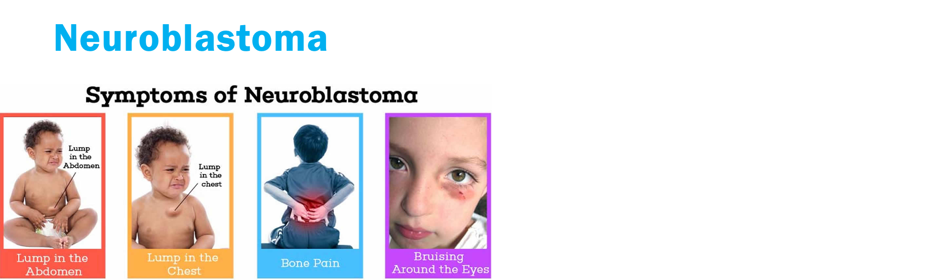
Neuroblastoma - Diagnosis
Biopsy of tumor, bone marrow biopsy, urine catecholamines, CT, MRI, PET
Neuroblastoma - Management
Surgical removal, chemotherapy
50-95% survival rate depending on risk group (low-risk to high-risk)
Wilms Tumor
Most common renal malignancy (95% diagnosed before age 10)
Most are solitary tumors surrounded by pseudocapsule (not an inherent part of an organ's structure; it is a protective border or layer of compressed tissue and fibers that forms around a slow-growing mass, tumor, or cyst as it pushes against surrounding healthy tissue)
Metastasis is rare
Once suspected… abdominal palpation should be limited; if capsule ruptured, tumor spreads rapidly + bleeding occurs
Wilms Tumor - Presentation
Firm, smooth abdominal mass, abdominal distention (can be one sided), abdominal pain, hematuria (blood in urine; tumor around kidney), fever, hypertension
Wilms Tumor - Diagnosis
Abdominal US, CT, MRI
Wilms Tumor - Management
Stage I and II - resection and chemotherapy (may loose kidney)
Stage III and IV - radiation also
90% survival rate
Diagnosing Pediatric Cancer
Thorough history of child and family
Current symptoms the child is having
Head-to-toe exam
Vital signs
Initial screening tests:
CBC with diff
Coagulopathies (PT, PTT, INR)
CMP (check to see if kidney and liver are okay for medications)
Blood cultures
Lumber puncture (checks cancer cells in CSF; sedation)
Imaging - xray, US, CT scan, MRI
Bone marrow aspiration (sedated)
Treatment Modalities for Pediatric Cancer
Surgery
Tumor debulking, tumor resection, CVL placement, etc…
Chemotherapy
Radiation
Biotherapy
BMT/HSCT
Supportive therapy
Cancer - Surgery: Postop Nursing Care
Admin pain/nausea meds and antibiotics as ordered
May have epidural (site checks) and/or PCA
Assess surgical site - bleeding (increases risk), signs of infection
Frequent physical assessments and monitoring VS closely
Strict I&O’s
Alert provider immediately of critical lab values
Incentive spirometry
Early ambulation
Radiation Therapy
Damages or kills cells using gamma or particle radiation
Affects all rapidly growing cells
Can be curative, palliative, or adjuvant therapy
Provide comfort measures and treat adverse affects as they occur
A/E: fatigue, nausea/vomiting, mucositis (mucosa inflammed), myelosuppression (bone marrow suppression), damage to skin
Phonton beam radiation = goes through to the other side (look for burns; skin assessment)
Proton beam = doesn’t pass through
Chemotherapy
Need training to admin
Alter or destroy cancer cells based on specific points of the cell cycle
Chemo drugs separated into classes based on actions or effect on cell cycle
A/E are determined by the regimen that is given
Myelosuppression, infection, constipation, tooth damage (soft toothbrush), hearing loss, vision changes, altered growth, neuropathy
3 phases of treatment:
Induction - short and intensive, lasts a month
Intensification (consolidation) - intensive, lasts a few months
Maintenance - post-consolidation; decreases intensive, lasts 2 years
Common Side Effects of Cancer Therapies
Infection
Pain
Anemia or blood loss (no rectal temps and IM injections)
GI symptoms: anorexia, N/V, diarrhea, constipation
Damage to oral mucosa
Damage to skin
Alopecia
Neutropenic Precautions
Absolute neutrophil count (< 1000 = neutropenia (risk of infection))
Private room, closed door, regular room cleaning
Frequent hand hygiene
Avoid rectal temps, enemas, and invasive procedures like catheterization
No fresh fruits/veggies, flowers, or plants in room
Visitor restrictions
CHG baths daily
Mouth care
Cancer - Infection Prevention
Regular assessments for potential infection(s)
Pain
Keep mucosa moist (oral hydration, ice chips if not NPO, petroleum jelly to lips)
Salt water rinses/mouthwash (no alcohol) q1-2h for hygiene
Soft toothbrush (prevents gum irritation/bladder)
Mucositis = magic mouthwash (order)
Avoid spicy/acidic foods, pain meds (order)
Admin prophylactic antibiotics as ordered (increases risk of other infections)
Promote and enforce proper hand hygiene
Promote nutrition (decreased taste) and periods of rest
Educate caregivers surrounding steps to prevent infection
Limit visitors, avoid sick contacts and crowds, encourage vaccinations (no live vaccines) once approved by provider
Monitor for signs of infection (no rectal temps) and notify oncologist or bring child to ED if febrile
Neutropenic precautions if indicated
Hematopoietic Stem Cell Transplant (HSCT)
Bone marrow transplant (BMT)
Necessary for leukemias (body making abnormal cells → giving them so their body can make normal cells)
Purging of abnormal/all bone marrow cells followed by transplant
Can be autologous or allogenic
Extensive evaluation and preparation
Evaluation for transplant
Family planning
CVL placement
HLA (human leukocyte antigen) matching
Stem cell harvest
Myeloablative therapy
Prepare family for long hospitalization (need caregiver, no visitors)

HSCT - Pre-Transplant Care
Prevention of infection:
Isolation in negative pressure room
Limit visitors (caregivers)
Admin prophylactic antibiotics and antifungals
Frequent hand hygiene
Regular CVL site care
Daily CHG bathing
Admin blood products PRN (d/t anemia)
Provide child and family education and support
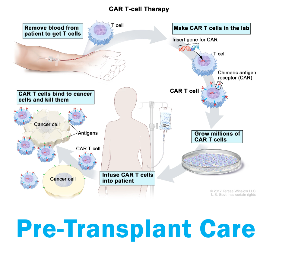
Prevention + Monitoring of Graft-Versus-Host-Disease (GVHD)
Signs: maculopapular rash, diarrhea, abdominal pain
Administer prophylactic antibiotics
Monitor for maculopapular rash (skin checks)
Administer steroids if development of GVHD occurs
Manage GI symptoms (zofran; diarrhea → need electrolytes)
Infection prevention
Blood products PRN
Admin G-CSF (type of growth factor; used after chemo to help WBCs recover after treatment) for autologous HSCT recipients

Tumor Lysis (Breakdown) Syndrome
Therapy begins
Rapid lysis of tumor cells
Can be chemo, radiation, or glucocorticoids
Intracellular contents released into systemic circulation
Potassium increases
Phosphate increases
Uric acid increases
Metabolic consequences
Hyperkelamia (increased potassium)
Hyperphosphatemia (increased phosphate)
Hyperuricemia (increases uric acid)
Hypocalcemia (decreases calcium
AKI
Tumor Lysis Syndrome - Clinical Manifestations
N/V/D
Anorexia
Lethargy/syncope
Muscle cramps (increased K+ + uric acid)
Dysrhythmias
Hematuria
Seizures
Tumor Lysis Syndrome - Prevention
Admin allopurinol prophylactically
IV fluids at 2x maintenance (d/t AKI)
Frequent monitoring of serum electrolytes, urine output, and serum uric acid
Tumor Lysis Syndrome - Treatment
Electrolyte replacement as needed
Hemodialysis for severe symptoms
Pediatric Cancer Survivorship
Clinic
Survivors are followed for many symptoms and late effect of treatments
Anxiety, depression, stress
Monitored for symptoms of secondary cancers or relapse
Transition into adult care when age appropriate
All patients have a survivorship care plan with info about entire treatment regimen and follow-up recommendations
Patients and families should be given resources for support groups
Watch growth of child

Cancer - Supportive Care for EOL
EOL care is focused on comfort, minimizing suffering
Symptom management
Pharmacologic, nonpharmacologic, or both
Are in a lot of pain, makes them tired, determine goals
Preparation for death (hospice/palliative)
Family decide on location
Discuss changes that occur during process
Assist family in obtaining memories and keepsakes: hair, handprints, photographs
Encourage cultural practices
Bereavement
Check in with family periodically
Provide info for caregivers and siblings when appropriate
Grief counseling
The process is on the family’s timeline (as much as possible)
Palliative goal → live as well as possible for as long as possible
When death occurs → give family as long as they need
Anterior Pituitary Disorders - Growth Hormone Deficiency
S/S
Short stature
Poor growth
Delayed puberty and facial development
Diagnosis
Rule out other reasons (genetic, FTT)
Wrist x-ray to assess “bone age”
End-organ blood testing (IGF-1)
Treatment
Daily SQ GH injections (earlier is better)
Address underlying causes (tumor, head injury)
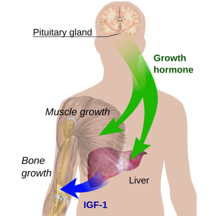
Anterior Pituitary Disorders - Precocious Puberty
Gonadotropin-releasing hormone (GnRH)
Normal puberty
Girls 10-12
Boys 11-14
Precocious puberty
Before 7-8 in girls
Before 9 in boys
In young patients - concern for pituitary issue
Secondary sex characteristics - pubic and axillary hair, breast and testicular development from premature release of sex hormones
May also see voice changes, rapid growth spurt, acne, body odor
Always needs medical evaluation
Diagnosis
Hormone levels
Imaging
Treatment
Medical therapy
GnRH agonist to work on feedback loop to stop GnRH release
Address underlying cause
Emotional support

Posterior Pituitary Disorders
ADH - antidiuretic hormone released by posterior pituitary
Causes kidneys to retain water
Disorders can cause fluid/electrolyte imbalances
ICP affects posterior pituitary (post brain injury) → concern for DI (diabetes insipidus → “dry inside”) and SIADH (syndrome of inappropriate ADH hormone → “soaked inside”)


Thyroid Disorders
Metabolism control center (how food is converted to energy)
Affects EVERYTHING
Weight gain or loss
HR
Temp
Speed of digestion

Congenital Hypothyroidism
Tested on Newborn Screen
Can cause irreversible intellectual disability and FTT if not treated early
Requires lifelong daily levothyroxine given in the morning
Coarse facies = facial abnormalities

Diabetes
Normal blood glucose 70-110 mg/dL

Diabetes - High Blood Sugar
Caused by carbohydrate intake, infection, stress, steroid intake, growth spurts, etc…
Fatigue, 3 Ps (increased urination, thirst, hunger), can progress to DKA
Treated with insulin or other meds
Causes damage over time
Diabetes - Low Blood Sugar
Caused by fasting, insulin overdose, exercise, etc…
S/S: shaky, sweaty, headache, irritability, can progress to changes in LOC and even seizure
Treated with rapid acting carbs (orange juice, glucose tabs) or IM glucagon if unconscious
Can cause rapid decompensation - treat FIRST

Type I Diabetes
Autoimmune destruction of pancreas = insulin deficiency
Peak diagnoses at age 4-7 and 10-14
Urinalysis, BG, A1C useful in diagnostics; many present in DKA
Fasting glucose (8 hours) > 125 on 2 separate occasions
Random glucose > 200
A1C > 6.5
Major S/S: 3 Ps, weight loss, bedwetting (in previous potty trained kid)
Type I Diabetes - Management
Frequent blood glucose checks (before meals, at bedtime, if symptomatic)
Lifelong insulin dependence (nasal-bolus)
SQ injections
SQ pump
Carb counting
Patient/family education is important!!
Interdisciplinary team
Type I Diabetes - Treatment
Tight BG management with minimal lows
Goal A1C < 7.5
Allow kids in run higher BG than adults
Hypoglycemia recognition can be more difficult
Immediate risk of hypoglycemia
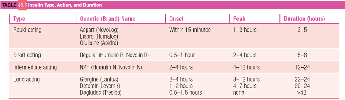
Insulin
Often take long acting once daily then rapid acting with meals based on carb intake and sliding scale
Will be ordered uniquely to each child by provider
Type II Diabetes
Characteristics
Insulin resistance (pancreas is producing insulin but cells are resistant)
Not autoimmune
Less likely linked to DKA
More common adults although peds diagnoses increasing
S/S
3 Ps
Obesity
HTN and hyperlipidemia
Management
BG control
Lifestyle adjustments!!
Exercise and healthy diet
BG control
Cook at home
May add metformin, GLP-1, or insulin depending on A1C
Pediatric Diabetes Considerations
TEST
Infants - educate the family, watch for lethargy or poor eating
Toddlers - picky eaters, temper tantrums, routine
Preschoolers - may begin to participate, awareness of DM
School-age - socialization, more participation
Adolescent - body image, independence, peer acceptance, consistency
Diabetic Ketoacidosis (DKA)
Blood glucose > 250 (usually more like 400-800)
Acidosis (pH < 7.3, usually closer to 7; bicarb < 15)
Ketones in blood and urine
D/t keto diet, prolonged N/V, eating d/o
Medical emergency!!!

DKA Management
ICU admission
IV fluids
IV insulin
Don’t want to drop BG too quickly (electrolyte shifts)
Assess LOC, blood sugar, K+
Hypoglycemia
BG < 70
Priority treatment
Can cause LOC, seizure, brain damage, and death if not treated

Hypoglycemia - Management
Mild - 15 g fast acting carbs and recheck in 15 mins
Severe - glucagon injection
Educate patient to always have these on hand
