C2.2 Neural Signalling x C3.1 Integration of Body Systems
 Call Kai
 Learn
 Practice Test
 Spaced Repetition
 Match
 Flashcards
 Knowt Play
1/44

There's no tags or description
Looks like no tags are added yet.
45 Terms
CNS and PNS
central nervous system comprises the brain and spinal cord (nerve tissue running from the base of the brain)
peripheral nervous system includes nerves outside the brain and spinal cord
State the function of a neuron and its respective cell parts: dendrites, axon and cell body
Neuron (nerve cells):
Specialized cells
Basic structural and functional units of the nervous system.
Transmit nerve impulses
Axon:
A long cytoplasmic extension from the cell body of a neuron;
Transmits impulses away from the cell body to other neurons or cells of an effector;
Dendron / dendrites:
short, highly branched extensions of a neuron;
It receives impulses from other neurons;
Transmits impulses towards the cell body
Myelin Sheath:
double layer of phospholipid bilayer
mixture of mainly lipids and some proteins
formed by the wrapping of schwann cells

Nodes of Ranvier:
Parts where the axon membrane is exposed (not covered by myelin sheath)
Function of Myelin sheath and nodes of ranvier (saltatory conduction):
Allows for saltatory conduction: the rapid jumping of nerve impulses (action potentials) from one node of Ranvier to the next along a myelinated axon.
Speed up rate of transmission of nerve impulses (~100 m/s).
Myelinated neurons account for only about one-third of the neurons in the body.
Organisation of nerves
Nerve fibres are the long threadlike extension (axon plus supporting cells) of a neuron that conducts electrical impulses.
NEW!
A nerve is a bundle of neurons and supportive tissues surrounded by a protective sheath.
Each bundle has several coverings of protective sheath as shown below:
Endoneurium: tissue surrounding each individual nerve fibre.
Perineurium: smooth connective tissue surrounding each bundle of nerve fibres (fascicle).
Epineurium: fibrous tissue surrounding a number of bundle of nerve fibres.


Fun little note:
A nerve contains really long, stretched-out axons that run the entire length of that specific nerve pathway, rather than thousands of short neurons stacked end-to-end like bricks.
However, because these cellular extensions are incredibly thin, a single nerve is made up of thousands of these long axons bundled together, not just a few.
Myelinated vs unmyelinated nerve fibres
In myelinated nerve fibres, Schwann cells wrap around and form myelin around each axon.
In unmyelinated nerve fibres, multiple axons are embedded in the cytoplasm of Schwann cells that do not produce myelin.

Define nerve impulse
A nerve impulse is an electrical signal
brief changes in the distribution of electrical charge across the plasma membrane of the neuron
3 different types of neurons and their relationship
Sensory neuron:

cytoplasmic fibre running to the cell body from receptors to dendrons
axon carry impulses away from the cell body
long dendron and long axon
Relay neuron:
also known as intermediate or interneuron
have numerous short fibres
occurs in the CNS
connects sensory to motor neurons
Motor neuron:
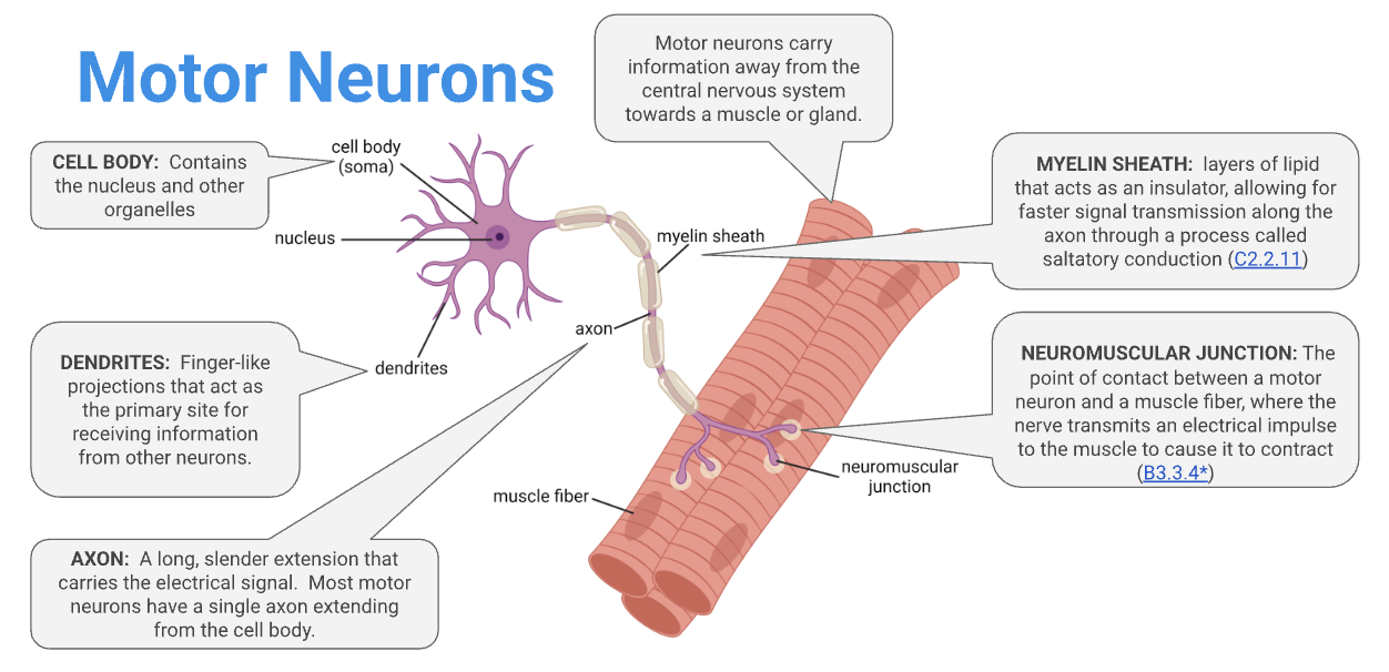
have many fine dendrites which bring impulses towards the cell body
a single long axon that carry impulses away from the cell body
carry impulses from CNS to a muscle or gland
Relationship

Why is the membrane considered polarised and what for?
WHY:
For a neuron to send an impulse, the neuron membrane must be "polarized." (it is also polarised at resting state)
A polarized membrane is a membrane that has
a positive electrical charge on one side
and a negative charge on the other side.
Almost all cells exhibit some degree of polarity.
If microelectrodes are placed inside and outside the cell, the amount of polarization can be measured as the voltage difference between the inside and outside of the cell membrane.
Define membrane potential
The difference in electrical charge between the inside and outside of the cell / across the plasma membrane is called the "membrane potential."
Cytoplasm is more negative than tissue fluid
Membrane potential difference ≈ −50mV and −100mV
The axon membrane is able to generate large changes in its membrane potential (neurons are excitable)
Define resting potential and how it is maintained
It refers to the electrical potential of a neuron that is not conducting an impulse
the inside of the cell more negative than outside at rest.
the membrane potential of a resting neuron is -70mV
Most Na+ / sodium ions are on the outside of the cell, most K+ ions are inside the cell
1st (sodium-potassium pumps):
Establish by active transport of both K+ and Na+ by sodium-potassium pumps.
3 Na+ ions pumped out, 2 K+ ions pumped in, creating concentration gradients for both ions.
2nd (leaky ion channels):
Ions follow their concentration gradient and diffuse passively and slowly (somewhat cancelled out by the sodium-potassium pump and resting potential is still maintained)
more Na+ / sodium ions outside → net movement inside

more K+ ions inside → net movement outside
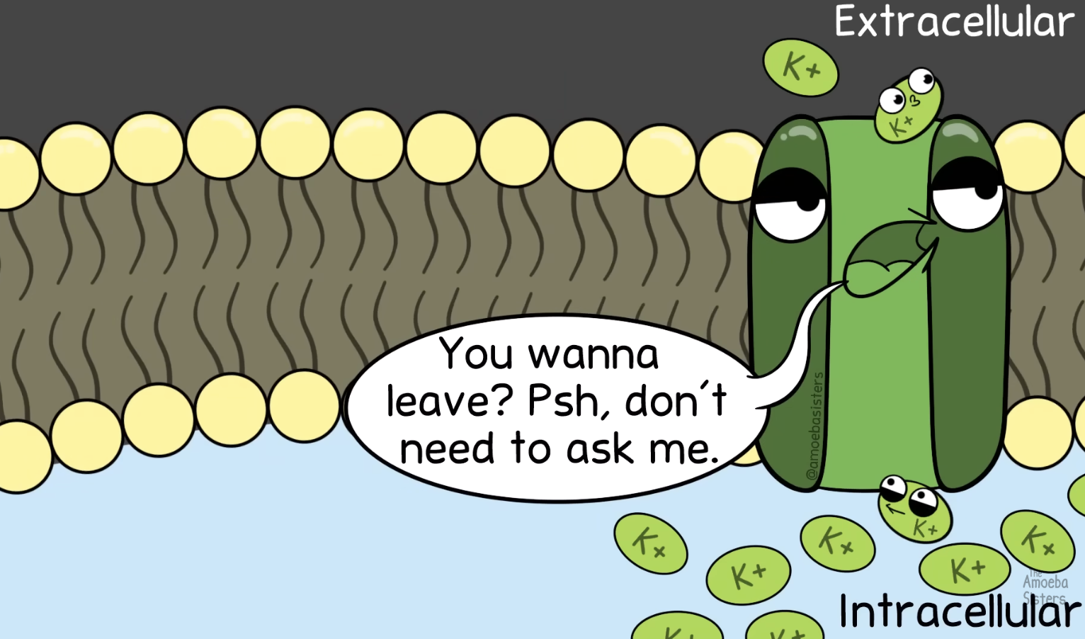
The axon membrane has many more K+-specific ion channels than Na+-specific ion channels
For every 1 Na+-specific ion channel present, there are 50 K+-specific ion channels
WHY? Results in more positive ions leaking out than leaking in, maintaining the resting potential by keeping the inside more negatively charged.
There is a net outward movement of K+ and a net inward movement of Na+
3rd (Other negative stuff inside the cell):
Organic anions / proteins present inside a cell are negatively charged.
Define action potential
Action potential is
the potential difference across the plasma membrane of a neuron when stimulated.
it is initiated by the reverse distribution of charges on both sides of the membrane due to the movement of Na+and K+
changing the membrane potential of the inside of part of the neuron from -70mV to +40mV
it is an all or nothing sequence of changes
“All or nothing” principle
Stimuli have widely different strengths - only a stimulus that is at or above a minimum intensity (threshold potential) can initiate an action potential.
“All or nothing” response: if stimulus is lower than the threshold potential, the influx of sodium ions will be quickly reversed to re-establish the resting potential.
The sequence of changes are:
Resting potential
Stimulus
Threshold
Depolarization
Repolarization
Refractory period
Function:
Nerve impulses are action potentials that are propagated along the neuron
Explain the different stages of action potential and self-propogation
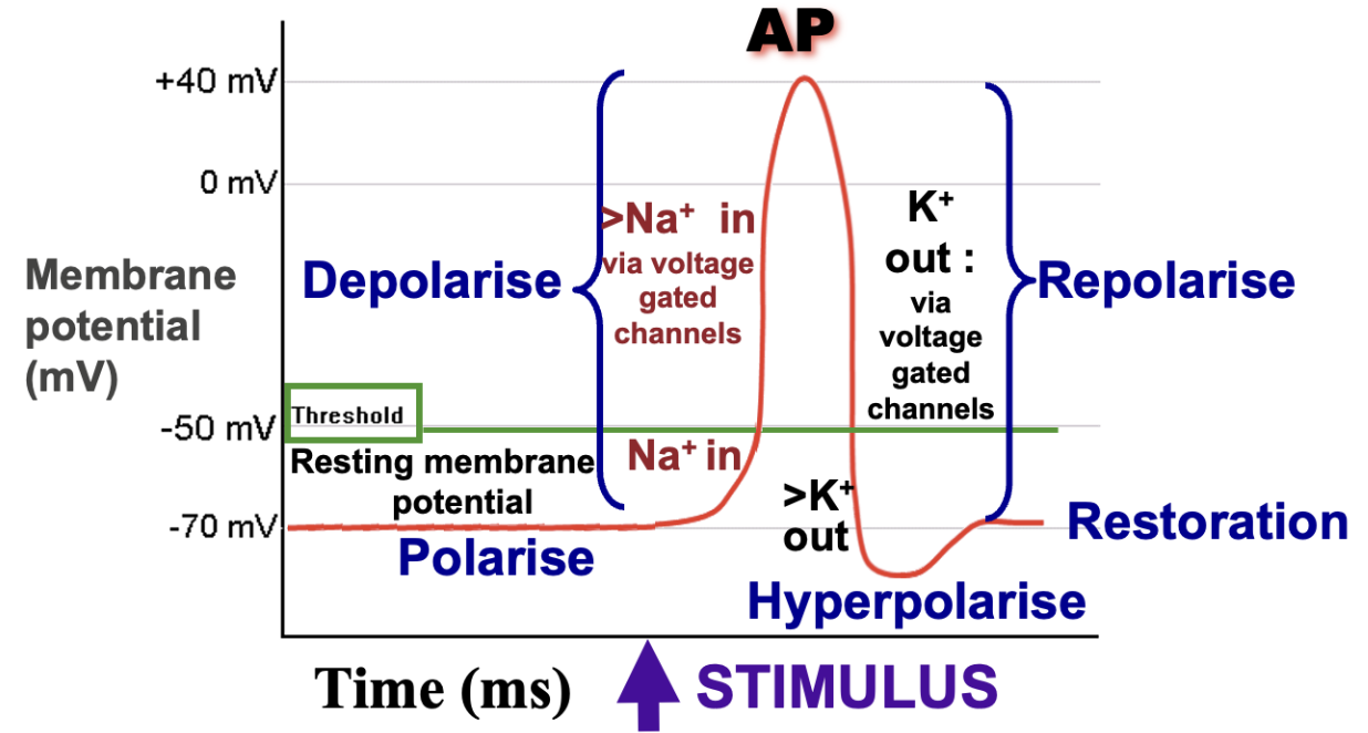
Resting potential – Before the action potential occurs, the neuron should be in a state of rest (approx. –70 mV)
Triggered by a stimulus,
Depolarisation – A rising spike corresponds to the depolarisation of the membrane via sodium influx (inwards) (up to roughly +30 mV)
Voltage-gated Na+ channels open → Na+ ions rush into the cell changing its membrane potential from negative to positive
Na+ ions follow their concentration gradients (high → low) and are attracted to the negatively charged proteins inside the cell
if the stimulus is strong enough, the threshold is reached and ACTION POTENTIAL is initiated
When the membrane potential reaches around +30mV, (more positive than surroundings)
Repolarisation – A falling spike corresponds to repolarisation via potassium efflux (outwards) (undershoots to approx. –80 mV)
Voltage-gated Na+ channels are inactivated → no more Na+ ions influx
BUT, voltage-gated K+ channels open → K+ ions rush OUT the cell changing its membrane potential from positive to negative
K+ ions follow their concentration gradients (high → low) moves out, bringing the membrane potential back down to a negative
Although the inside of the neuron is now relatively negative, there are more potassium ions outside and more sodium ions inside, opposite of what it was originally.
Hyperpolarisation — Undershoots, membrane potential is more negative than resting potential
Restoration – Returns to the level of the resting potential
Voltage-gated K+ channels close
restored by the action of the Na+/K+ pump which was continuously working, it is just that there is no more efflux of K+ions.
Explain how the movement of sodium ions propagates an action potential along an axon
depolarisation at one point of the axon form local currents, which trigger the opening of ion channels in the next segment of the axon
Na+ ions flow to the adjacent areas, causing these areas to be less negative
Leading to depolarisation which opens the sodium ion channels
Areas of the membrane that have recently depolarised will not be depolarised again as they are in refractory period
This causes depolarisation to spread along the length of the axon as a unidirectional ‘wave’


Outline the cause and consequence of the refractory period after depolarization
The period of time following a nerve impulse before the neuron is able to fire again (dictating the max. rate a neuron can fire)
Cause:
In a normal resting state, sodium ions are predominantly outside the neuron and potassium ions mainly inside (resting potential)
Hyperpolarisation
Following depolarisation (sodium influx) and repolarisation (potassium efflux), this ionic distribution is largely reversed
Before a neuron can fire again, the resting potential must be restored via the action of the sodium-potassium pump
Consequence:
Even though Na+ diffuses via local current in both directions, an action potential will be only be propagated forward.
Outline the correlation between conduction speed of nerve impulses and axon diameter
positive correlation with the conduction speed of nerve impulses (bigger diameter → higher conduction speed)
because the resistance to flow is decreased with a wider axon diameter.
Explain the difference in nerve impulse speed for myelinated and unmyelinated fibers
Saltatory conduction in myelinated neurons
insulates the axon
ions no longer need to move in and out along the entire length of the axon, but rather only at the nodes of ranvier
reduces ATP usage, (required for the sodium potassium pump)
increases the speed of the conduction of the electrical impulse
Define synapse, recall its main features and explain why signals are unidirectional
Is a junction between cells in the nervous system
3 types:
Between neurons
Between receptor cells and sensory neurons
Between motor neurons and muscle fibres /glands (aka effector)
triggers contraction
Main features

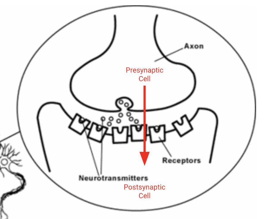
Presynaptic neuron sends chemical signals to the postsynaptic cells at the synapses and releases neurotransmitters into the synaptic cleft
The synaptic cleft is the space between an axon terminal of the presynaptic neuron and the postsynaptic cell
The postsynaptic cell receives chemical signals from the presynaptic cell at the synapse.
A signal can only pass in one direction across a typical synapse due to the fact that the presynaptic cell releases a chemical neurotransmitter and the postsynaptic cells has the receptors for the neurotransmitter.
State the role of neurotransmitters
Neurotransmitters are small chemical messengers typically produced in the synaptic knob (tip of axon terminal)
Held in tiny vesicles before release
Release from the presynaptic knob, across the synaptic cleft to bind with receptors at the postsynaptic membrane → ensures transmission of impulses is only in a single direction.
Acetylcholine as a neurotransmitter
What it do:
commonly released at neuromuscular junctions and binds to receptors on muscle fibres to trigger muscle contraction
also commonly released within the autonomic nervous system to promote parasympathetic responses (‘rest and digest’)
promotes calmness, slowing down heart rate, lowers stress
Process:
Acetylcholine is created in the axon terminal by combining choline with an acetyl group
Acetylcholine is stored in vesicles within the axon terminal until released via exocytosis in response to a nerve impulse
Acetylcholine activates a post-synaptic cell by binding to specific receptors
Acetylcholine must be continually removed from the synapse, as overstimulation can lead to fatal convulsions and paralysis
Acetylcholine is broken down into its two components
by the synaptic enzyme acetylcholinesterase (AChE)
choline is returned to the presynaptic neuron where it is coupled with another acetate to reform acetylcholine

Neonicotinoids (a class of synthetic neurotoxic insecticides chemically related to nicotine)
Why are neonicotinoids used in insecticides?
ACh: (acetylcholine, neurotransmitter carrying messages from the brain to the body through nerve cells)
They bind to Ach receptor in cholinergic synapses in the CNS of insects.
Ach receptor is blocked so Ach cannot bind
Synaptic transmission is prevented (message blocked)
Paralysis occurs
Acetylcholinesterase does not break down neonicotinoids, thus the binding is irreversible. This makes neonicotinoids very effective insecticides.

Cocaine (stimulant drug) and how its effects come about
It acts on dopamine, serotonin and norepinephrine receptors
Cocaine binds to and blocks specific reuptake transporters (specialised proteins) located on the membrane of the presynaptic neuron
meant to take up the neurotransmitters for reuse
This prevents the neurotransmitters from being taken back up into the presynaptic cell
Instead they repeatedly bind to and activate the complementary receptors on the postsynaptic neuron.
This causes a continuous influx of ions (such as Na⁺),
The postsynaptic neuron is continuously excited, due to the repetitive firing of action potentials.
This heavily amplifies the downstream electrical signals in the brain's reward pathways, ultimately resulting in the characteristic feelings of euphoria.
Outline the mechanism of synaptic transmission occurring at a presynaptic cell
Outline the mechanism of synaptic transmission occurring at a post-synaptic cell.
Steps 1-5:
Also state that calcium functions as a chemical signal triggering exocytosis of neurotransmitter from a presynaptic cell. (step 3)
Steps 6-9:
Often the neurotransmitter receptor (transmembrane) is a ligand-gated channel.
Binding of the neurotransmitter to the receptor leads to the opening of an ion channel within the postsynaptic cell membrane.
Ions to move via facilitated diffusion into the postsynaptic cell, causing the membrane potential to change.
Different neurotransmitters bind to different receptors, resulting in different ions being moved around.
Depending on which ions move, the neurotransmitter can either
have an excitatory effect (in pic.)
Influx of ions (e.g Na+) depolarizes the postsynaptic membrane (EPSP - Excitatory postsynaptic potential)
triggering an action potential in the postsynaptic membrane
have an inhibitory effect
Influx of negative ions (Cl-) or efflux of positive ions (K+) hyperpolarises the postsynaptic membrane (IPSP - Inhibitory postsynaptic potential)
Drives the neuron away from the AP threshold
preventing the generation of an action potential


“All or nothing” event elaborated
The action potential is an all-or-nothing event: if the threshold potential has not reached through the additive effect of postsynaptic potentials (summation), then the action potential will not be generated.
When the NET effect of excitatory action potentials and inhibitory action potentials exceed the threshold potential (becomes postive), an action potential will be generated.
Temporal summation: several impulses may arrive at the synapse in quick succession from a single axon, causing an action potential.
Spatial summation: impulses from different axons arrive at the synapse, causing an action potential.
Define emergent property (ft. consciousness)
Multicellullar organisms are capable of completing functions that unicellular organisms could not undertake – this is due to the collective actions of individual cells combining to create new synergistic effects
e.g consciousness
an example of emergent properties that are consequences of interaction between individual neurons.
Consciousness is not confined to one region of the brain, but various cells and pathways are engaged depending on the stimulus.
intricately linked to the cerebral hemispheres
Researched using
MRI uses a powerful magnetic field to produce detailed images
fMRI a non-invasive method that detects and maps brain activity with high resolution → transformed into 3D images that work out the spatial organisation
detect an increase in red blood cell oxygenation to the site of special neural activity during periods of intense activity.
Constrast Nervous system and Endocrine system
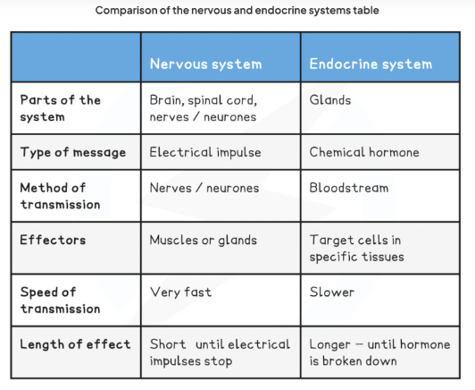
Note:
Pancreas is directly behind the stomach and in front of the spine.
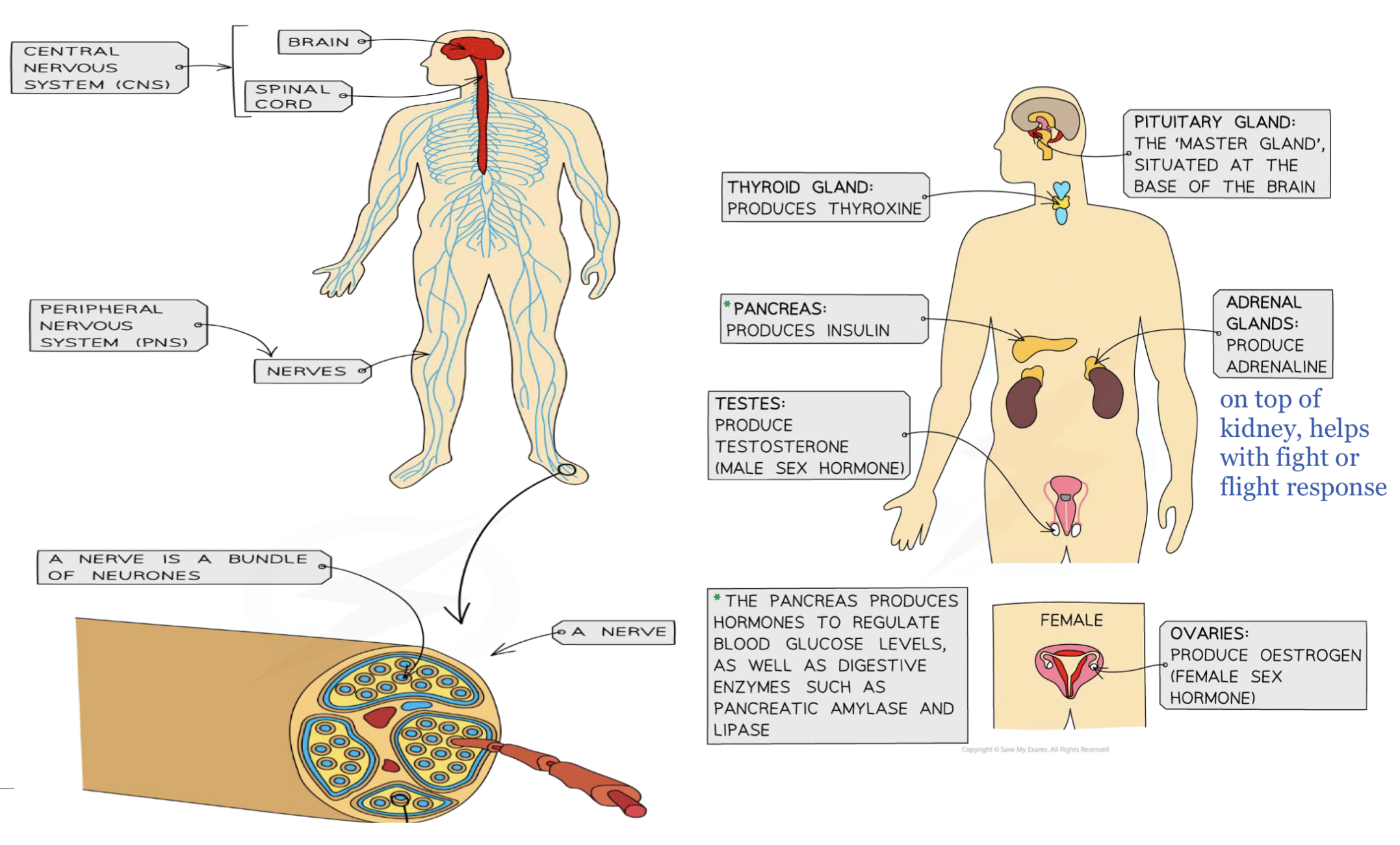
Define circadian rhythm and outline the role of melatonin
Circadian rhythms:
are physical, mental and behavioural changes that follow a 24-hour cycle.
rhythms are controlled by a biological clock (SNC) in the brain and coordinated with the cycle of light and dark.
however, it will still continue even if an organism is placed experimentally in continuous light or continuous darkness
extra: suprachiasmatic nucleus SCN (cells in the eye sense wavelengths of light and send a neural impulse to the suprachiasmatic nucleus, the pacemaker of the circadian rhythm → rhythmic firing of neurons → allows for the synchronisation of cells throughout the body)
Role of melatonin:
Melatonin is an amine hormone produced by the pineal gland (center of brain) in response to darkness to coordinate the timing of circadian rhythms and sleep (stimulated by the SNC).
Light = X, less melatonin (LL)
Retinal cells detect light and send impulses to the hypothalamus to reduce melatonin production.
Low melatonin level prepares body for being awake vice versa
Dark = more melatonin
The amount of melatonin circulating in the blood is low during the day and high at night
By association,
Winter depression (seasonal affective disorder) is associated with higher than normal melatonin levels.
For people experiencing jet lag, it takes several days before patterns of “sleep and activeness” return to normal.

Epinephrine and its effects
Epinephrine (adrenaline) is a peptide hormone secreted by the adrenal glands when the body is under stress / prepares for “fight or flight” response.
Main effects are as follows:
Increase heart rate to deliver more oxygen and glucose for aerobic respiration by muscle cells.
Break down glycogen in liver to glucose, which is then released into the bloodstream so that more glucose is used for aerobic respiration by muscle cells that are contracting intensely.
Outline the role of the hypothalamus as a link between nervous and endocrine systems.

Hypothalamus:
part of the nervous system
a brain structure well-supplied with blood capillaries and special neurons.
controls the autonomic nervous system
is a source of hormones for the pituitary gland
although it is part of CNS it makes its own hormones
maintains homeostasis
It links the nervous system to the endocrine system via the pituitary gland:
It monitors the body using two inputs: nerve signals from sensory receptors and chemical levels in the bloodstream.
Nervous System Role: It coordinates the autonomic nervous system to manage automatic functions (like wake-sleep cycles) and maintain overall homeostasis (internal balance).
Endocrine System Role: It acts as a master control center, producing hormones that dictate exactly what the pituitary gland releases.
How the hypothalamus controls the pituitary gland
The pituitary gland (part of endocrine sys.) sits directly below the hypothalamus (connected by a narrow stalk)
It has two distinct parts. The hypothalamus controls each part differently:
The Anterior Lobe (Front): Controlled via blood
The Mechanism: Special neurosecretory cells in the hypothalamus release hormones into a local network of blood vessels (the portal vein) that runs directly to the anterior pituitary.
The Effect: These hypothalamic hormones act as "stop or go" signals, either stimulating or inhibiting the anterior pituitary from releasing its own hormones (which control growth, reproduction, and metabolism).
The Posterior Lobe (Back): Controlled via nerves
The Mechanism: Instead of using blood vessels, the hypothalamus fires nerve impulses down direct neuronal connections into the posterior pituitary.
The Effect: The posterior pituitary does not make its own hormones. It simply stores and releases two specific hormones manufactured in the hypothalamus: oxytocin and antidiuretic hormone (ADH).
List types of sensory receptors and outline the function of sensory neurons (input)

Functions of __and __:
A neural pathway begins with a receptor
A receptor is a specialised cell that can detect changes in the environment that cause a stimulus
Receptor cells convert energy in one form (such as light, heat or sound) into energy in an electrical impulse within a sensory neuron
transmits the signal to spinal cord (receives info abt pain, temp, touch etc.) and cerebral hemisphere (CNS) for processing when the action potential has been reached.
Sensory neurones are located in the skin and sense organs. Sensory neurones have receptors activated by -
At a conscious level:
Light → Photoreceptors
respond to wavelengths of light
located within the retina of the eye for visual information
Chemicals → Chemoreceptors
respond to chemical signals
found in the tongue for tasting, but also aids in smell, blood pH and blood glucose concentration
Temperature → Thermoreceptors
located in the skin for detection of temperature changes
Physical forces → Mechanoreceptors
respond to mechanical stimulation (touch, pressure, vibration and sound)
located in the inner ear which are sensitive to sound vibrations
At unconscious level:
Osmoreceptors
respond to changes in the osmotic pressure of body fluids and blood plasma
located in the carotid arteries and hypothalamus which detect the water content of the blood
Baroreceptors
respond to mechanical stretch of blood vessel walls due to pressure changes in blood
located in the carotid arteries and the aorta
Proprioceptors
located in muscles and joints and provide information on balance and movement
Describe structure and function of the brain and label the parts on a diagram
Function:
The human brain acts as an integration & coordination system for the control of body systems
Structure:
It processes sensory information received from the body and relays motor responses to effector organ
human brain is organised into clearly identifiable sections:
major external structures
cerebral cortex
cerebellum
brainstem
medulla oblongata
*pons
Internal structures
hypothalamus
pituitary gland
*corpus callosum

State the location, structure, function of the cerebral hemispheres
Cerebral hemispheres:
human brain is divided into 2 halves
left cerebral hemispheres → controls muscles on the right of body
right cerebral hemispheres → controls muscles on the left of body
surface has folding with deep grooves. (more S.A)
foldings correlate with intelligence to a certain extent (more S.A more brain cells)
Cerebral cortex (outer layer):
is the surface of the hemispheres covered by grey matter
densely packed with unmyelinated neurons
which have a mass of dendrites and large numbers of synapses. (grey-pinkish hue)
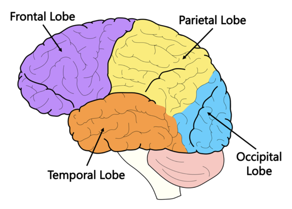
organised into two cerebral hemispheres and composed of four distinct lobes:
frontal lobe controls motor activity and tasks associated with the dopamine system (memory, attention, etc.)
parietal lobe is responsible for touch sensation (tactility) as well as spatial navigation
temporal lobe is involved in auditory processing and language comprehension
occipital lobe is the visual processing centre of the brain and is responsible for sight perception
Subcortex (inner layer):
the bulk of the cerebral hemispheres consists of white matter
made up of myelinated neurons.
myelin is milky white in colour hence the name

Locate the cerebellum (coral-like structure) and state its functions
Location:
a separate structure at the base of the brain, underneath the cerebral hemispheres
Composition:
Cerebellum (part of the hindbrain) makes up 10% of the brain’s volume but contains 50% of the neurons in our brains.
has an external surface layer of grey matter
Function:
maintenance of balance and posture (involuntary)
Cerebellum receives input from receptors and adjusts commands to motor neurons to move the body to maintain balance. (fine-tune but does not initiate motor controls).
coordination of voluntary movements
coordinates timings and force of different muscle groups to produce fluid movements
speech and writing etc.
facilitates motor memory
learn and remember how to coordinate muscles to perform a specific movement through practice
riding a bike, walking etc.
Locate the brain stem and state its functions
Location and structure:
is the posterior part of the brain that connects to the spinal cord (which relays signals to and from the body)
The brainstem includes the pons, medulla oblongata (often referred to as the medulla) and the midbrain
Function:
The brainstem (via the medulla) controls automatic and involuntary activities (breathing, swallowing, heart rate, etc.)
Spinal cord
It is a column of nerves that runs from the base of the skull downwards, connecting the brain with the rest of the body.
Function:

only coordinates UNCONSCIOUS processes, especially reflexes.
occurs when awake or asleep
involuntary - can not intentionally cause or prevent a process
coordinated by the spinal cord and sometimes the brain
tricky bcus some actions can be both conscious and unconscious such as blinking and swallowing.
responsible for bringing sensory information to the CNS from the body and enables motor (muscular) information to be sent out
Structure:
White matter (outer layer)
contains mainly the myelinated axons of neurons
transmit signals from
sensory receptors to the brain
Brain to other organs
Grey matter
contains the unmyelinated neurons, its cell bodies and synapses
involved in spinal cord integration processes which create a reflex response (removing hand from hot surfaces)
and other unconscious processes e.g peristalsis (food movin’)
Input and output
Chain (one-way signal):
Sensory receptors → sensory neurons → CNS → motor neuron → effector organ
Receptors
A neural pathway begins with a receptor
a specialised cell that can detect changes in the environment that cause a stimulus
convert energy in one form into energy in an electrical impulse within a sensory neuron
often found in sense organs
Stimulated receptors
When receptors cells are stimulated they are depolarised (+)
If the stimulus is very weak, the cells are not sufficiently depolarised and the sensory neuron is not activated to send impulses
If the stimulus is strong enough, an action potential is initiated in the sensory neuron and the impulse is transmitted to the spinal cord and the cerebral hemispheres (CNS)
Output through motor neurons
Cerebrum within the brain uses the information to process movements within the body as needed
Motor neurons are used to carry action potentials to muscles to initiate the movement required
All motor neurons terminate within a muscle within a neuromuscular junction (like synapses)
located between the terminal branches of a motor neuron and a muscle cell
multiple neuromuscular junctions spread across several muscle fibres within the muscle
Describe the perception of pain by neurons with free nerve endings and link it to the reflex arc and overrule by brain
Free nerve endings are unencapsulated dendrites (bare) of a sensory neuron in the skin (can receive + ions freely)
These nerve endings have transient receptor potential (TRP) channels that are positively charged
open in response to a stimulus (e.g. high temperature, acid, specific chemicals such as capsaicin in chili and pepper, mechanical sensation)
and allow positively charged ions to diffuse into the axon to generate an action potential.
The action potential passes along the sensory neuron, which connects to other neurons till the impulse reaches the brain, where it is perceived as pain.
Reflex arc:

In a reflex arc, a specific stimulus produces the same immediate, quick and involuntary response called a reflex action.
protects the body from potential harm by triggering immediate reactions to dangerous situations bypassing the brain.
the brain still receives the sensory input, it is just not required for the reflex action to occur
Pain reflex arc:

The pain reflex arc has
a single relay neuron in the grey matter of the spinal cord
Free nerve endings in a sensory neuron as the pain receptor.
Process:
Stimulation: A pain receptor in the skin detects a painful stimulus
Sensory neuron: An electrical impulse is transmitted along the sensory neuron
Spinal cord: The sensory neuron enters the spinal cord and synapses with a relay neuron
Motor neuron: The relay neuron synapses with a motor neuron, which leaves the spinal cord
Muscle: The motor neuron synapses with a muscle, causing it to contract and remove the limb from the pain stimulus
But the brain has the ability to override the reflex action:
Many neurons connect reflex arcs with the brain (control centre) so that impulses originating in a reflex arc also travels to the brain
Descending Impulses: The brain acts as a higher control centre, sending voluntary nerve impulses down descending tracts in the spinal cord to meet the reflex arc.
Impulses arrive at the synapse between the relay neuron and the motor neuron
Inhibitory Neurotransmitters: The brain's signals release inhibitory neurotransmitters, causing hyperpolarization (IPSPs) of the postsynaptic motor neuron.
Prevents the motor neuron from reaching the threshold potential
Blocking the impulse to the effector organ (muscle) and modifying the response.
Hence, reflex actions may be overruled by the brain and the response is modified.
Described the structures and functions of baroreceptors in regulating heart rate
The medulla oblongata receives input from the sensory neurons located in the walls of the aorta and carotid arteries.
Aorta and carotid arteries have receptors that monitor blood pressure and blood pH and oxygen levels.

Baroreceptors (stretch receptors)
in carotid arteries, the aorta and the right atrium
detect changes in blood pressure and send impulses to the cardiovascular centre in the medulla to regulate the stroke volume and heart rate accordingly.
Chemoreceptors
in carotid arteries, the aorta and the brain monitor the blood CO2, O2 and pH levels.
Increasing CO2 / low pH / low O2 level stimulates the chemoreceptors to signal the heart beat faster so that more blood rich in CO2 can be pumped to the lungs quickly for removal.
Outline the source and effect of epinephrine on heart rate.
Outline factors that will increase heart rate.
Outline factors that will decrease heart rate.
Innervation of the heart:
The innervation of the heart refers to the network of nerves that connect to the heart and are responsible for modulating the heart rate and stroke volume.
There are two major nerves that innervate the heart:
1. The sympathetic cardiac nerve:
Conditions:
low blood pressure
high blood CO2 level
The cardiovascular centre (part of the medulla) is activated
Sends a signal down the sympathethic nerve
Which releases the neurotransmitter norepinephrine
It increases the firing rate of sinoatrial (SA) node, causing the heart to increase frequency and force of the contraction.
2. The vagus nerve:
Conditions:
High blood pressure
Low blood CO2 level
The cardiovascular centre (part of the medulla) is activated
Sends a signal down the parasympathetic nerve
Which releases the neurotransmitter acetycholine
It decrease the firing rate of sinoatrial (SA) node, causing the heart to decrease frequency and force of the heart contraction.
Breathing (as an involuntary action)
Breathing rate is controlled by the respiratory centre located in the medulla of the brain consisting of 2 groups of nerve cells:
inspiratory centre
sends impulses to increase rate and depth of breathing
expiratory centre
sends impulses to inhibit the inspiratory centre and stimulate expiration.
Function of both centres:
Alternating impulses from these two centres cause rhythmic breathing by reflex action.
Allows breathing rates can be adjusted unconsciously
E.g. during increased physical activities.
E.g
When blood CO2 level increases → more carbonic acid is formed → blood pH level decreases
This stimulates the chemoreceptors in the medulla, carotid arteries and aorta to send impulses to the inspiratory centre.
This centre sends impulses to the intercostal muscles and diaphragm, increasing their contraction rates.
Hence, a more rapid breathing rate causes more CO2 to be exhaled from the lungs.
BUT,
Breathing rates can be adjusted consciously with messages sent from cerebral hemispheres.
E.g. during shouting or singing.
ENS and table showing different types of nervous systems
The enteric nervous system (ENS) is part of the autonomic nervous system (ANS)
serves the gastrointestinal system.
extends from the lower third of the oesophagus through to the rectum.
Peristalsis is waves of contraction and relaxation of the circular and longitudinal muscles of the gut wall that propel food along.
The swallowing of food and egestion of faeces are under voluntary control by the CNS, but peristalsis is under involuntary control by ENS.
ENS ensures that movement of material through the gut is coordinated.
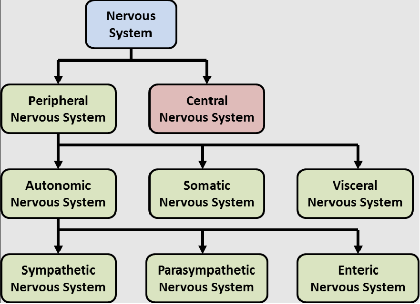
Define tropisms, contrast positive vs negative tropism and outline the benefits of positive phototropism
a growth response of plants in which the direction of growth is determined by the direction of an external stimulus
Direction of growth
Positive: towards stimulus
Negative: away from stimulus
Different types of tropism
Phototropism
Benefits (+) phototropism
Plant grows towards light source
Increased photosynthesis
Adaptation such that leaves are orientated towards the sunlight
positioned to capture the maximum amount of light energy for photosynthesis
Outcompete neighbouring vegetation
ensures survival and reproductive sucess
Controlled by a phytohormone, auxin
Define phytohormones (plant hormones)
naturally occurring organic signaling chemicals (different types of phytohormones) which are categorised into
growth promoter stimulating
Plant growth
Auxin (causes plant to bend towards light source, promoting the lengthening of stems)
Plant development
Plant reproduction
growth inhibitor mediating
stress responses, dormancy, deterioration
produced in extremely low concentrations
Function of a phytohormone can differ depending on
Specific tissue it acts on
Its concentration
Other phytohormones present
State two roles of the hormone auxin.
Describe the mechanism of movement of auxin into and between plant cells.

Auxin’s distribution is critical for controlling plant growth patterns.
plants maintain concentration gradients of auxins by transporting them in specific directions → regulates growth .
Cellular Movement:
Auxin enters plant cells via passive diffusion.
However, cannot diffuse out freely
Thus, its directional movement requires specialized membrane-bound pump proteins called auxin efflux carriers (specifically known as PIN proteins).
PIN proteins can be localized to a specific side of the cell membrane
auxin can be actively transported from cell to cell → building up a high concentration in a targeted area.
The Phototropic Response
photoreceptors (phototropins) in the shoot tip detect directional light
→ trigger the lateral redistribution of auxin
→ PIN efflux proteins are relocated to the shaded side of the shoot cells.
→ use active transport to pump auxin out of the cells, causing it to accumulate heavily on the shaded side of the stem.
→ high concentration of auxin on the shaded side stimulates cell elongation (via the loosening of cell walls), causing the shoot to bend towards the light source.
Effect of auxin on plant cell: cell growth/elongation
Auxin increases proton pump activity, which causes cells to pump H+ out of the cell & into the cell wall (apoplast, the bigger structure).
the higher the concentration 𝐻+ ions, the lower the pH value becomes
pH in the cell wall decreases, activating expansins (enzymes) that break cross-bridges between cellulose fibres in the cell wall.
Cellulose fibres loosen. Water (fromx outside) enters the cell by osmosis, causing the cell to expand as turgor pressure inside the cell pushes against the cell wall.

Interaction between auxin & cytokinin regulate shoot and root development
Root tips produce cytokinin, which is transported to shoots.
Shoot tips produce auxin, which is transported to roots.
Function of interaction:
Interaction between these phytohormones help to ensure that root and shoot growth are integrated.
High Auxin : Low Cytokinin → Promotes Root development
Low Auxin : High Cytokinin → promotes shoot development
Ethylene and fruit ripening
Fruit ripening is controlled by a positive feedback loop involving the gaseous plant hormone ethylene.
Ethylene (ethene), a gas, is a phytohormone which promotes fruit ripening
During fruit ripening
flesh becomes softer
acids and starch are converted to sugar and the skin colour changes
scents are produced to advertise the fruit to animals
rate of respiration increases sharply
Ethylene can diffuse to the same fruit or to other fruits
Ethylene stimulates ripening, and ripening fruits produce ethylene, causing more ripening
This is an example of positive feedback, where one change leads to an increased change away from the original condition
Function:
This results in rapid and synchronized ripening
attracts dispersers
a larger quantity → stronger visual and olfactory signal → more dispersers attracted
satiation of predators
ripening all at once → plant provides more seeds than predators can consume → ensuring some seeds survive
