OT881 Anatomy Module 2: Proximal Upper Extremity
 Call Kai
 Learn
 Practice Test
 Spaced Repetition
 Match
 Flashcards
 Knowt Play
1/58

Earn XP
Description and Tags
Comprehensive vocabulary flashcards covering joint classifications, stability-mobility trade-offs, osteology of the shoulder and arm, muscle compartments, rotator cuff function, and the organization and branches of the brachial plexus.
59 Terms
What is a Joint (Arthrosis)?
A site where two skeletal elements come together; also referred to as an articulation.
Solid Joints
Joints held together by connective tissue without a joint cavity, allowing little to no movement; examples include the sutures of the skull and the pubic symphysis.

Synovial Joints? What is purpose of synovial fluid?
Specialized joints structured for movement, consisting of an articular cavity containing synovial fluid. Freely movable, makes up the majority of the joints in the body. Typically found at end of long bones, examples: elbow, hip, knee, etc.
Synovial fluid acts as a lubricant and nutrient source for articular cartilage.

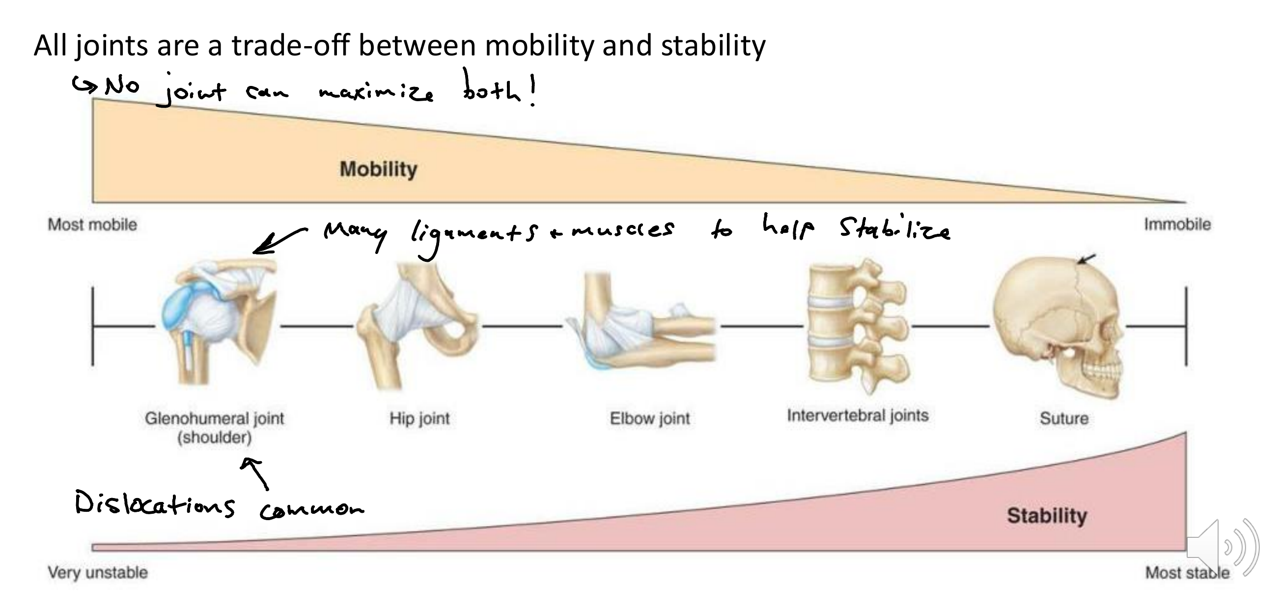
Joint Stability vs. Mobility Trade-off
The inverse anatomical relationship across all joints, ranging from the most mobile and very unstable (e.g., glenohumeral joint) to the immobile and most stable (e.g., skull sutures).
Hinge Joint definition and movement?
A type of synovial joint that permits uniaxial motion, exemplified by the elbow joint. Flexion/extension
Saddle Joint def and movement?
A type of synovial joint characterized by opposing saddle-shaped surfaces, exemplified by the thumb carpometacarpal joint. Flexion/extension, abduction/adduction, circumduction.
Ball and Socket Joint def and movement?
A highly movable multiaxial synovial joint where a spherical head articulates with a cup-like depression, exemplified by the glenohumeral (shoulder) and hip joints. Flexion/ext, abd/add, circumduction.
Pivot Joint
A synovial joint that permits rotational movement around a single axis, exemplified by the atlantoaxial articulation in the neck.
Plane (Gliding) Joint
A synovial joint with flat articulating surfaces that permits sliding or gliding movements, exemplified by intercarpal joints in the wrist.
Condyloid (Ellipsoid) Joint def and movement?
A biaxial synovial joint permitting movement in two planes, exemplified by the radiocarpal (wrist) joint. Flexion/ext, add/abd, circumduction.
Muscle Origin
The fixed, proximal anatomical attachment point of a skeletal muscle that remains stationary during contraction.
Muscle Insertion
The attachment point of a muscle (typically distal) that moves toward the fixed origin when the muscle contracts.
Actin and Myosin Filaments
The microscopic protein filaments found within muscle fibers that interact at the sarcomere level to generate contractile force. Contraction of muscle is actin anc myosin filaments siding over each other, causing muscle to shorten.

Upper Limb Skeleton
A framework comprised of 32 bones in each upper limb: 2 in the pectoral girdle, 1 in the arm, 2 in the forearm, 8 in the wrist, and 19 in the hand.
Pectoral Girdle
The skeletal arch formed by the clavicle (collarbone) articulating with the scapula (shoulder blade), anchoring the upper limb to the axial skeleton.
Clavicle
The collar bone; an S-shaped bone that articulates medially with the manubrium of the sternum and laterally with the acromion of the scapula.
Holds the shoulder away from the thorax; without it, the shoulder would collapse inward, reducing movement significantly!
Scapula
The shoulder blade; a flat triangular bone possessing the coracoid process, spine, acromion, glenoid fossa, supraspinous fossa, infraspinous fossa, and subscapular fossa.

Proximal Humerus Landmarks
Key proximal anatomical features of the arm bone including the head, anatomical neck, greater tubercle, lesser tubercle, and the intertubercular groove.

Sternoclavicular Joint - what kind of joint, and movement?
The articulation formed between the medial end of the clavicle and the manubrium of the sternum.
Synovial saddle joint - elavation/depression, protraction, retraction, rotation of clavicle

Acromioclavicular Joint - what kind of joint and movement?
The synovial joint formed by the articulation between the acromion of the scapula and the lateral end of the clavicle.
Plane synovial joint - allows small gliding movements bwn clavicle and scapula

Glenohumeral Joint - type of joint and movement?
A multiaxial synovial ball-and-socket joint formed between the glenoid fossa of the scapula and the head of the humerus, permitting flexion/extension, abduction/adduction, medial/lateral rotation, and circumduction.
Humerus head is 4x bigger than the glenoid cavity, allowing for greater mobility but sacrificing stability
Scapulothoracic Joint - what kind of joint, and what does it help with?
A functional articulation (not a true anatomical joint) between the convex posterior thoracic wall and the concave anterior surface of the scapula.
Provides dynamic stability to shoulder, supports glenohumeral joint with elevation and abduction.

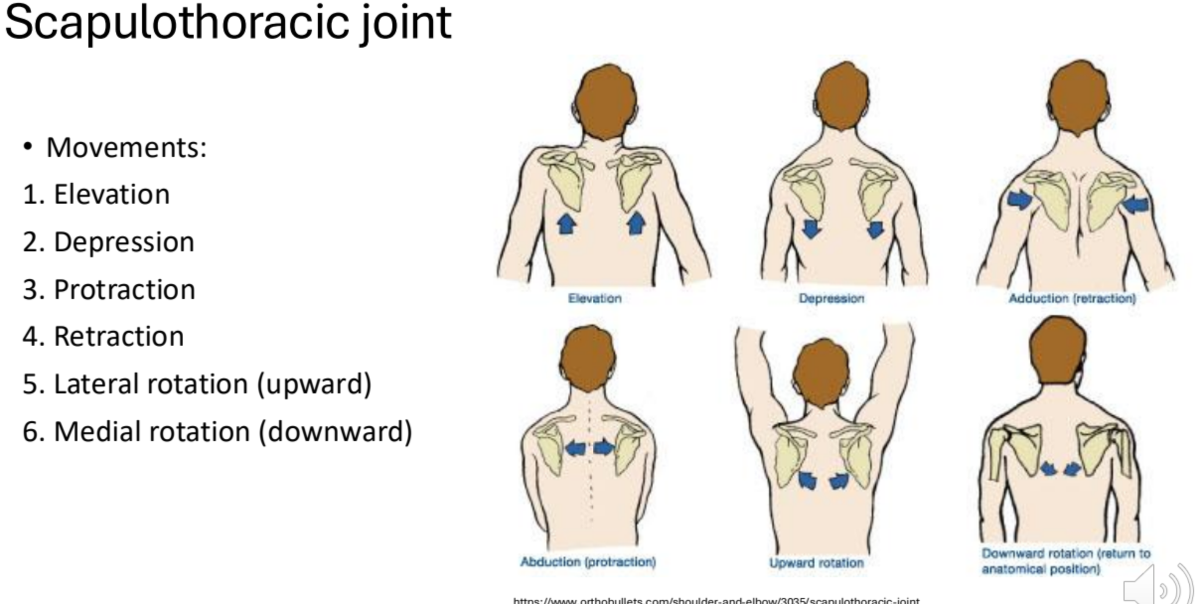
Scapulothoracic Movements
The six cardinal motions of the scapula: elevation, depression, protraction (abduction), retraction (adduction), upward (lateral) rotation, and downward (medial) rotation.
Scapulohumeral Rhythm
The kinematic coordination of shoulder elevation characterized by a 2:1 degree ratio of glenohumeral movement to scapulothoracic movement during abduction.
ST joint starts to laterally rotate and move with GH when GH joint exceeds 30 degrees in abduction.

Pectoralis Major - where is it found, and what movements does it perform?
Most superficial shoulder muscle. An anterior chest muscle that performs flexion, adduction, and internal (medial) rotation of the humerus at the glenohumeral joint.

Subclavius - location, function?
A small anterior muscle located beneath the collarbone that functions to stabilize and depress the clavicle.
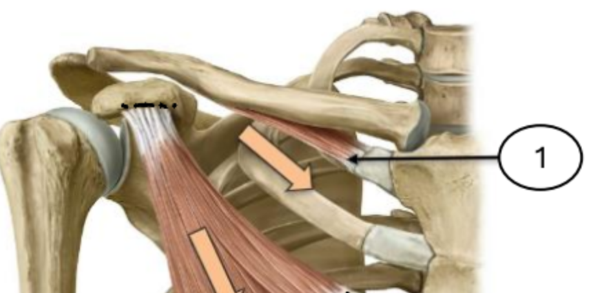
Pectoralis Minor - location, function?
A deep anterior thoracic muscle situated beneath the pectoralis major that draws scapula down and forward.

Serratus Anterior - location, function?
Finger like muscles lining the anterolateral chest wall that protracts the scapula and holds it against the thoracic cage.

Trapezius - location, function?
Most superficial back muscle.
Elevation, retraction, upward rotation of scapula.

Latissimus Dorsi - location, function?
One of largest back muscles.
Extension, adduction, internal rotation of arm at GH joint (similar to teres major)

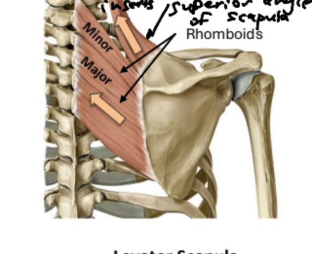
Levator Scapulae - location, function?
A posterior neck muscle that elevates the superior angle and medial border of the scapula.

Rhomboid Major and Minor - location, function?
Deep posterior upper back muscles extending from the vertebral column to the medial border of the scapula that act to retract the scapula towards midline.
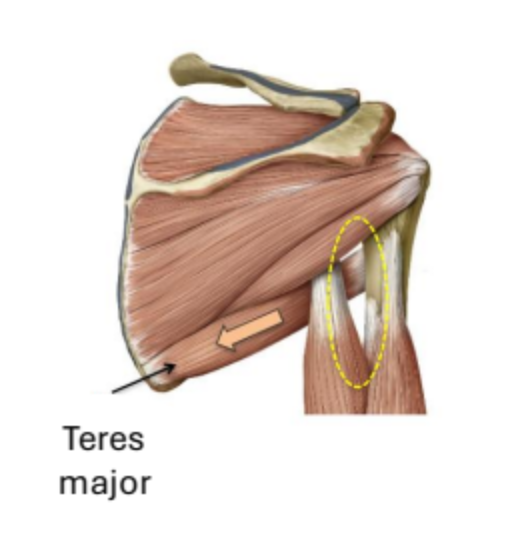
Teres Major - location, function?
A thick posterior shoulder muscle running from the inferior angle of the scapula to the humerus.
extension, adduction, and internal rotation of the arm at the GH joint (same as latissimus dorsi).
Rotator Cuff Muscles (SSIT)
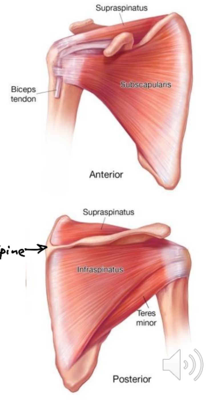
A stabilizing group of four muscles—Subscapularis, Supraspinatus, Infraspinatus, and Teres Minor—that blend with and reinforce the capsule of the glenohumeral joint.
Subscapularis - function?
Anterior rotator cuff muscle that performs internal rotation of the humerus and prevents posterior gliding of the humeral head.
Supraspinatus - function?
Superior rotator cuff muscle that initiates arm abduction before the deltoid takes over and prevents superior dislocation of the humeral head.
Infraspinatus - function?
Posterior rotator cuff muscle that performs external rotation, assists in abduction, and prevents anterior (forward) dislocation of the humerus.
Teres Minor - function?
Posterior rotator cuff muscle that executes external rotation of the arm and prevents anterior (forward) dislocation of the humeral head.
Deltoid - function of each fiber?
The principal lateral muscle of the shoulder.
Anterior fibers - GH flexion, internal rotation
Middle fibers - GH abduction (primary fcn of deltoid)
Posterior fibers - GH extension, external rotation

Biceps Brachii - location and function?
An anterior compartment arm muscle with two heads. Crosses two joints (GH and elbow), so acts on both!
primary forearm supinator, elbow flexor, and accessory glenohumeral flexor.

Coracobrachialis - location and function?
Anterior arm muscle stretching from the coracoid process to the shaft of the humerus; functions in flexion of the glenohumeral joint.

Brachialis - location and function?
Anterior arm muscle spanning from the anterior shaft of the humerus to the ulnar tuberosity; acts as a primary flexor of the elbow joint.

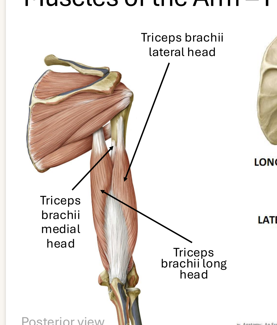
Triceps Brachii
A three-headed muscle of the posterior arm.
acts to extend the elbow joint, slight humeral extension & abduction
Spinal Nerves
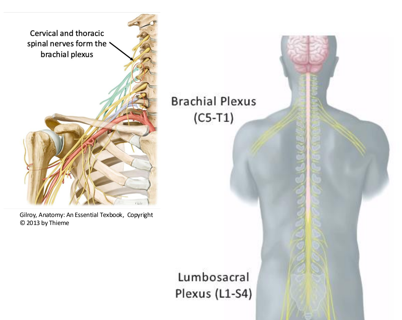
The 31 pairs of mixed sensory and motor nerves emerging symmetrically along regions of the spinal cord.
Brachial Plexus
A mixed somatic nerve network formed by the anterior (ventral) rami of spinal nerves C5 through T1 providing motor and sensory innervation to the pectoral girdle and upper limb.
Subdivisions of the Brachial Plexus? Hint: Real Thirsty? Drink Cold Beer
The anatomical organization of the brachial plexus into 5 Roots, 3 Trunks, 6 Divisions, 3 Cords, and 5 Terminal Branches (remembered by the mnemonic 'Read The Darn Cadaver Book!' or 'Real Thirsty? Drink Cold Beer!').

Musculocutaneous Nerve
A terminal branch of the brachial plexus (C5−C7) that supplies motor innervation to the muscles of the anterior compartment of the arm (biceps brachii, brachialis, coracobrachialis).
Axillary Nerve
A terminal branch of the brachial plexus that provides motor innervation to the deltoid and teres minor muscles.
Radial Nerve
A terminal branch of the brachial plexus (C5−C8, T1) providing motor innervation to the posterior compartments of the arm and forearm.
Median Nerve
A terminal branch of the brachial plexus (C6−C8 , T1) supplying muscles of the anterior compartment of the forearm and thenar (thumb) muscles.
Ulnar Nerve
A terminal branch of the brachial plexus (C7−C8, T1) providing primary motor innervation to the intrinsic muscles of the hand.
Axilla
The pyramidal passage region beneath the glenohumeral joint through which neurovascular structures, including the axillary artery and brachial plexus cords, transit to and from the upper limb.
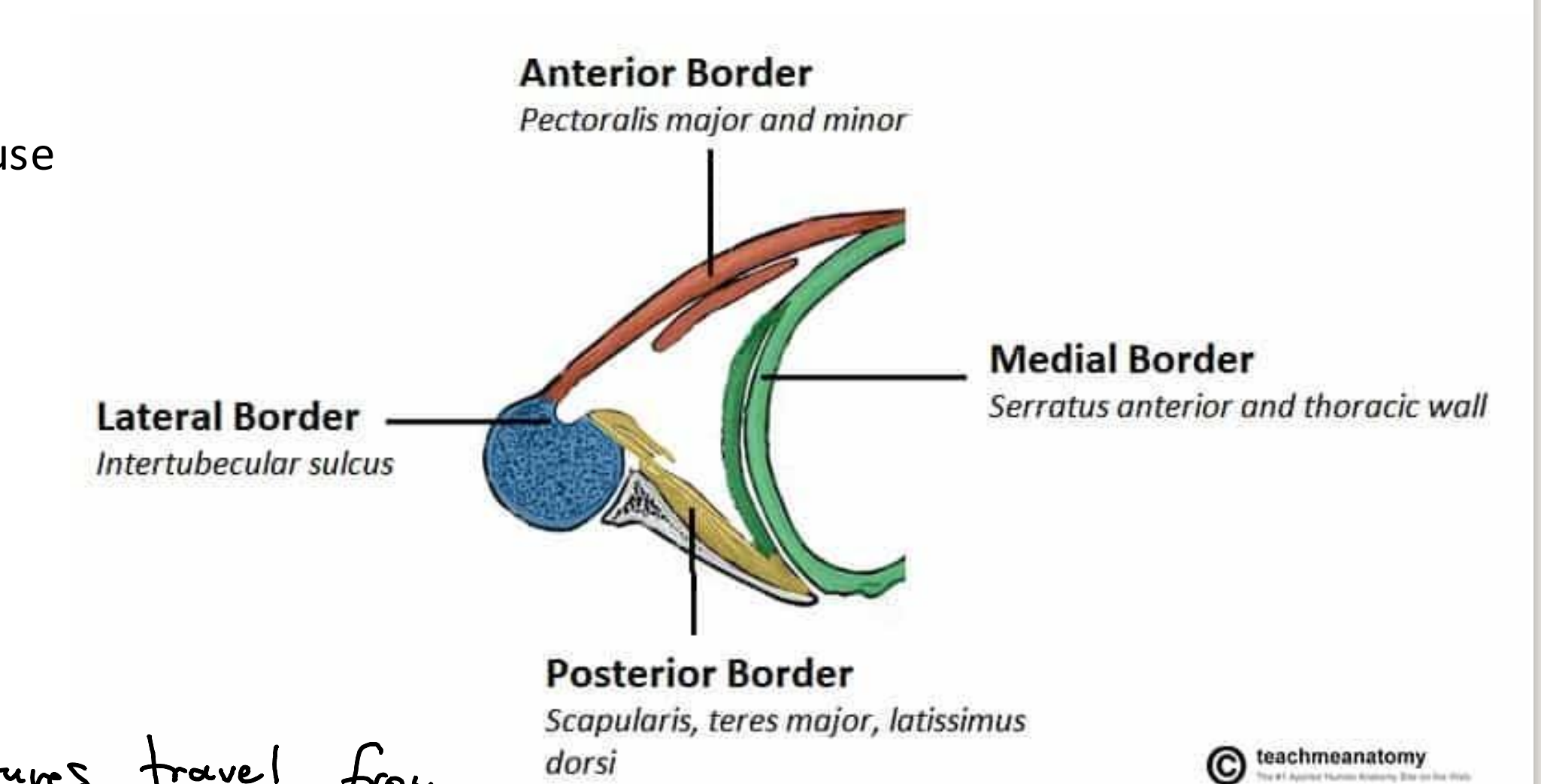
Borders of the Axilla
The anatomical boundaries of the axilla: anterior border (pectoralis major and minor), posterior border (subscapularis, teres major, latissimus dorsi), medial border (serratus anterior and thoracic wall), and lateral border (intertubercular sulcus of humerus).

What are the 4 different categories of synovial joints?
Uniaxial - movement in one plane along one axis; pivot, hinge joint
Non-axial - sliding rather than rotation around an axis; plane joint, gliding joint
Biaxial - movement in 2 planes around 2 axis; saddle joint
Multiaxial - movement in 3+ planes; ball and socket joint
What is the connection point between the upper limb and the axial skeleton?
Pectoral girdle
Where does the inferior angle of the scapula typically lie on the posterior side of the thorax?
Around the 7th thoracic vertebrae
Which muscles use the greater + lesser tubercles of the humerus as their attachement point?
Rotator cuff muscles, to stabilize the shoulder joint
What occurs anatomically in a shoulder dislocation?
Head of humerus pops out of glenoid fossa of scapula, shifts anteriorly/posteriorly
What are the 5 terminal branches (nerves) of the brachial plexus?
Musculocutaneous, Axillary, Median, Radial, Ulnar.
