BIO 329 EXAM 2
 Call Kai
 Learn
Learn Practice Test
 Spaced Repetition
 Match
Match Flashcards
1/235

There's no tags or description
Looks like no tags are added yet.
Name | Mastery | Learn | Test | Matching | Spaced | Call with Kai | Chat |
|---|

236 Terms
Describe the structure of a generic nucleotide, including its three parts, and explain how nucleotide monomers are added to each other to form a nucleic acid strand, and define the 5’ and 3’ end of a nucleic acid strand
A nucleotide has three parts:
Pentose sugar (ribose in RNA; deoxyribose in DNA)
Phosphate group attached to the 5′ carbon
Nitrogenous base (A, G, C, T, U) attached to the 1′ carbon
Polymerization (How nucleotides link)
Nucleotides join via phosphodiester bonds between the 3′‑OH of one sugar and the α‑phosphate of the incoming nucleotide.
This creates a 5′ → 3′ sugar‑phosphate backbone.
5′ and 3′ Ends
5′ end: free phosphate on the 5′ carbon.
3′ end: free hydroxyl on the 3′ carbon.
Polynucleotide sequences are always written 5′ → 3′.
Contrast DNA and RNA structure
Feature | DNA | RNA |
|---|---|---|
Sugar | Deoxyribose | Ribose (2′‑OH) |
Bases | A, G, C, T | A, G, C, U |
Strands | Double‑stranded | Single‑stranded |
Stability | More stable | Less stable (2′‑OH increases reactivity) |
Differentiate between purines and pyrimidines, and name the five bases present in nucleic acids
Purines (double‑ring): Adenine (A), Guanine (G)
Pyrimidines (single‑ring): Cytosine (C), Thymine (T), Uracil (U)
Five bases in nucleic acids: A, G, C, T, U.
Characterize the structure of DNA double helix
DNA is a right‑handed double helix (B‑form):
Two antiparallel strands (one 5′→3′, one 3′→5′).
Bases face inward; sugar‑phosphate backbone faces outward.
Complementary base pairing: A–T, G–C
Describe how hydrogen bonding drives complementary base pairing
A–T pairs form 2 hydrogen bonds
G–C pairs form 3 hydrogen bonds
Hydrogen bonding ensures specific pairing and contributes to helix stability
Explain how hydrogen bonding and van der Waals forces stabilize the structure of the double helix
Two major stabilizing forces:
1. Hydrogen Bonds
-Hold complementary bases together (Watson–Crick pairing)
2. Base Stacking (van der Waals / π‑π interactions)
-Hydrophobic bases stack tightly, creating stabilizing van der Waals interactions
Base stacking is the dominant contributor to helix stability.
List the 3 functions of the double stranded DNA molecule
Stores genetic information (stable long‑term archive)
Allows accurate replication via complementary base pairing
Supports transcription by providing templates for RNA synthesis
(General functions supported by DNA structural descriptions in sources.)
Explain the two forces stabilizing the DNA structure
Two major forces stabilize DNA structure: hydrogen bonding and base‑stacking interactions.
Together, they keep the double helix aligned, compact, and energetically favorable
1. Hydrogen Bonds Between Complementary Bases
Hydrogen bonds form between specific base pairs:
A–T pairs form two hydrogen bonds
G–C pairs form three hydrogen bonds
These bonds:
Ensure specificity (Watson–Crick pairing).
Hold the two strands together in a predictable, repeatable pattern.
Contribute significantly to duplex stability, especially in GC‑rich regions, which have more hydrogen bonds.
Hydrogen bonding is consistently identified as one of the key stabilizing interactions in DNA duplexes
2. Base‑Stacking (van der Waals / π–π Interactions)
The nitrogenous bases are flat, aromatic rings that stack on top of each other inside the helix
This stacking produces:
van der Waals interactions
π–π electron cloud overlap
Hydrophobic stabilization (bases avoid water by stacking)
Base‑stacking:
Is the dominant stabilizing force in DNA
Provides continuous stabilization along the helix axis
Helps maintain the uniform helical shape and prevents strand separation
Recent biophysical studies quantify base‑stacking as a major contributor to duplex stability
Summary Table
Force | What It Does | Why It Matters |
|---|---|---|
Hydrogen bonds | Link complementary bases (A–T, G–C) | Provide specificity and directional pairing |
Base‑stacking interactions | Stack aromatic bases via van der Waals/π–π forces | Major contributor to helix stability and shape |
Describe prokaryotic genome structure and DNA packaging
Prokaryotes have one circular chromosome
DNA is located in the nucleoid, not membrane‑bound
Packaging involves supercoiling and DNA‑binding proteins (not histones)
Describe the structure of eukaryotic genomes, and characterize the structure of a nucleosome, 30 nm fiber
Eukaryotic DNA is linear, found in the nucleus, and packaged with histones into chromatin
Nucleosome
DNA wrapped ~1.65 turns around a histone octamer (H2A, H2B, H3, H4)
Forms the “beads‑on‑a‑string” 10‑nm fiber
30‑nm Fiber
Higher‑order folding of nucleosomes into a thicker fiber (solenoid or zig‑zag)
(General chromatin structure supported by eukaryotic DNA packaging descriptions.)
Contrast eukaryotic DNA packaging during eukaryotic interphase and metaphase
Stage | Packaging |
|---|---|
Interphase | Chromatin is partially condensed; includes euchromatin (open) & heterochromatin (condensed) |
Metaphase | Chromatin is maximally condensed into visible chromosomes for segregation |
(General chromatin condensation differences supported by eukaryotic chromosome descriptions.)
Describe the anatomy of a eukaryote chromosome, including the location and function of telomere and centromere
Telomeres: repetitive sequences at chromosome ends; protect DNA from degradation and prevent end‑to‑end fusion
Centromere: constricted region; attachment site for kinetochore; essential for chromosome segregation.
(Chromosome structure supported by eukaryotic genome descriptions.)
Contrast the structure and function of euchromatin and heterochromatin
Type | Structure | Function |
|---|---|---|
Euchromatin | Loosely packed | Active transcription; accessible DNA |
Heterochromatin | Densely packed | Gene silencing; structural stability |
(General chromatin functional distinctions supported by chromatin descriptions.)
Explain how interphase chromosomes contain both highly condensed and more extended forms of chromatin
Interphase chromosomes contain both:
Highly condensed heterochromatin (centromeres, telomeres, silenced regions)
More extended euchromatin (actively transcribed genes)
This mixed organization allows simultaneous gene expression and structural maintenance
(General chromatin organization supported by eukaryotic genome descriptions.)
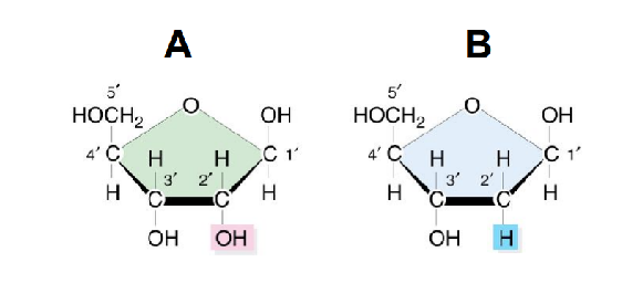
Which of these sugars is in DNA?
B

5’ carbon of the sugar
Condensin function differs from cohesin because:
a) Condensin compacts DNA into loops, cohesin maintains chromatid cohesion
b) Condensin recruits histone acetyltransferases, cohesin recruits histone methyltransferases
c) Condensin attaches to replication forks, cohesin attaches to centromeres
d) Condensin acts only in interphase, cohesin acts only in mitosis
a) Condensin compacts DNA into loops, cohesin maintains chromatid cohesion
In a DNA double helix,
Option A
the two DNA strands are identical.
Option B
purines pair with purines.
Option C
thymine pairs with cytosine.
Option D
the two DNA strands run antiparallel.
D - the two DNA strands run antiparallel
Mitotic chromosomes were first visualized with the use of very simple tools: a basic light microscope and some dyes. What characteristic of mitotic chromosomes reflects how they were named?
color
The inactivation of one X chromosome is established by the directed spreading of heterochromatin. The silent state of this chromosome is __________ in the subsequent cell divisions.
maintained
What makes up the backbone of DNA?
sugar-phosphate
The octameric histone core is composed of four different histone proteins, assembled in a stepwise manner. Once the core octamer has been formed, DNA wraps around it to form a nucleosome core particle. Which of the following histone proteins does NOT form part of the octameric core?
Option A
H4
Option B
H2A
Option C
H3
Option D
H1
D - H1
In the 1940s, proteins were thought to be the more likely molecules to house genetic information. What was the primary reason that DNA was not originally believed to be the genetic material?
DNA was found to contain only four different chemical building blocks
Which of the following structural characteristics is NOT normally observed in a DNA duplex?
Option A
purine–pyrimidine pairs
Option B
external sugar–phosphate backbone
Option C
uniform left-handed twistOption D
antiparallel strands
C - uniform left handed twist
Which of the following DNA strands can form a DNA duplex by pairing with itself at each position?
Option A
5´-AAGCCGAA-3´
Option B
5´-AAGCCGTT-3´
Option C
5´-AAGCGCAA-3´
Option D
5´-AAGCGCTT-3´
D
What is the basic unit of the eukaryotic chromosome structure?
nucleosome
Certain genes that were active in early development become subsequently repressed when development is complete. Where would these genes be found in an interphase chromosome?
heterochromatin
What type of macromolecule helps package DNA in eukaryotic chromosomes?
proteins
What functional group is found at the 5’ end of a DNA strand?
phosphate
Which of the following best describes the mechanism by which chromatin-remodeling complexes “loosen” the DNA wrapped around the core histones?
Option A
They use energy derived from ATP hydrolysis to change the relative position of the DNA and the core histone octamer.Option B
They chemically modify the DNA, changing the affinity between the histone octamer and the DNA.
Option C
They remove histone H1 from the linker DNA adjacent to the core histone octamer.
Option D
They chemically modify core histones to alter the affinity between the histone octamer and the DNA.
A - They use energy derived from ATP hydrolysis to change the relative position of the DNA and the core histone octamer
How did Chargaff’s findings contradict the tetranucleotide hypothesis proposed by Phoebus Levene?
They demonstrated that DNA is composed of an equal ratio of purines to pyrimidines, rather than repeating identical tetranucleotides
Which of the following chemical groups is NOT used to construct a DNA molecule?
Option A
five-carbon sugar
Option B
phosphate
Option C
nitrogen-containing base
Option D
six-carbon sugar
D - six carbon sugar
The classic “beads-on-a-string” structure is the most decondensed chromatin structure possible and is produced experimentally. Which chromatin components are NOT retained when this structure is generated?
Option A
linker histonesOption B
linker DNA
Option C
nucleosome core particles
Option D
core histones
A - linker histones
How do changes in histone modifications lead to changes in chromatin structure?
They help recruit other proteins to the chromatin
Specific regions of eukaryotic chromosomes contain sequence elements that are absolutely required for the proper transmission of genetic information from a mother cell to each daughter cell. Which of the following is NOT known to be one of these required elements in eukaryotes?
Option A
protein-coding regionsOption B
origins of replication
Option C
telomeres
Option D
centromeres
A - protein coding regions
Although the chromatin structure of interphase and mitotic chromosomes is very compact, DNA-binding proteins and protein complexes must be able to gain access to the DNA molecule. Chromatin-remodeling complexes provide this access by…
using the energy of ATP hydrolysis to move nucleosomes
Heterochromatin spreading during DNA activation is often restricted by:
Boundary elements (insulators) that block histone-modifying enzyme propagation
Which best describes the distinct role of cohesin compared to condensin during chromosome organization?
Option A
Both cohesin and condensin compact chromosomes by forming supercoils in chromatin
Option B
Cohesin holds sister chromatids together, while condensin drives large-scale chromosome compactionOption C
Condensin holds sister chromatids together, while cohesin condenses chromatin loops
Option D
Cohesin prevents histone acetylation, while condensin promotes histone methylation
B - Cohesin holds sister chromatids together, while condensin drives large-scale chromosome compaction
During mitotic chromosome condensation, condensin action depends on:
ATP hydrolysis to drive loop extrusion of chromatin
Why is ATP hydrolysis essential for nucleosome repositioning?
It provides energy for chromatin remodelers to break histone–DNA contacts transiently
Explain how a DNA double helix provides a template for its own replication, and describe the resulting daughter helices in terms of their sequence and the distribution of parental and newly synthesized DNA strands
Each strand of the DNA double helix is complementary to the other (A pairs with T, G with C). During replication, the two parental strands separate, and each serves as a template for synthesis of a new complementary strand. The result is two daughter helices, each containing one parental strand and one newly synthesized strand—the definition of semiconservative replication
Recount the experiment that revealed the semiconservative nature of DNA replication
Meselson and Stahl (1958) grew E. coli in heavy nitrogen (^15N), then transferred them to ^14N medium. DNA was analyzed by density‑gradient centrifugation. After one generation, DNA showed intermediate density (one heavy strand + one light strand). After two generations, DNA showed intermediate and light bands, confirming semiconservative replication and ruling out conservative and dispersive models
Recall where on a chromosome DNA synthesis begins, and explain what characterizes these nucleotide sequences in simple cells such as bacteria and yeasts
Replication begins at origins of replication—specific DNA sequences where helicase can initiate unwinding
Bacteria: typically one origin (oriC), AT‑rich to ease strand separation
Yeast: multiple Autonomously Replicating Sequences (ARS), also AT‑rich and bound by origin‑recognition proteins
Compare the direction in which replication forks move with the direction in which the new DNA strands are synthesized
Replication forks move bidirectionally outward from each origin. DNA polymerase synthesizes DNA only in the 5′→3′ direction, even though forks themselves progress in both directions
Compare the bonds that link together nucleotides in a DNA strand with the bonds that hold together the two strands of DNA in a double helix
Within a strand: phosphodiester bonds link nucleotides between the 3′‑OH of one sugar and the 5′‑phosphate of the next
Between strands: hydrogen bonds between bases (A–T: 2 bonds; G–C: 3 bonds)
Outline how deoxyribonucleoside triphosphates provide the energy for DNA synthesis
Each incoming deoxyribonucleoside triphosphate (dNTP) loses two phosphates (pyrophosphate) during incorporation. Hydrolysis of the high‑energy phosphate bonds drives formation of the new phosphodiester bond, powering polymerization
Recount how DNA polymerase contributes to the accuracy of DNA replication
DNA polymerases have 3′→5′ exonuclease proofreading activity. When an incorrect nucleotide is added, the polymerase stalls, removes the wrong base, and resumes synthesis. Additional accuracy comes from mismatch repair after replication
Describe the primers required for DNA replication and compare how they are used in synthesizing the leading and lagging strands
Replication requires RNA primers synthesized by primase
Leading strand: needs one primer; synthesized continuously
Lagging strand: needs many primers; synthesized discontinuously as Okazaki fragments
Explain how primers are removed and replaced to produce a continuous newly synthesized DNA strand
RNA primers are removed by exonucleases (RNase H or polymerase activities). DNA polymerase fills the gaps with DNA, and DNA ligase seals remaining nicks to create a continuous strand
Name five proteins that form part of the replication machine and state the role each plays in DNA replication
Helicase – unwinds the double helix
Primase – synthesizes RNA primers
DNA polymerase – extends DNA from primers; proofreads
Sliding clamp – increases polymerase processivity
Topoisomerase – relieves torsional stress ahead of the fork
(Other valid examples: ligase, single‑strand binding proteins, telomerase.)
Describe the problem created by a moving replication fork, and explain how this is resolved by the DNA topoisomerases
As helicase unwinds DNA, positive supercoils accumulate ahead of the fork, creating torsional strain. Topoisomerases cut one or both strands, allow controlled rotation to relieve tension, and reseal the DNA, preventing fork stalling or breakage
Summarize the “end replication problem” and explain how telomerase solves this dilemma
Linear chromosomes cannot fully replicate their ends because DNA polymerase cannot replace the final RNA primer on the lagging strand. This causes progressive telomere shortening. Telomerase, a reverse transcriptase, extends telomeres using its RNA template, allowing complete replication and protecting chromosome ends
List some of the causes of DNA damage
Common causes include:
UV radiation
Ionizing radiation
Reactive oxygen species
Chemical mutagens
Replication errors
Environmental toxins
(General DNA damage causes discussed in replication/repair sections.)
Name some of the types of damage that can alter DNA
Examples include:
Base modifications (oxidation, alkylation)
Thymine dimers (UV‑induced)
Single‑strand breaks
Double‑strand breaks
Mismatched bases
Bulky adducts
List the three main steps involved in repairing damage that affects only one strand of the DNA double helix
Recognition and excision of damaged bases or nucleotides
DNA synthesis to fill the gap using the undamaged strand as template
Ligation to restore strand continuity
Explain how the mismatch repair system recognizes and corrects replication errors
Mismatch repair proteins detect distortions in the helix caused by mispaired bases. The system identifies the newly synthesized strand (often by nicks), removes a segment containing the mismatch, and DNA polymerase resynthesizes the correct sequence
Contrast nonhomologous end joining and homologous recombination as mechanisms for repairing double-stranded DNA breaks
NHEJ: Directly ligates broken ends; fast but error‑prone, may lose nucleotides
HR: Uses a homologous template (usually the sister chromatid); accurate, but restricted to S/G2 phases
Describe the consequences of a failure to repair damaged DNA
Unrepaired damage can lead to:
Mutations
Chromosomal rearrangements
Replication fork collapse
Cell cycle arrest
Apoptosis
Cancer and aging‑related decline
(General consequences supported by DNA repair discussions.)
DNA replication is described as semiconservative because after
replication:
A. Each daughter DNA molecule contains only newly synthesized
strands
B. One daughter DNA molecule contains both parental strands
C. Each daughter DNA molecule contains one parental strand and
one newly synthesized strand
D. Parental DNA is completely degraded during replication
C - Each daughter DNA molecule contains one parental strand and
one newly synthesized strand
The replication fork is characterized by:
A) Bidirectional DNA synthesis
B) DNA transcription
C) One DNA polymerase acting on both strands simultaneously
D) Inhibition of helicase
A - Bidirectional DNA synthesis
The leading strand is synthesized:
A) Discontinuously in Okazaki fragments
B) Continuously in the 5′→3′ direction
C) Continuously in the 3′→5′ direction
D) Only in prokaryotes
B - Continuously in the 5′→3′ direction
The lagging strand is synthesized:
A) In short fragments initiated by multiple primers
B) Continuously
C) Only by helicase
D) In the 3′→5′ direction directly
A - In short fragments initiated by multiple primers
Homologous recombination requires:
A) Sister chromatid or homologous chromosome as a template
B) Ligase only
C) RNA primer synthesis
D) Telomerase RNA
A - Sister chromatid or homologous chromosome as a template
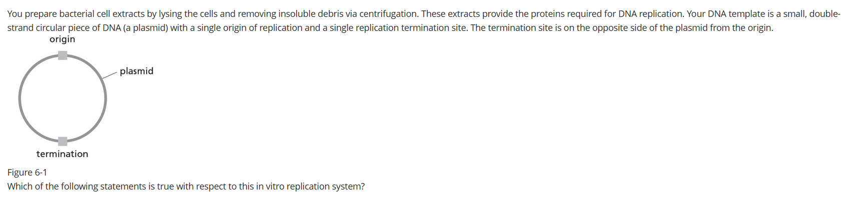
Option A
There will be only one leading strand and one lagging strand produced using this template.Option B
The leading and lagging strands compose one-half of each newly synthesized DNA strand.
Option C
The DNA replication machinery can assemble at multiple places on this plasmid.
Option D
One daughter DNA molecule will be slightly shorter than the other.
B
Which of the following statements correctly explains what it means for DNA replication to be bidirectional?
Option A
The replication fork can open or close, depending on the conditions.
Option B
The DNA replication machinery can move in either direction on the template strand.
Option C
Replication-fork movement can switch directions when the fork converges on another replication fork.
Option D
The replication forks formed at the origin move in opposite directions.
D
Which of the following statements is NOT an accurate statement about thymine dimers?
Option A
Thymine dimers can cause the DNA replication machinery to stall.
Option B
Thymine dimers are covalent links between thymidines on opposite DNA strands.
Option C
Prolonged exposure to sunlight causes thymine dimers to form.
Option D
Repair proteins recognize thymine dimers as a distortion in the DNA backbone.
B
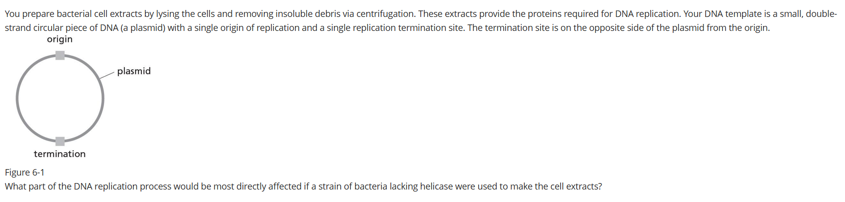
Option A
initiation of DNA synthesis
Option B
Okazaki fragment synthesis
Option C
leading-strand elongation
Option D
lagging-strand completion
A
The DNA duplex consists of two long covalent polymers wrapped around each other many times over their entire length. The separation of the DNA strands for replication causes the strands to be “overwound” in front of the replication fork. How does the cell relieve the torsional stress created along the DNA duplex during replication?
Topoisomerases break the covalent bonds of the backbone, allowing the local unwinding of DNA ahead of the replication fork
Which of the following statements about the newly synthesized strand of a human chromosome is true?
Option A
It was synthesized from a single origin solely by continuous DNA synthesis.
Option B
It was synthesized from a single origin by a mixture of continuous and discontinuous DNA synthesis.
Option C
It was synthesized from multiple origins solely by discontinuous DNA synthesis.
Option D
It was synthesized from multiple origins by a mixture of continuous and discontinuous DNA synthesis.
D
How many replication forks are formed when an origin of replication is opened?
two

A
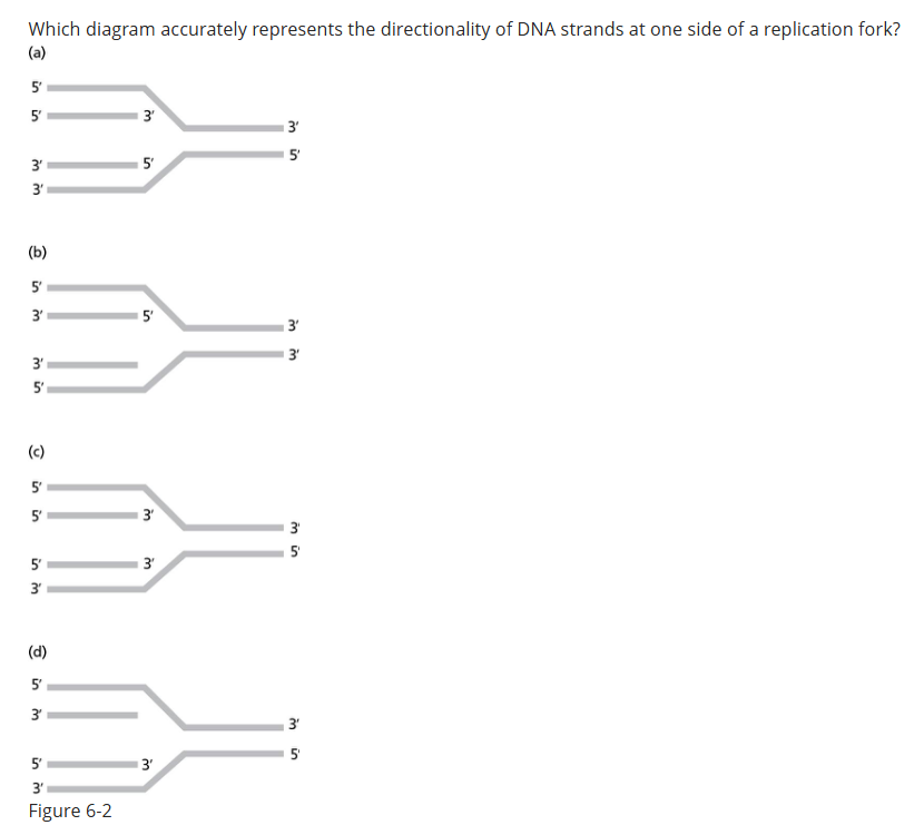
D
The repair of mismatched base pairs or damaged nucleotides in a DNA strand requires a multistep process. Which choice below describes the known sequence of events in this process?
Option A
DNA damage is recognized, the newly synthesized strand is identified by an existing nick in the backbone, a segment of the new strand is removed by repair proteins, the gap is filled by DNA polymerase, and the strand is sealed by DNA ligase.
Option B
DNA repair polymerase simultaneously removes bases ahead of it and polymerizes the correct sequence behind it as it moves along the template. DNA ligase seals the nicks in the repaired strand.
Option C
DNA damage is recognized, the newly synthesized strand is identified by an existing nick in the backbone, a segment of the new strand is removed by an exonuclease, and the gap is repaired by DNA ligase.
Option D
A nick in the DNA is recognized, DNA repair proteins switch out the wrong base and insert the correct base, and DNA ligase seals the nick.
A

Option A
initiation of DNA synthesisOption B
Okazaki fragment synthesis
Option C
leading-strand elongation
Option D
lagging-strand completion
B

Helicase
DNA polymerase can add nucleotides to both the 3' and 5' ends of a growing DNA strand.
True
False
False
What is the correct definition of excision repair?
Option A
Repair of a single damaged nucleotide
Option B
Repair of a damaged oligonucleotide
Option C
Removal of a single damaged nucleotide
Option D
Removal of a damaged oligonucleotide
C
Okazaki fragments are used to elongate what?
the lagging strand away from the replication fork
Which enzyme is responsible for unwinding the DNA double helix during replication?
Helicase
Which enzyme is responsible for sealing the single strand nick between the nascent chain and Okazaki fragments on the lagging strand?
DNA ligase
Which enzyme is responsible for synthesizing the leading strand during DNA replication?
DNA polymerase III
Which enzyme is responsible for initiating synthesis of RNA primers during DNA replication?
Primase
Since the first nucleotides cannot be linked in a newly synthesized strand in DNA replication, ___________ is required
an RNA primer
What is the primary function of the "Mismatch Repair" pathway?
To correct errors in base pairing that occur during DNA replication
Which strand grows continuously towards the replication fork?
leading strand
DNA replication is a semi-conservative process.
True
False
True
What is the primary mechanism by which DNA repair systems identify incorrect base pairs during replication?
Exonuclease activity
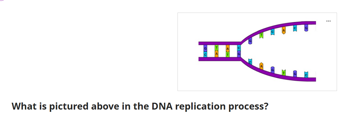
replication fork
What is the basis for inheritance?
DNA replication
What is the directionality of DNA?
Complementary strands run in opposite directions
What is the DNA replication model according to Herbert Taylor
semi conservative
Outline the Central Dogma, explain why it does not apply for all genes
Takeaway: The central dogma describes information flow DNA → RNA → protein, but many genes do not encode proteins
Central Dogma: DNA is transcribed into RNA; RNA is translated into protein
Exceptions:
Some genes encode functional RNAs (tRNA, rRNA, snRNA, miRNA, lncRNA) that are never translated
Reverse transcription
Explain the regulatory advantage of having and RNA “middle step” between DNA and the proteins DNA encodes
Takeaway: RNA provides a flexible, controllable intermediate that allows cells to regulate gene expression efficiently
Key advantages:
Amplification: One DNA gene → many RNA copies → many proteins
Temporal control: RNA can be rapidly synthesized or degraded
Spatial control: mRNA can be transported to specific cellular regions
Quality control: RNA processing (capping, splicing, polyadenylation) allows regulation before translation
Contrast the structure of a DNA and RNA nucleotide, including the structure of the pentose sugar and the type of nitrogenous bases used by RNA and DNA
Takeaway: DNA uses deoxyribose + A,G,C,T; RNA uses ribose + A,G,C,U
Sugar difference:
DNA: deoxyribose (no 2′‑OH)
RNA: ribose (has 2′‑OH)
Base difference:
DNA: A, G, C, T
RNA: A, G, C, U (uracil replaces thymine)
Explain the importance of the 2’ hydroxyl group in RNA
Takeaway: The 2′‑OH makes RNA more reactive, less stable, and able to form complex structures
Functions:
Enables intramolecular base‑pairing → secondary structures
Makes RNA chemically reactive, allowing catalytic RNAs (ribozymes)
Causes instability, enabling rapid turnover (useful for regulation)
State the functions of mRNA, tRNA, rRNA, and snRNAs, snoRNAs, miRNA, and lncRNA
Takeaway: mRNA is translated; all others are functional RNAs
mRNA: Encodes amino acid sequence; template for translation
tRNA: Transfers amino acids to ribosome
rRNA: Forms ribosome structure; catalyzes peptide bond formation
snRNA: Splicing of pre‑mRNA
snoRNA: Modifies rRNA
miRNA: Post‑transcriptional gene regulation
lncRNA: Regulatory roles in gene expression
Outline the process of RNA interference and explain its potential in medicine
Takeaway: RNAi uses small RNAs (miRNA/siRNA) to silence gene expression
Process:
dsRNA processed into siRNA/miRNA
Loaded into RISC complex
RISC binds complementary mRNA → degradation or translation inhibition
Medical potential:
Silencing disease‑causing genes (e.g., oncogenes)
Antiviral therapies
Precision gene regulation
(General RNAi mechanism inferred from miRNA function in sources.)
Outline the 3 major steps of transcription
Takeaway: Initiation → Elongation → Termination
Initiation: Transcription factors bind promoter; RNA polymerase recruited
Elongation: RNA polymerase reads DNA 3′→5′ and synthesizes RNA 5′→3′
Termination: Polymerase stops at termination sequence
Contrast promoter, start site, and termination sequence
Takeaway: Promoter = binding site; start site = +1; termination = stop signal
Promoter: Upstream DNA sequence where transcription factors & RNA polymerase bind
Start site (+1): First nucleotide transcribed
Termination sequence: Signals RNA polymerase to stop
Outline the RNA synthesis by RNA polymerase, including which direction this synthesis happens, and which DNA strand is read by RNA polymerase
Takeaway: RNA polymerase reads template strand 3′→5′ and synthesizes RNA 5′→3′
Reads template (antisense) strand
RNA sequence matches coding strand except U replaces T