Anatomy Lab: Heart and Major Vessel Identification
 Call Kai
 Learn
 Practice Test
 Spaced Repetition
 Match
 Flashcards
 Knowt Play
1/28

There's no tags or description
Looks like no tags are added yet.
29 Terms

Apex of Heart
Pointed inferior tip of the heart formed by the left ventricle, pointing downward and to the left. [Image search: base and apex of heart]

Auricles (on atria)
Ear-like, wrinkled muscular pouches attached to the outer upper walls of the right and left atria. [Image search: right and left auricle heart]

Atria (Left and Right)
Two superior receiving chambers of the heart; Right receives deoxygenated blood from venae cavae, Left receives oxygenated blood from pulmonary veins. [Image search: heart chambers atria ventricles]

Ventricles (Left and Right)
Two inferior pumping chambers; Left ventricle has a much thicker muscular wall (myocardium) than the right ventricle. [Image search: right and left ventricles heart]

Tricuspid Valve (or right AV valve)
Three-cusped valve between the right atrium and right ventricle; anchored by chordae tendineae. (Tip: "Tri" on the "Ri"ght). [Image search: tricuspid valve heart]

Aorta
Largest artery in the body; arches superiorly out of the left ventricle to distribute oxygenated blood systemically. [Image search: aorta heart model]

Aortic Semilunar Valve
Three-pocket half-moon valve located at the exit of the left ventricle into the ascending aorta; lacks chordae tendineae. [Image search: aortic semilunar valve]

Pulmonary Trunk (R & L Pulmonary Arteries)
Large vessel exiting the right ventricle anteriorly and splitting into the right and left pulmonary arteries (colored blue on models because it carries deoxygenated blood). [Image search: pulmonary trunk heart]
Pulmonary Veins
Four vessels (2 left, 2 right) entering the posterior left atrium (colored red on models because they carry oxygenated blood from the lungs). [Image search: pulmonary veins heart]

Pulmonic/ Pulmonary Semilunar Valve
Three-pocket half-moon valve at the exit of the right ventricle into the pulmonary trunk; lacks chordae tendineae. [Image search: pulmonary semilunar valve]

Bicuspid Valve (or Left AV valve or mitral valve)
Two-cusped valve between the left atrium and left ventricle; anchored by chordae tendineae. [Image search: bicuspid mitral valve heart]

Interatrial Septum
Thin muscular wall separating the right atrium from the left atrium. [Image search: interatrial septum heart]

Interventricular Septum
Thick muscular wall separating the right ventricle from the left ventricle. [Image search: interventricular septum heart]
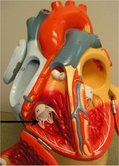
Chordae Tendineae
White fibrous "heart strings" connecting the cusps of the tricuspid and bicuspid (AV) valves to the papillary muscles. [Image search: chordae tendineae heart]

Pectinate Muscles (in Atria)
Parallel, comb-like muscular ridges located on the internal anterior wall of the right atrium and inside both auricles. [Image search: pectinate muscles heart]

Papillary Muscles (in Ventricles)
Cone-shaped muscular projections inside the ventricular walls that anchor the chordae tendineae to prevent valve prolapse. [Image search: papillary muscles heart]

Ligamentum Arteriosum
White fibrous cord connecting the underside of the aortic arch to the pulmonary trunk (remnant of fetal ductus arteriosus). [Image search: ligamentum arteriosum]

Fossa Ovalis
Oval-shaped shallow depression on the right side of the interatrial septum (remnant of fetal foramen ovale). [Image search: fossa ovalis heart]

Coronary Sinus
Large blue venous channel on the posterior surface of the heart (in the coronary sulcus) that collects deoxygenated blood from the myocardium and empties into the right atrium. [Image search: coronary sinus heart]

Right Coronary Artery (RCA)
Red vessel emerging from the aorta and traveling along the right coronary sulcus (groove between right atrium and right ventricle). [Image search: right coronary artery]

Left Coronary Artery (LCA)
Short red vessel emerging from the left side of the aorta behind the pulmonary trunk before splitting into the anterior interventricular (LAD) and circumflex branches. [Image search: left coronary artery]

Anterior Interventricular Branch of LCA
Also called the LAD (Left Anterior Descending); red vessel running down the anterior interventricular sulcus toward the apex of the heart. [Image search: anterior interventricular artery]
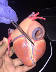
Posterior Interventricular Branch of RCA
Also called the PDA; red vessel running down the posterior interventricular sulcus toward the apex of the heart. [Image search: posterior interventricular artery]

Great Cardiac Vein
Large blue vein running alongside the anterior interventricular artery on the front of the heart, wrapping around to drain into the coronary sinus. [Image search: great cardiac vein]

Posterior Interventricular Vein
Also called the middle cardiac vein; blue vein running alongside the posterior interventricular artery on the back of the heart into the coronary sinus. [Image search: posterior interventricular vein]
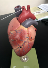
Brachiocephalic A.
First and largest branch off the aortic arch (on the right side); quickly splits into the right common carotid and right subclavian arteries. [Image search: aortic arch branches]

Left Common Carotid A.
Second (middle) branch off the aortic arch; travels straight up the left side of the neck. [Image search: aortic arch branches]

Left Subclavian A.
Third (leftmost) branch off the aortic arch; travels laterally under the left clavicle toward the left arm. [Image search: aortic arch branches]
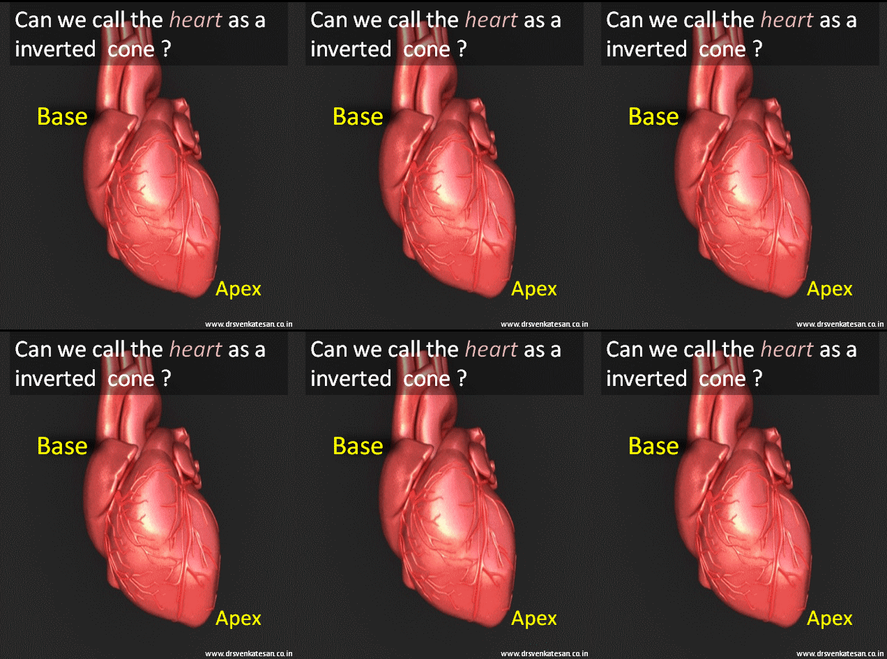
Base of Heart
The base of the heart is the broad, flat section located opposite the apex, primarily composed of the left atrium and part of the right atrium.