Intro to pathology and Cell Injury and Cell Death
 Call Kai
 Learn
 Practice Test
 Spaced Repetition
 Match
 Flashcards
 Knowt Play
1/122

There's no tags or description
Looks like no tags are added yet.
123 Terms
Pathology is the study of:
Links between disease and basic science (pathos=disease)
A disease is:
A physical or functional disorder of normal body systems that places an individual as risk for adverse consequences
General disease diagnosis and treatment
Health care providers diagnose disease with a combination of tools, and then treatment is given to improve prognosis
Etiology Definition
Cause of disease
Pathogenesis definition
Mechanism of disease development
Morphologic/Pathogenic change definition
Alteration in cells and organs, they are characteristic of a disease or diagnostic of an etiologic process
Pathophysiology definition
Functional changes associated with or resulting form disease or injury. Can be seen in signs and symptoms
Example of pathological process affecting various tissues differently
Squamous cell carcinoma:
In skin=indolent (painless)
In esophagus= life-threatening
Example of pathologic process causing different diseases in different organs
Myocardial infarction (MI)=due to ischemia
Cerebrovascular indecent (CVA)=due to ischemia
2 classes of etiologic factors:
Genetic and acquired
Genetic etiology factor
Inherited mutations and disease-associated gene variants or polymorphisms
Acquired etiology factor
Infectious, nutritional, chemical, physical
Most common disorders are caused by:
Multifactorial effects, arise from effects of various external factors on a genetically susceptible individual
Example of acquired disorders:
Traumatic ulcer, syphilis, coronary atherosclerosis with thrombosis, caries, cataracts
Examples of genetic disorders:
Amelogenesis imperfecta, Marfan syndrome, Crouzon syndrome, APC gene mutation, familial Polyposis
Pathogenesis, the mechanism of development is:
Sequence of events in response of cells/tissues to etiologic event, form initial stimulus to disease expression
Examples of different general pathogenesis:
cancer needing multiple mutations to be cancerous
Smoking and alcohol
radiation exposure
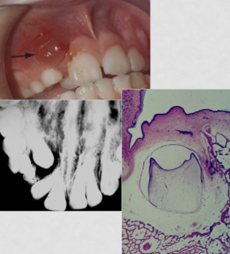
Photo of odontogenic cyst
Odontogenic cyst pathogenesis:
Develops by accumulation of fluid between the reduced enamel epithelium and the tooth crown
Odontogenic cyst functional consequences:
Pericoronal (around tooth crown) radiolucency, loss of bone, failure of teeth to erupt, need for ortho/OS (to restore occlusion)
Retinoblastoma can be caused by:
Inheriting a mutation in the Rb gene
The field of diagnostic pathology has expanded to include what methods?
Molecular biologic and immunologic approaches for analyzing disease states
Morphology of kidney disease:
Granular surface, decreased function, smaller size, high urine protein
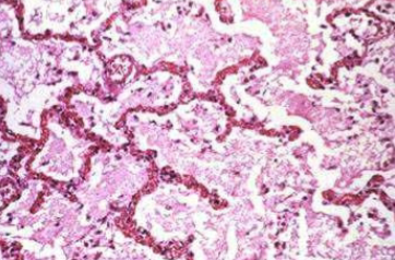
(Lung) chronic passive venous congestion
3 Categories that providers use to make diagnoses
clinical diagnosticians identify diseases through pt history and physical exam
Pathologists examine cells/tissues that were removed from the body to identify diseases
Radiologists image the intact body to identify disease
Virtually all forms of diseases start with:
Molecular structural alterations in cells
Tissue with functional abnormalities lead to:
Clinical manifestations (symptoms, signs) of disease and its progress (clinical course and outcome)
What is the most commonly mutated protein in cancer
p53
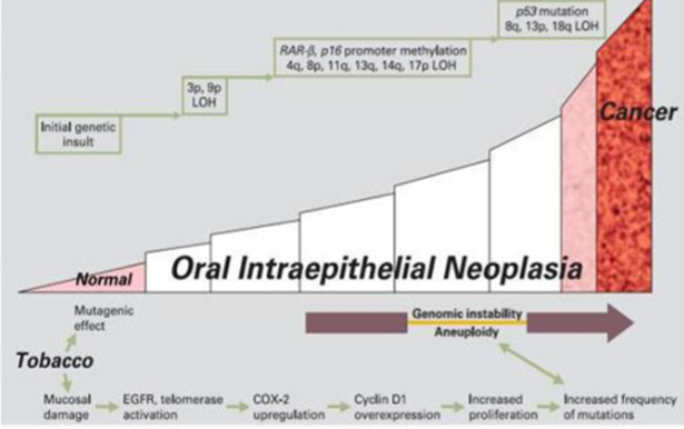
Etiologic agents (smoking) —> pathogenesis —> pathologic changes —> clinical manifestations (HPV wart)
Many proliferation genes are upregulated in:
HSNCC, Head and neck squamous cell carcinoma
Symptom definition
Experienced, reported by the patient
Sign definition
Discovered by doctor during examination or by in vitro examination
Call injury results when:
Limits of adaptive responses are exceeded
Cells are exposed to damaging insults, deprived of critical nutrients
compromised by mutations effecting cellular function
Increased load of normal myocyte leads to:
Respond to increased load with: hypertrophy
increased BP, required more mechanical effort of myocardial cells=enlarge
Normal myocyte responds to cell injury with:
A reversibly injured myocyte undergoes functional alterations:
cytoplasmic changes
cellular swelling
fat accumulation
Leads to reduced blood flow and cell death
Causes of cell injury
oxygen deprivation
physical agents
chemical and infectious agents
tapeworms and fungi
radiation and extreme cold

Sequential development of biochemical and morphologic changes in cell injury
Range of function and structure depends on:
genetic program of metabolism (receptors to allow substrates)
differentiation (stem vs mature)
specialization of cell
availability of metabolic substrates (like iron)
What is cellular adaptation
A cell adapting to an altered or new steady state
They’re acceptable changes to the environment by modifying metabolism or growth pattern.
2 Types of physiologic adaption
Metabolic and structural
Metabolic physiologic adaptation
Fine regulation of metabolic function that’s not easily detectable as a structural change
ex: FA mobilization in fasting
Structural physiologic adaptation
Increase in cellular activity (size and #)
Decreased cellular activity (size and #)
Alteration of cell morphology (differentiation)
Atrophy definition
Decrease in size and function
Hypertrophy definition
Increase in size
Hyperplasia definition
Increase in number of cells
Metaplasia definition
Reversible change from one cell type to another (altered differentiation)
Dysplasia definition
Abnormal changes in cellular components (usually a cancer precursor)
Pathologic causes of Atrophy
Atrophy of disease, loss if innervation, less blood supply, aging
Physiologic causes of atrophy
Uterine atrophy after giving birth
By puberty the thymus starts to shrink and become replaces by fat

Left=atrophic muscle cell
Right= normal muscle cell
Hypertrophy definition
Increased functional demand on tissue met by increase in cell size (more structural proteins made, no new cells)
Hyperplasia definition
Increased functional demand on tissue met by increase in cell number
Hypertrophy physiologic properties
Stimulated by hormones (like uterus and breast in pregnancy)
Increase in workload (exercise)=skeletal muscle hypertrophy
Hypertrophy pathologic properties
Increased workload is associated with changes in gene expression (heart muscle adaptation) atrial/brain natriuretic factors
Alpha-myosin replaced by beta-myosin heavy chain
TGF-beta, etc

Photo of hypertrophied uterus (recently pregnant)
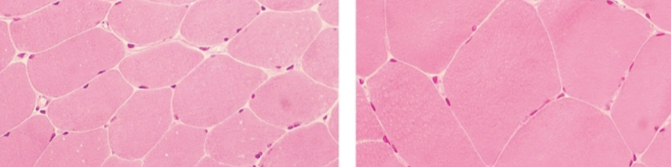
Left=regular skeletal muscle
Right= hypertrophy of skeletal muscle in response to exercise
Beyond the limit of compensation, adaptive change can cause:
Congestive heart failure, apoptosis, and/or necrosis
Physiologic cause of hyperplasia examples:
Hormonal: female breasts during puberty and pregnancy
Compensatory: Liver after partial hepatectomy (liver regrowth)
Pathologic cause of hyperplasia(may be precancerous):
excessive hormonal stimulation causes abnormal cell division
prostate cells increase in umber=enlarged prostate
excess estrogen causes uterus lining to thicken
excessive growth factors= excessive multiplying of cells
injury=growth factors to repair=fibroblast recruitment= new tissue
Some viruses can cause abnormal cell growth
HPV: causes warts and epithelial growths like papillomas
some high-risk HPV can contribute to precancerous changes
Benign prostatic hyperplasia (BPH)
Non-cancerous, age-related enlargement of the prostate gland

Metaplasia mechanism
A result of reprogramming stem cells (reserve cells) that are signaled by: cytokines, growth factors, and extracellular factors
(—>induce TF—>cascade of genes—>differentiated metaplastic cell)
Columnar squamous metaplasia example
In uterine cervix inflammation, normal columnar cells are replaced with squamous cells (to better withstand environment and irritation)
Barrett esophagus metaplasia example
Normal squamous intestinal cells convert to columnar cells (esophageal mucosa reacting to chronic acid inflammation, GERD) PRECANCEROUS change
Reversible injury involves(hydropic degeneration):
membrane blebbing/cell swelling
mitochondrial swelling
ribosomal detachment
loosen attachments
fatty change in liver
Irreversible injury involves:
lysosomal rupture/disappearance
large, bizarre mitochondria
calcium deposits
membrane break

Left=normal kidney tubules and epithelial cells
Middle=reversible, early ischemic injury
Right= Necrosis, irreversible injury, of epithelial cells
Reversible cell injury effect on tissues/organs:
Pallor (compression of capillaries), increase weight (from cellular swelling), and increase turgor (from increase in size)

Left=normal mitochondria
Right= Ischemic cell mitochondria (swollen and round), REVERSIBLE
2 causes of sublethal liver cell damage
Hydropic degeneration in paraquat poisoning and fatty change in alcoholism
Cell response to injury depends on:
Type, duration, and severity of injury
Consequences of injury depends on:
Type, state, and adaptability of injured cell
Cellular sites with more vulnerability to injury:
Cell membrane, aerobic respiration, protein synthesis, genetic apparatus
When do morphologic changes become apparent?
Only after derangement of some critical biochemical system
2 Principle types of cell death:
Necrosis and apoptosis
Necrosis morphologic changes
Progressive injury leads to breakdown of plasma membrane, organelles, and nucleus and leakage of contents, and inflammation
Apoptosis morphologic changes
As chromatins condense and membrane blebs, cellular fragmentation occurs and apoptotic bodies are phagocytosed
Morphologic signs suggestive of apoptosis in kidney biopsy:
Cellular shrinkage and nuclear condensation
Signs of lung necrosis:
bronchiolar lumen containing fibrin, neutrophils, cellular debris, and extravasated RBCs (inflammatory cells)
bronchiolar epithelial cells contain intracytoplasmic eosinophilic inclusion (suggest viral infection)
2 types of damage that can cause inadequate ATP production:
Hypoxia or mitochondrial damage
Anaerobic ATP production is limited by:
Accumulation of lactate, and is usually quickly exhausted
What causes cloudy swelling?
Lack of ATP—>failure of membrane pumps—> swelling on internal membrane systems
Outcome of ischemia:
Ischema=less blood blow=less O2=Less ATP to tissues=pumps stop working=membrane damage=necrosis
Examples of ischemia cell damage
damage to cytoskeleton (from swelling)
free radical injury, lipid breakdown
entry of extracellular Ca and H2O
leakage of enzymes/proteins
lysosomal rupture
Coagulative necrosis
Protein denaturation, from ischemic in most tissue except the brain
Liquefactive necrosis
Hydrolytic enzymatic digestion of tissues, mostly from infections. Ischemic brain infarcts develops from lack of blood flow not from infection.
Caseous necrosis
Typical of Tuberculosis (TB), cheese-like, see in lung
Hemorrhagic necrosis
Dead tissue is replaced by extravasated blood (venous block)
Fat necrosis:
Enzymatic: lipases released in pancreatitis
Traumatic: breast fat is replaced by lipid-laden macrophages
Gangrenous necrosis:
Ischemic most commonly found in skin and soft tissues of feet
Fibrinoid necrosis
Immune deposits common in vessel walls
Gummatous necrosis
Restricted necrosis in spirochetal infection, seen in syphilis
Coagulative vs liquefactive necrosis
Coagulative: Dead tissue is preserved for a few days, localized area of coagulative necrosis is an infarct (early in process, hold shape)
Liquefactive: Digestion of dead cells makes a viscous liquid, like from many infections. Creamy yellow necrotic material (leukocyte presence)=pus (dead cells begin digestion)

Caseous necrosis, TB granulomas

Liquefactive necrosis in brain of patient with stroke

Liquefactive necrosis in an abscess

Acute pancreatitis causing enzymatic fat necrosis (in alcoholic pt with elevated serum amylase levels)

Gangrenous necrosis: atherosclerotic—>ischemia
Apoptosis in induced by:
Tightly regulated suicide system where cells destined to die activate intrinsic enzymes that degrade cells’ genomic DNA and nuclear/cytoplasmic proteins (most apoptosis is intrinsic)
Apoptosis cellular mechanism
Breaks up cells into plasma membrane bound fragments. Plasma membrane remains intact, surface components produce signals for phagocytes to find.
Physiologic situations where phagocytosis is important:
removal of supernumerary cells during development
involution (shrinkage) of hormone-dependent tissue on hormone withdrawal
cell turnover in proliferating clel populations
elimination of potentially harmful self-reactive lymphocytes to prevent immune reactions against self
death of host cells that have served their purpose