USMDO Custom Cards
 Call Kai
 Learn
 Practice Test
 Spaced Repetition
 Match
 Flashcards
 Knowt Play
1/234

There's no tags or description
Looks like no tags are added yet.
235 Terms
Anterior (ventral)
toward the front
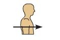
Posterior (dorsal)
toward the back

Superior (cranial)
toward the head

Inferior (caudal)
toward the tail

Proximal
Closer to the point of attachment

Distal
Father away from the point of attachment

Medial
Closer to the midline

Lateral
Farther away from the midline
Superficial
Closer to the surface

Deep
Farther below the surface

Axial region
head, neck, trunk

Appendicular
Upper and lower limbs/appendages

Upper limb
arm

Lower limb
leg

Cephalic
head

Occipital
back of head

Cervical
neck

Thoracic
chest

Sternal
sternum
Abdominal
abdomen
Pelvic
pelvis
Palmar
palm
Inguinal
groin
Pubic
genital region
Pedal
foot
Dorsal
back
Vertebral
spinal column
Lumbar
lower back/loin
Sacral
between hips
Gluteal
buttock
Popliteal
back of knee
Sural
calf
Plantar
sole of foot
Manual
hand
Cranial
skull
Frontal
forehead
Occular
eye
Otic
ear
Nasal
nose
Buccal
cheek
Oral
mouth
Mental
chin
Axillary
armpit
Acromial
top of shoulder
Brachial
upper arm
Antecubital
front of elbow
Antebrachial
forearm
Carpal
wrist
Pollex
thumb
Metacarpal
palm
Digital
fingers/toes
Coxal
hip
Femoral
thigh
Patellar
kneecap
Crural
lower leg
Tarsal
ankle
Metatarsal
top of foot
Hallux
big/great toe
Sagittal plane
vertical division of the body into right and left portions

Midsagittal/median plane
divides the body into equal right and left sides

Parasagittal plane
Divides body into unequal right and left sides
Frontal (coronal) plane
divides the body into anterior and posterior parts
Transverse (horizontal) plane
divides the body into superior and inferior parts
Oblique plane
passes through the body at an angle
Cavity
- fluid-filled space within the body
- protects internal organs, allows them to move and expand as necessary
Dorsal body cavity
- contains the cranial cavity and spinal column
- continuous and filled with cerebrospinal fluid (CSF); bathes and protects both brain and spinal cord
Cranial cavity
within skull; protects brain
Vertebral (spinal) cavity
within vertebral column; protects spinal cord
Ventral body cavity
- thoracic and abdominopelvic cavities
- separated into two divisions by diaphragm
Thoracic cavity
- contains pleural cavities, mediastinum, pericardial cavity
- superior to diaphragm
Pleural cavities
each surround either left or right lung
Mediastinum
between pleural cavities; houses heart, great vessels, trachea, and esophagus; not within serous membrane
Pericardial cavity
within mediastinum; within serous membrane that surrounds heart
Abdominopelvic cavity
abdominal cavity and pelvic cavity
Abdominal cavity
spans from diaphragm to bony pelvis
Pelvic cavity
area within bony pelvis
Peritoneal cavity
abdominal subcavity found within serous membrane
Right upper quadrant (RUQ)
- upper left side
- contains most of biliary system, including gallbladder on far right, half of pancreas and most of liver and bile ducts
Right lower quadrant (RLQ)
- lower left side
- contains appendix, half of uterus, half of bladder, half of small intestine, half of large intestine
Left upper quadrant (LUQ)
- upper right side
- spleen, stomach, 1/3 of liver
Left lower quadrant (LLQ)
- lower right side
- contains half of uterus, half of bladder, half of small intestine, half of large intestine
Right hypochondriac region
liver, gallbladder
top right
Epigastric region
located above the stomach
top center
Left hypochondriac region
diaphragm, spleen
top left
Right lumbar region
ascending colon of large intestine
middle right
Umbilical region
centermost region, includes the umbilicus
middle center
Left lumbar region
descending colon of large intestine
middle left
Right iliac region
cecum, appendix
bottom right
Hypogastric region
located below the stomach, bladder, uterus
bottom center
Left iliac region
small intestines, descending colon, sigmoid colon
bottom left
Serous membranes
Thin sheets of tissue; form certain cavities found in ventral cavity
appear to be two membranes when sectioned; actually consists of a single, continuous layer of tissue; folds over itself to create a double-layered structure
Serous fluid
watery, slippery lubricant
fills space between membrane layers
prevents friction
Visceral layer
in contact with underlying organ
Parietal layer
outermost layer attached to surrounding structures
Retroperitoneal organs
organs that lie outside of and behind parietal peritoneum (kidneys)
X-Ray
uses ionizing radiation
Computerized tomography (CT) scan
uses ionizing radiation; 3-D image is computer generated from data
bones and soft tissue, visualize slices of body
Magnetic resonance imagine (MRI)
body is placed within a magnetic field; computer compiles data producing a 3-D image
powerful magnet where the magnetic field is used to align the magnetization of some atomic nuclei in the body, and radio frequency magnetic fields are applied to systematically alter the alignment of this magnetization
Ultrasound
high frequency sound waves, substances of different densities reflect back waves, transducer records those reflected waves
Positron Emission Tomography (Pet Scan)
radioactive tracers travel though blood to organs and tissues, PET detects signals from tracer. Can pick up metabolically active tissues (cancer diagnosis)