OTR 704 2.6 Knee, Ankle, & Foot

0.0(0)
Studied by 23 people
call kaiCall Kai
learnLearn
examPractice Test
spaced repetitionSpaced Repetition
heart puzzleMatch
flashcardsFlashcards
GameKnowt Play
Card Sorting

1/135

encourage image

There's no tags or description

Looks like no tags are added yet.

136 Terms

1
New cards
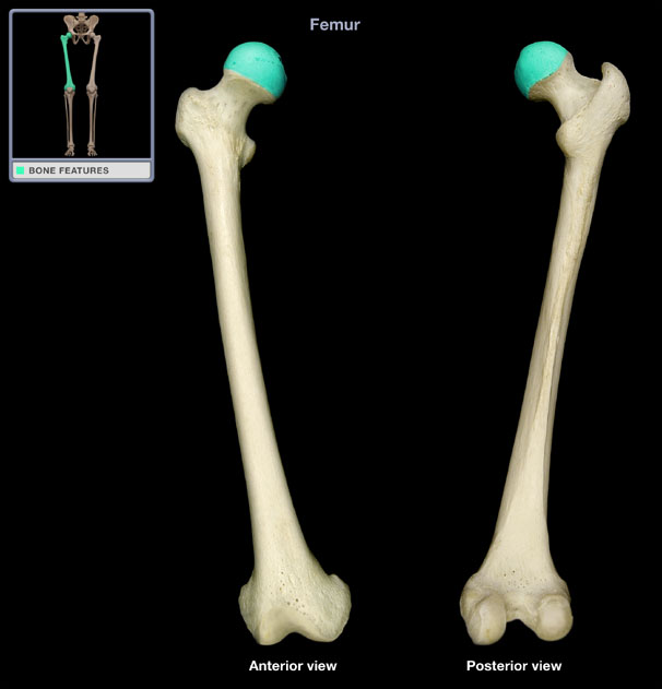
<p>What structure is shown?</p>

What structure is shown?

Femur

2
New cards
<p>What structure is shown?</p>

What structure is shown?

Femoral head

3
New cards
<p>What structure is shown?</p>

What structure is shown?

Neck of the femur

4
New cards
<p>What structure is shown?</p>

What structure is shown?

Greater trochanter

5
New cards
<p>What structure is shown?</p>

What structure is shown?

Lesser trochanter
6
New cards
<p>What structure is shown?</p>

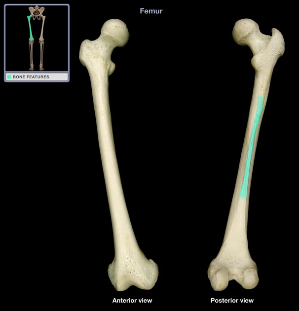
What structure is shown?

Linea aspera

7
New cards
<p>What structure is shown?</p>

What structure is shown?

Medial epicondyle

8
New cards
<p>What structure is shown?</p>

What structure is shown?

Lateral epicondyle

9
New cards
<p>What structure is shown?</p>

What structure is shown?

Medial condyle

10
New cards
<p>What structure is shown?</p>

What structure is shown?

Lateral condyle

11
New cards
<p>What structure is shown?</p>

What structure is shown?

Intercondylar fossa

12
New cards
<p>What structure is shown?</p>

What structure is shown?

Patella

13
New cards
<p>What structure is shown?</p>

What structure is shown?

Apex

14
New cards
<p>What structures are shown?</p>

What structures are shown?

Medial and lateral facets

15
New cards
<p>What structure is shown?</p>

What structure is shown?

Tibia

16
New cards
<p>What structure is shown?</p>

What structure is shown?

Medial condyle

17
New cards
<p>What structure is shown?</p>

What structure is shown?

Lateral condyle

18
New cards
<p>What structure is shown?</p>

What structure is shown?

Tibial tuberosity

19
New cards
<p>What structure is shown?</p>

What structure is shown?

Medial malleolus

20
New cards
<p>What structure is shown?</p>

What structure is shown?

Fibula

21
New cards
<p>What structure is shown?</p>

What structure is shown?

Fibular head

22
New cards
<p>What structure is shown?</p>

What structure is shown?

Lateral malleolus
23
New cards
<p>What structure is shown?</p>

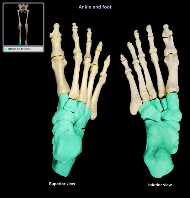
What structure is shown?

Tarsals

24
New cards
<p>What structure is shown?</p>

What structure is shown?

Talus

25
New cards
<p>What structure is shown?</p>

What structure is shown?

Calcaneus

26
New cards
<p>What structure is shown?</p>

What structure is shown?

Cuboid

27
New cards
<p>What structure is shown?</p>

What structure is shown?

Navicular

28
New cards
<p>What structure is shown?</p>

What structure is shown?

Medial cuneiform

29
New cards
<p>What structure is shown?</p>

What structure is shown?

Intermediate cuneiform
30
New cards
<p>What structure is shown?</p>

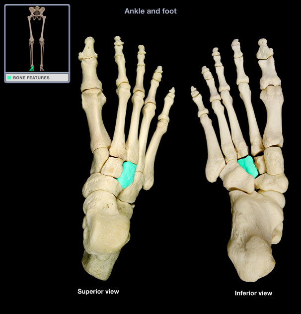
What structure is shown?

Lateral cuneiform

31
New cards
<p>What structure is shown?</p>

What structure is shown?

Metatarsals

32
New cards
<p>What structure is shown?</p>

What structure is shown?

Proximal phalanges

33
New cards
<p>What structure is shown?</p>

What structure is shown?

Middle phalanges

34
New cards
<p>What structure is shown?</p>

What structure is shown?

Distal phalanges

35
New cards

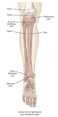
Bones of the lower extremity

  • Femur

  • Tibia & fibula

  • Patella

  • Bones of the foot — tarsals, metatarsals, & phalanges

<ul><li><p>Femur</p></li><li><p>Tibia &amp; fibula</p></li><li><p>Patella</p></li><li><p>Bones of the foot — tarsals, metatarsals, &amp; phalanges</p></li></ul><p></p>
36
New cards

Femur

Structural support for the thigh

  • Muscle attachments at bony landmarks: medial and lateral epicondyles, medial and lateral condyles, intercondylar fossa


<p><strong>Structural</strong> support for the thigh</p><ul><li><p>Muscle attachments at bony landmarks: medial and lateral epicondyles, medial and lateral condyles, intercondylar fossa</p></li></ul><p></p>
37
New cards

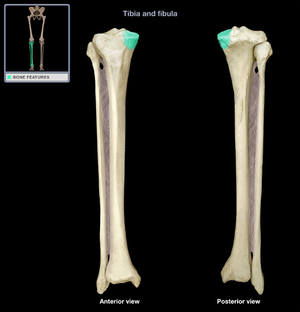
Tibia

Primary weight-bearing bone of the lower leg; direct link between the knee & ankle

  • Muscle attachments at bony landmarks: tibial plateau, medial and lateral condyles, tibial tuberosity, medial malleolus


<p>Primary <strong>weight-bearing</strong> bone of the lower leg; direct link between the knee &amp; ankle</p><ul><li><p>Muscle attachments at bony landmarks: tibial plateau, medial and lateral condyles, tibial tuberosity, medial malleolus</p></li></ul><p></p>
38
New cards

Fibula

Bone of the lower leg that bears little weight

  • Muscle attachments at bone landmarks: head of fibula, lateral malleolus


<p>Bone of the lower leg that <strong>bears little weight</strong></p><ul><li><p>Muscle attachments at bone landmarks: head of fibula, lateral malleolus</p></li></ul><p></p>
39
New cards

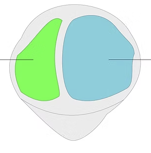
Patella

Sesamoid bone embedded in the quadriceps tendon; stabilizes the anterior knee

  • Bony landmarks: apex, medial & lateral facets


<p><strong>Sesamoid bone</strong> embedded in the quadriceps tendon; <strong>stabilizes the anterior knee</strong></p><ul><li><p>Bony landmarks: apex, medial &amp; lateral facets</p></li></ul><p></p>
40
New cards

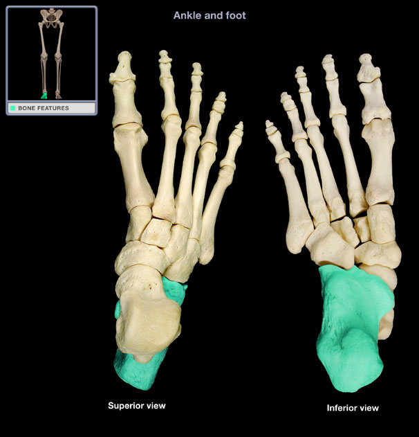
Bones of the foot

  • Tarsals (7) — talus, calcanceous, navicular, cuboid, cuneiforms (medial, intermediate, lateral)

  • Metatarsals (5) — 1st metatarsal sesamoid bones

  • Phalanges — proximal, middle, and distal


<ul><li><p><strong>Tarsals (7)</strong> — talus, calcanceous, navicular, cuboid, cuneiforms (medial, intermediate, lateral)</p></li><li><p><strong>Metatarsals (5)</strong> — <em>1st metatarsal sesamoid bones</em></p></li><li><p><strong>Phalanges </strong>— proximal, middle, and distal</p></li></ul><p></p>
41
New cards

Arches of the foot

  • Lateral longitudinal — pinky toe to heel

  • Medial longitudinal — great toe to heel

  • Transverse — just proximal to toes


<ul><li><p><strong>Lateral longitudinal</strong> — pinky toe to heel</p></li><li><p><strong>Medial longitudinal</strong> — great toe to heel</p></li><li><p><strong>Transverse</strong> — just proximal to toes</p></li></ul><p></p>
42
New cards

Atypical high arch

Aka pes cavus; excessive supination/inversion or genu varus

<p>Aka <strong>pes cavus;</strong> excessive <strong>supination/inversion</strong> or genu<strong> varus</strong></p>
43
New cards

Low/flatter arch

Aka pes planus; excessive pronation/eversion or genu valgus

<p>Aka <strong>pes planus;</strong> excessive <strong>pronation/eversion</strong> or genu<strong> valgus</strong></p>
44
New cards

Hallux valgus

Great toe turned laterally toward the other toes (bunion)
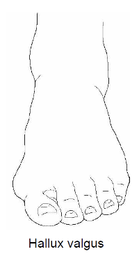
<p>Great toe turned <strong>laterally</strong> toward the other toes (bunion)</p>
45
New cards

Claw toes

Extended MTP, flexed PIP & DIP

<p>Extended MTP, flexed PIP &amp; DIP</p>
46
New cards

Hammer toes

Extended MTP, flexed PIP, & extended DIP

<p>Extended MTP, flexed PIP, &amp; extended DIP</p>
47
New cards

Joints of the LE

Joints of the LE are designed to absorb & withstand compressive forces during weight-bearing activities

  • Tibiofemoral (knee) joint

  • Patellofemoral joint

  • Tibiofibular joint

  • Talocrural joint (ankle joint #1)

  • Subtalar and transverse tarsal joints (ankle joint #2)

  • Intertarsal joints

  • Metatarsophalangeal (MTP) joints

  • Interphalangeal (IP) joints


48
New cards

Tibiofemoral joint

Knee joint; a biaxial, hinge joint

  • Supports the weight of the trunk & upper body

  • Movements: flexion/extension and internal/external rotation

  • Screw-home mechanisms — “locks” & “unlocks” the knee


<p><strong>Knee joint;</strong> a biaxial, <strong>hinge joint</strong></p><ul><li><p>Supports the weight of the trunk &amp; upper body</p></li><li><p>Movements: <strong>flexion/extension and internal/external rotation</strong></p></li><li><p>Screw-home mechanisms — “locks” &amp; “unlocks” the knee</p></li></ul><p></p>
49
New cards

Knee rotation (screw-home mechanism)

  • “Locks” the knee in closed-pack position for maximal stability; femoral condyles with tibial fossas

    • Tibia rotates laterally and femur rotates medially in full extension

  • Reverse movements “unlocks” the knee

    • Requires activation of the popliteus muscle to medially rotate the tibia (and lateral rotation of the femur)


<ul><li><p>“<u>Locks</u>” the knee in closed-pack position for maximal stability; femoral condyles with tibial fossas</p><ul><li><p><strong>Tibia rotates laterally</strong> and femur rotates medially in full extension</p></li></ul></li><li><p>Reverse movements “<u>unlocks</u>” the knee</p><ul><li><p>Requires activation of the <strong>popliteus muscle</strong> to <strong>medially rotate the tibia</strong> (and lateral rotation of the femur)</p></li></ul></li></ul><p></p>
50
New cards

Tibiofemoral joint supporting structures

  • Anterior & posterior cruciate ligaments (ACL & PCL) — prevents anterior/posterior translation of the tibia

  • Medial & lateral collateral ligaments (MCL & LCL) — prevents medial/lateral translation of the tibia

  • Medial & lateral meniscus — cushions & increases surface area


<ul><li><p><strong>Anterior &amp; posterior cruciate ligaments (ACL &amp; PCL) </strong>— prevents anterior/posterior translation of the tibia</p></li><li><p><strong>Medial &amp; lateral collateral ligaments (MCL &amp; LCL) </strong>— prevents medial/lateral translation of the tibia</p></li><li><p><strong>Medial &amp; lateral meniscus</strong> — cushions &amp; increases surface area</p></li></ul><p></p>
51
New cards

Why are women more susceptible to injuries to the supporting structures of the knee?

Strength programs often lead to muscle imbalance for women, with strong quadriceps muscles and weak hamstrings; also due to anatomical differences (e.g., greater angle from pelvis/hip to knee)

52
New cards

Knee varus

Disruption (injury or laxity) of LCL causes the knee position to be altered in the frontal plane

  • Inward angulation of the tibia

  • Lateral shift of the knee complex

  • Genus varus/varum = bow-leggedness


<p>Disruption (injury or laxity) of <strong>LCL</strong> causes the knee position to be altered in the frontal plane</p><ul><li><p><strong>Inward angulation</strong> of the tibia</p></li><li><p><strong>Lateral shift</strong> of the knee complex</p></li><li><p><strong>Genus varus/varum =</strong> bow-leggedness</p></li></ul><p></p>
53
New cards

Knee valgus

Disruption (injury or laxity) of MCL causes the knee position to be altered in the frontal plane

  • Outward angulation of the tibia

  • Medial shift of the knee complex

  • Genus valgus/valgum = knock-kneed


<p>Disruption (injury or laxity) of <strong>MCL</strong> causes the knee position to be altered in the frontal plane</p><ul><li><p><strong>Outward angulation</strong> of the tibia</p></li><li><p><strong>Medial shift</strong> of the knee complex</p></li><li><p><strong>Genus valgus/valgum =</strong> knock-kneed</p></li></ul><p></p>
54
New cards

Genu recurvatum

Hyperextension of the knee

55
New cards

Tibial torsion (knee rotation)

Tibia rotates relative to the femur; approximately 20-25° normal

  • Improper positioning (sitting) can create more torsion


56
New cards

Medial tibial torsion

Knees anterior & inward; feet lateral → can lead to increased valgus

<p><strong>Knees</strong> <strong>anterior &amp; inward;</strong> feet lateral → can lead to <strong>increased valgus</strong></p>
57
New cards

Lateral tibial torsion

Knees anterior & outward; feet medial → can lead to increased varus

<p><strong>Knees anterior &amp; outward;</strong> feet medial → can lead to <strong>increased varus</strong></p>
58
New cards

Patellofemoral joint

Gliding articulation between the femur and patella

  • Knee flexion causes the patella to glide inferiorly and laterally

  • Knee extension causes the patella to glide superiorly and medially
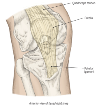

<p><strong>Gliding</strong> articulation between the femur and patella</p><ul><li><p>Knee <strong>flexion</strong> causes the patella to glide <strong>inferiorly</strong> and laterally</p></li><li><p>Knee <strong>extension</strong> causes the patella to glide <strong>superiorly</strong> and medially</p></li></ul><p></p>
59
New cards

Tibiofemoral joints

Proximal AND distal articulations between the tibia & fibula; very slight movement, contribute primarily to ankle stability

<p>Proximal AND distal articulations between the tibia &amp; fibula; <strong>very slight movement,</strong> contribute primarily to <strong>ankle stability</strong></p>
60
New cards

Talocrural joint

Ankle joint #1; articulation between the superior talus and medial & lateral malleoli of the tibia/fibula

  • Uniaxial, hinge joint

  • Movements = dorsiflexion/plantar flexion

  • Closed-pack position = maximal dorsiflexion


<p>Ankle joint #1; articulation between the superior talus and medial &amp; lateral malleoli of the tibia/fibula</p><ul><li><p>Uniaxial, <strong>hinge </strong>joint</p></li><li><p>Movements = dorsiflexion/plantar flexion</p></li><li><p>Closed-pack position = maximal dorsiflexion</p></li></ul><p></p>
61
New cards

Subtalar and transverse tarsal joint

Ankle joint #2; subtalar is between inferior talus & calcaneus and transverse tarsal is between hindfoot & midfoot tarsals

  • Movements = inversion/eversion

  • Functionally important for navigation of uneven terrain, balance


<p>Ankle joint #2; <strong>subtalar</strong> is between inferior talus &amp; calcaneus and <strong>transverse tarsal</strong> is between hindfoot &amp; midfoot tarsals</p><ul><li><p>Movements = inversion/eversion</p></li><li><p>Functionally important for navigation of uneven terrain, balance</p></li></ul><p></p>
62
New cards

Intertarsal joints

Articulation between tarsal bones and between tarsals and metatarsals

<p>Articulation between tarsal bones and between tarsals and metatarsals</p>
63
New cards

Metatarsophalangeal (MTP) joints

Ellipsoid joint; allows for flexion/extension and some abduction/adduction

<p><strong>Ellipsoid joint; </strong>allows for flexion/extension and some abduction/adduction</p>
64
New cards

Interphalangeal (IP) joints

Hinge joints; allows for flexion/extension
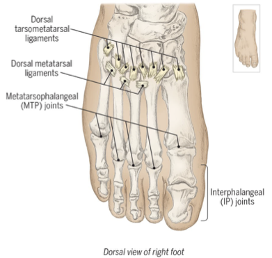
<p><strong>Hinge joints;</strong> allows for flexion/extension</p>
65
New cards

Plantar fascia

Connects calcaneus to base of toes and supports arches of the foot

  • Primary shock absorber during walking/running (weight-bearing)

  • Inflammation → plantar fasciitis


<p>Connects <strong>calcaneus to base of toes</strong> and <strong>supports arches</strong> of the foot</p><ul><li><p>Primary <strong>shock absorber</strong> during walking/running (weight-bearing)</p></li><li><p>Inflammation → <strong>plantar fasciitis</strong></p></li></ul><p></p>
66
New cards

Semimembranosus action

Flexion & internal (medial) rotation of the knee; extension & internal rotation (assists) of the hip; tilts pelvis posteriorly

**Hamstring

<p><span style="font-family: Calibri, sans-serif; line-height: 107%;"><strong>Flexion</strong> &amp; <strong>internal (medial) rotation </strong>of the knee; <strong>extension </strong>&amp; internal rotation (assists) of the hip; tilts pelvis posteriorly</span></p><p><span style="font-family: Calibri, sans-serif; line-height: 107%;">**Hamstring</span></p>
67
New cards

Semimembranosus origin

Ischial tuberosity

68
New cards

Semimembranosus insertion

Posterior aspect of medial condyle of tibia

69
New cards

Semimembranosus innervation

Sciatic (tibial branch) L4 & L5, S1 & S2

70
New cards

Semitendinosus action

Flexion & internal (medial) rotation of the knee; extension & internal rotation (assists) of the hip; tilts pelvis posteriorly

**Hamstring

<p><span style="font-family: Calibri, sans-serif; line-height: 107%;"><strong>Flexion</strong> &amp; <strong>internal (medial) rotation </strong>of the knee; <strong>extension </strong>&amp; internal rotation (assists) of the hip; tilts pelvis posteriorly</span></p><p><span style="font-family: Calibri, sans-serif; line-height: 107%;">**Hamstring</span></p>
71
New cards

Semitendinosus origin

Ischial tuberosity

72
New cards

Semitendinosus insertion

Proximal, medial shaft of the tibia at the per anserinus tendon

73
New cards

Semitendinosus innervation

Sciatic (tibial branch) L4 & L5, S1 & S2

74
New cards

Biceps femoris action

Flexion & external (lateral) rotation of the knee; extension & external rotation (assists) of the hip; tilts pelvis posteriorly

**Hamstring
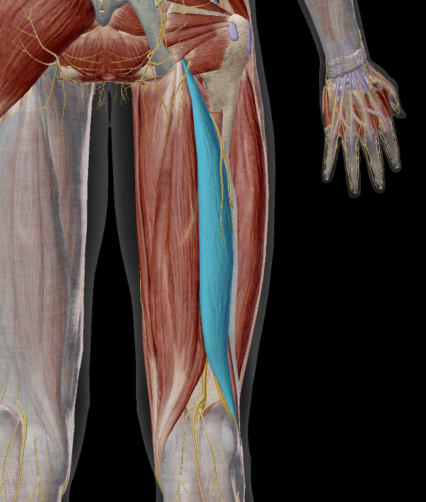
<p><span style="font-family: Calibri, sans-serif; line-height: 107%;"><strong>Flexion</strong> &amp; <strong>external (lateral) rotation </strong>of the knee; <strong>extension </strong>&amp; external rotation (assists) of the hip; tilts pelvis posteriorly</span></p><p><span style="font-family: Calibri, sans-serif; line-height: 107%;">**Hamstring</span></p>
75
New cards

Biceps femoris origin

Long head: ischial tuberosity

Short head: lateral lip of linea aspera

76
New cards

Biceps femoris insertion

Head of the fibula

77
New cards

Biceps femoris innervation

Long head: Sciatic (tibial branch), L5, S1 to S3

Short head: Sciatic (fibular branch), L5, S1 & S2

78
New cards

Popliteus action

Internal (medial) rotation of the knee (“unlocks” the knee); assists in knee flexion

<p><span style="font-family: Calibri, sans-serif; line-height: 107%;"><strong>Internal (medial) rotation</strong> of the knee (“unlocks” the knee); assists in knee flexion</span></p>
79
New cards

Popliteus origin

Lateral condyle of the femur

80
New cards

Popliteus insertion

Proximal, posterior aspect of tibia

81
New cards

Popliteus innervation

Tibial L4 & L5, S1

82
New cards

Quadriceps femoris group action

All: Extension of the knee

Rectus femoris: flexion of the hip

**Rectus femoris; vastus medialis, intermedius, & lateralis

<p><span style="line-height: 107%;"><u>All</u>: <strong>Extension </strong>of the knee</span></p><p><span style="font-family: Calibri, sans-serif; line-height: 107%;"><u>Rectus femoris</u>: flexion of the hip</span></p><p><span style="font-family: Calibri, sans-serif; line-height: 107%;">**Rectus femoris; vastus medialis, intermedius, &amp; lateralis</span></p>
83
New cards

Rectus femoris origin

Anterior inferior iliac spine (AIIS)

<p><span style="line-height: 107%;">Anterior inferior iliac spine (AIIS)</span></p>
84
New cards

Vastus medialis origin

Medial lip of linea aspera

<p><span style="line-height: 107%;">Medial lip of linea aspera</span></p>
85
New cards

Vastus intermedius origin

Anterior & lateral shaft of the femur

<p><span style="line-height: 107%;">Anterior &amp; lateral shaft of the <strong>femur</strong></span></p>
86
New cards

Vastus lateralis origin

Lateral lip of linea aspera, gluteal tuberosity, & greater trochanter

<p><span style="line-height: 107%;">Lateral lip of linea aspera, gluteal tuberosity, &amp; greater trochanter</span></p>
87
New cards

Quadriceps femoris group insertion

Tibial tuberosity (via the patella & patellar ligament)

88
New cards

Quadriceps femoris group innervation

Femoral L2 to L4

89
New cards

Tibialis anterior action

Inversion of the foot; dorsiflexion of the ankle

<p><span style="font-family: Calibri, sans-serif; line-height: 107%;"><strong>Inversion</strong> of the foot; <strong>dorsiflexion</strong> of the ankle</span></p>
90
New cards

Tibialis anterior origin

Lateral condyle of the tibia; proximal, lateral surface of the tibia; interosseous membrane

91
New cards

Tibialis anterior insertion

Medial cuneiform & base of the 1st metatarsal

92
New cards

Tibialis anterior innervation

Deep fibular L4 & L5, S1

93
New cards

Extensor hallucis longus action

Extension of the 1st toe, dorsiflexion of the ankle, inversion of the foot

<p><span style="font-family: Calibri, sans-serif; line-height: 107%;"><strong>Extension </strong>of the 1<sup>st</sup> toe, <strong>dorsiflexion</strong> of the ankle,<strong> inversion</strong> of the foot</span></p>
94
New cards

Extensor hallucis longus innervation

Deep fibular L4 & L5, S1

95
New cards

Extensor digitorum longus action

Extension of the 2nd through 5th toes, dorsiflexion of the ankle, eversion of the foot

96
New cards

Extensor digitorum longus innervation

Deep fibular L4 & L5, S1

97
New cards

Gastrocnemius action

Flexion of the knee, plantar flexion of the ankle

<p><span style="font-family: Calibri, sans-serif; line-height: 107%;"><strong>Flexion</strong> of the knee, <strong>plantar flexion</strong> of the ankle</span></p>
98
New cards

Gastrocnemius origin

Condyles of the femur, posterior surfaces

99
New cards

Gastrocnemius insertion

Calcaneus via calcaneal tendon

100
New cards

Gastrocnemius innervation

Tibial S1 & S2