Behavioral Neuro Midterm study guide
 Call Kai
 Learn
 Practice Test
 Spaced Repetition
 Match
 Flashcards
 Knowt Play
1/58

There's no tags or description
Looks like no tags are added yet.
59 Terms
Structural divisions: 2
1. Central nervous system (CNS)
2. Peripheral nervous system (PNS)
Central nervous system
Composed of the brain and spinal cord. Responsible for processing, integration, and initiating responses.
peripheral nervous system
includes cranial and spinal nerves
-subdivided into somatic and autonomic systems
somatic system
voluntary control of skeletal muscles
autonomic system
involuntary control of smooth muscle, glands, viscera
divisions: sympathetic and parasympathetic
sympathetic division of autonomic NS
prepares body for emergency situations by increasing heart rate, dilating pupils, etc... (fight, flight, fear)
Parasympathetic System
maintains homeostasis and conserves energy (rest and digest)
Spinal cord function
serves as link between brain and body, mediates reflexes and simple motor functions (not higher cog)
Frontal lobe
Primary motor cortex (Area 4), premotor areas (6-8) for planning and initiation, executive functions, personality, speech (Broca's).
parietal lobe
Primary somatosensory cortex (Areas 3, 1, 2), superior parietal lobule for spatial awareness and constructional tasks.
temporal lobe
Auditory processing (Wernicke's area, Area 22), memory (hippocampus), emotion (amygdala).
occipital lobe
Area 17 (primary visual), Areas 18-19 (visual recognition)
left hemisphere lateralization and specialization
Language, analytical reasoning, sequential processing
right hemisphere lateralization and specialization
Pragmatics, prosody, visual-spatial functions, environmental orientation
Hemispheres lateralization and specialization
Hemispheres are equipotential at birth; specialization emerges by ~2 years
Contralateral organization: each hemisphere controls the opposite side
Topographical representation: specific cortical areas map to specific body regions
Functional networking: specialized yet integrated systems
Sensory-afferent
neuron cell bodies are in dorsal root ganglia (pseudounipolar)
dorsal pathways convey information (touch, proprioception)
dorsal horn processes sensory input.
motor-efferent
Ventral horn contains motor neurons
dermatomes
correspond to skin areas supplied by specific spinal nerves
Neuron structure:
• Dendrites receive signals; axon hillock initiates action potentials; axon terminals release neurotransmitters.
• Myelin (Schwann cells in PNS; oligodendrocytes in CNS) enables saltatory conduction.
• Nodes of Ranvier facilitate action potential propagation.
Electrical properties of neuron
• Resting membrane potential depends on high K⁺ inside and high Na⁺ outside.
• Irritability: ability to respond to a stimulus and generate an impulse.
• Conductivity: ability to transmit impulses.
• Absolute refractory period (~8 ms): neuron cannot refire.
Synaptic transmission
• Exocytosis: vesicles release neurotransmitters into the synaptic cleft.
• Neurotransmitters diffuse and bind to postsynaptic receptors, altering ion channels.
• Postsynaptic binding generates excitatory or inhibitory effects.
• Examples: acetylcholine at neuromuscular junction, receptor issues in myasthenia gravis, demyelination in MS.
Neuroplasticity
brain's ability to reorganize after injury
• Repeated motor/swallowing tasks strengthen synaptic efficacy (Hebbian learning).
• Underlies rehab for dysarthria, dysphagia, aphasia.
Limbic system (3)
amygdala
hippocampus
cingulate gyrus
amygdala
fear and aggression
part of limbic system
hippocampus
memory consolidation
part of limbic system
cingulate gyrus, thalamus, fusiform gyrus
emotion and recognition
part of limbic system
Visual processing pathways (2)
Dorsal-where: spatial location
ventral-what: object and face recognition
tactile agnosia
failure to recognize objects by touch
hemineglect
nondominant inferior parietal lesion
prosopagnosia
face recognition deficits
foramen magnum
transition point from brainstem
(hole in the base of the skull through which the spinal cord passes)
Spinal cord sections (5)
1. cervical
2. thoracic
3. lumbar
4. sacral
5. coccygeal
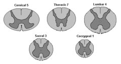
1. cervical section
slender gray matter and largest amount of white (all ascending/descending fibers pass through)
· Neck area- 12 SN
· Dorsal lemniscal column (gracilis and cuneatus)
· Corticospinal
· Lateral column- spinothalamic and spinocerebellar
2. thoracic
enlarged white matter (more processing
· Torso, chest cavity, lung- 12 SN
· Fasciculus gracilis: discriminative touch from lower body
· Fasciculus cuneatus: discriminative touch from upper body
· Lateral corticospinal tract, Spinothalamic tract, spinocerebellar
3. lumbar
larger dorsal and ventral horn (serves leg movement)
· 5 SN
· Dorsal lemniscal- gracilis- fine touch, proprioception, vibration
· Lateral corticospinal tract- volunt. Info to limbs
· Anterolateral system: spinothalamic and spinocerebellar tracts
conus medullaris
tapered end of spinal cord
cauda equina (horse's tail)
roots of nerves that extend from the end of the spinal cord
filum terminale
thin thread of fibrous tissue at end of conus medullaris, attaches to coccygeal ligament to stabilize
4. sacral
large proportion of gray matter (receive sensory)
· 5 SN
· Fasciculus gracilis- sensation from sacral to midthoracic level
· Lateral corticospinal tract- motor fibers
· Spinothalamic tract- pain, temperature (sensory)
5. coccygeal
1 SN
contralateral vs ipsilateral control
contralateral: opposite side
ipsilateral: same side
broadmann area: 4
primary motor cortex (precentral gyrus)
broadmann area: 44 + 45
broca's area association
associated w/ motor 4 (activate tongue, lips, expressive speech movement)
broadmann area 3,1,2
primary sensory cortex
broadmann area 41 +42
primary auditory cortex
broadmann area 22
wernicke's (superior temporal convolution)
associated w/ auditory 41+ 42 (receptive speech)
Broadmann area 17
primary visual cortex (occipital)
broadmann area 18, 19
occipital lobe
broadmann area 39, 40
39- angular gyrus
parietal lobe: reading, naming, writing
pseudounipolar neurons
a type of sensory neuron that has a single short process extending from its cell body (soma) which quickly splits into a T-shaped junction with two distinct axonal branches
bipolar neuron
a nerve cell that has a single dendrite at one end and a single axon at the other end
multipolar neuron
a neuron with one axon and many dendrites attached to its soma
saltatory conduction
signals travel quickly under myelin between nodes; speed depends on axon diameter and myelination
absolute refractory period
period of unresponsiveness; short but consistent- Na channels reset
exocytosis
release of substances out a cell by the fusion of a vesicle with the membrane.
postsynaptic receptors
The other side of the synapse. The neuron that contains receptors (usually on dendrites) ready to bind to the neurotransmitter released from the presynaptic neuron.
neuroplasticity
the brain's ability to change, especially during childhood, by reorganizing after damage or by building new pathways based on experience
functional networking
networks of neurons have special functions
topographical organization
location of peripheral sensory neuron maps directly onto specific cells in the cortex