OT 881 Anatomy Module 3: Distal UE
 Call Kai
 Learn
 Practice Test
 Spaced Repetition
 Match
 Flashcards
 Knowt Play
1/46

There's no tags or description
Looks like no tags are added yet.
47 Terms
What are the articulations of the humerus and radius?
Capitulum of the humerus articulates with the head of the radius
What are the articulations of the humerus and ulna during flexion/extension of the elbow?
During flexion of the elbow, the coronoid process of the ulna fits into the coronoid fossa of the humerus. During elbow extension, the olecranon process of the ulna articulates with the olecranon fossa of the humerus.

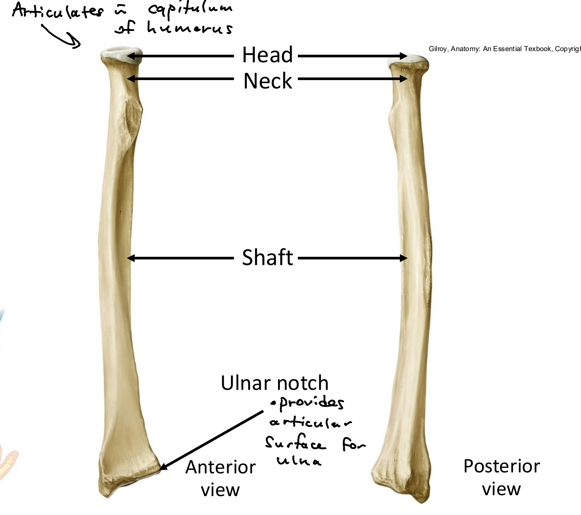
What are the anatomical structures of the radius?
Head, neck, shaft, ulnar notch
What are the anatomical structures of the ulna, and what does it look like?
Olecranon process, coronoid process, radial notch, trochlear notch, shaft, head. It looks like a wrench!

Which radioulnar joints are both synovial joints, and are responsible for pronation and supination of the forearm?
Proximal and distal radioulnar joints.
What are the 3 radioulnar joints, and what type of joints are they, and what are they stabilized by?
Proximal radioulnar joint - head of radius and radial notch of ulna. Synovial pivot joint stabilized by annular ligament.
Middle radioulnar joint (interosseus membrane) - between shafts of radius and ulna. Solid syndesmosis joint.
Distal radioulnar joint - head of ulna and ulnar notch of radius. Synovial pivot joint stabilized by ventral and dorsal radioulnar ligaments.

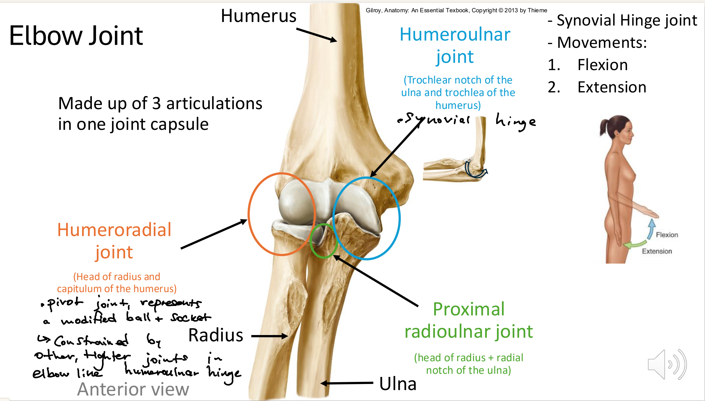
What are the 3 articulations of the elbow joint capsule?
Humeroradial joint - head of radius and capitulum of the humerus. Pivot joint (modified ball & socket)
Humeroulnar joint - trochlear notch of ulna and trochlea of the humerus. Synovial hinge joint (flexion & extension)
Proximal radioulnar joint - head of radius and radial notch of ulna. Synovial pivot joint
How many phalanges, metacarpals, and carpals in the hand?
14 phalanges, 5 metacarpals, 8 carpals
What are the names of the 8 carpal bones?
Scaphoid
Lunate
Triquetrium
Pisiform
Hamate
Capitate
Trapezium
Trapezoid
“So Long To Pinky, Here Comes The Thumb”

Which carpal bone is most commonly broken in wrist injuries?
Scaphoid fracture
What are the two types of interphalangeal joints, and what kind of joints are they?
Distal and proximal IPs; all synovial hinge joints
What are the ligaments supporting the MCPs called?
Collateral ligaments
What kind of joints are the metacarpophalangeal (MCP) joints, and what kind of movement are they responsible for?
Synovial condyloid. Rotational movements and flexion/extension.
What is the connection point of the carpo-metacarpal (CMC) joint I, and what kind of joint is it?
THUMB! Distal carpal (trapezium) and 1st metacarpal; synovial saddle joint (allows for greater thumb movement)
What kind of joints are the carpo-metacarpal (CMC) II-V, and what movement is permitted?
Gliding (plane) joints, limited gliding & slight flexion/extension.
What is an example of an impairment of the CMCs?
CMC osteoarthritis, causes difficulty using scissors, writing, performing pinch movement.
What kind of joints are the intercarpal and midcarpal joints, and what kind of motion occurs?
Synovial, plane/gliding joints; gliding motion that contributes to wrist movement.
What kind of motion is possible at the wrist joint?
Flexion/extension, radial/ulnar deviation (abduction/adduction), supination/pronation
What movement occurs at each joint in the hand (PIP/DIPs, MCPs, CMCs)

What are the 3 arches of the hand called, and what is the importance of them?
Longitudinal arch, proximal transverse arch, distal transverse arch. Important for precise grips for things like using cutlery and scissors. 3 arches work together to balance, stabilize and mobilize the hand.

What separates the forearm into its anterior and posterior compartments?
Interosseus membrane
How many compartments in the forearm?
3 - anterior compartment, posterior compartment, lateral compartment
What are the 4 muscles in the superior layer of the anterior (flexor) compartment of the forearm, and what movements are they responsible for? Hint: p/f/p/f
Pronator Teres - pronation
Flexor carpi radialus - flexion of wrist, abduction of wrist
Palmaris longus - vestigial, weak wrist flexor
Flexor carpi ulnaris - flexion of wrist, adduction of wrist
What is the common origin of all flexor muscles in the superior compartment of the anterior forearm?
Medial epicondyle of humerus
What muscle is found in the intermediate layer of the anterior compartment of the forearm, and what movement does it do?
Flexor digitorum superficialis - wrist flexion, digits at PIPs
What are the 3 muscles found in the deep layer of the anterior compartment of the forearm? Hint: 3 P’s
Flexor digitorum profundus - flexion of wrist & fingers
Pronator quadratus - pronation
Flexor pollicis longus - flexion of wrist & thumb
Which muscle flexes the PIP, and which flexes the DIP?
PIP - Flexor digitorum superficialis
DIP - Flexor digitorum profundus
Remember: superficialis split to let profundus pass
How many wrist flexors, wrist and finger flexors, and pronators in the anterior comp. of forearm?
3 wrist flexors - flexor carpi radialus, pulmaris longus, flexor carpi ulnaris
3 both wrist and finger flexors - flexor digitorum superficialis, flexor digitorum profundus, flexor pollicis longus
2 pronators - pronator teres, pronator quadratus
What are the 3 muscles in the superficial layer of the posterior (extensor) compartment of the forearm, and what are their movements? Hint: Double Dip Cherries (DDC)
Extensor digitorum - extension of wrist and fingers
Extensor digitorum minimi - extension of wrist and pinky
Extensor carpi ulnaris - extension of wrist, ulnar deviation
Common extensor origin: lateral epicondyle of the humerus
What are the 5 muscles in the deep layer of the posterior (extensor) compartment of the forearm, and what are their movements? Hint: Syrup And Peanuts Preferably Included
Supinator - supination (only one!)
Abductor pollicis longus - abduction of wrist and thumb
Extensor pollicis brevis - extension of wrist and thumb, abduction of thumb
Extensor pollicis longus - extension of wrist and thumb
Extensor indicis - extension of wrist an index finger
In the deep layer of the posterior compartment of the forearm, which muscle is sandwiched by which other 2 muscles?
Brevis sandwhich - Extensor pollicis brevis is sandwiched by the abductor pollicis longus and the extensor pollicis longus

Which 3 muscles are found in the lateral compartment of the forearm, and what do they do? Hint: Big Chocolate Chips
Brachioradialis - Flexion of forearm at elbow joint. Inserts at radius, does not cross wrist.
Extensor carpi radialis longus - extension and abduction of wrist
Extensor carpi radialis brevis - same as longus
What are the 4 thenar muscles (anterior muscles that move the thumb) and what do they do?
Adductor pollicis - adduction of thumb
Flexor pollicis brevis - flexion of thumb
Abductor pollicis brevis - abduction of thumb
Opponens pollicis - flexion of thumb for opposition
What are the 3 posterior muscles that move the thumb, and what do they do?
Abductor pollicis longus - abduction of thumb, assists w extension
Extensor pollicis longus - extension of thumb
Extensor pollicis brevis - extension of thumb
In the intrinsic hand muscles, what are the order of muscles from most superficial to deep?
Lumbricals - most superficial
Palmar interossei - 2nd most superfical
Dorsal interossei - deepest
How many interossei muscles are there, and what are the functions (palmar and dorsal)? Hint: PAD and DAB
The 7 interossei muscles pull fingers together and apart, full space between hand bones
Palmar interossei - 3 muscles, Adduct (PAD)
Dorsal interossei - 4 muscles, Abduct (DAB)
How many lumbricals are there in the hand, and what are the lumbrical functions?
4 slender wormlike muscles:
precision movement
flexion of MCP joint
extension of PIP and DIP joints (combined flexion helps with precision grip, prevents fingers from clawing)
originate from tendons of flexor digitorum profundus
Which nerve innervates the posterior forearm, and what are the functions of its two splits?
Radial nerve:
Superficial redial nerve for sensory innervation
Deep radial nerve for motor innervation of posterior forearm and hand

Which two nerves innervate the muscles of the anterior compartment of the forearm and hand?
Median nerve:
Most forearm muscles innervated by median nerve
Median nerve also innervates muscles of thumb and digits 1 + 2
Ulnar nerve:
Innervates 3 anterior flexors on pinky side
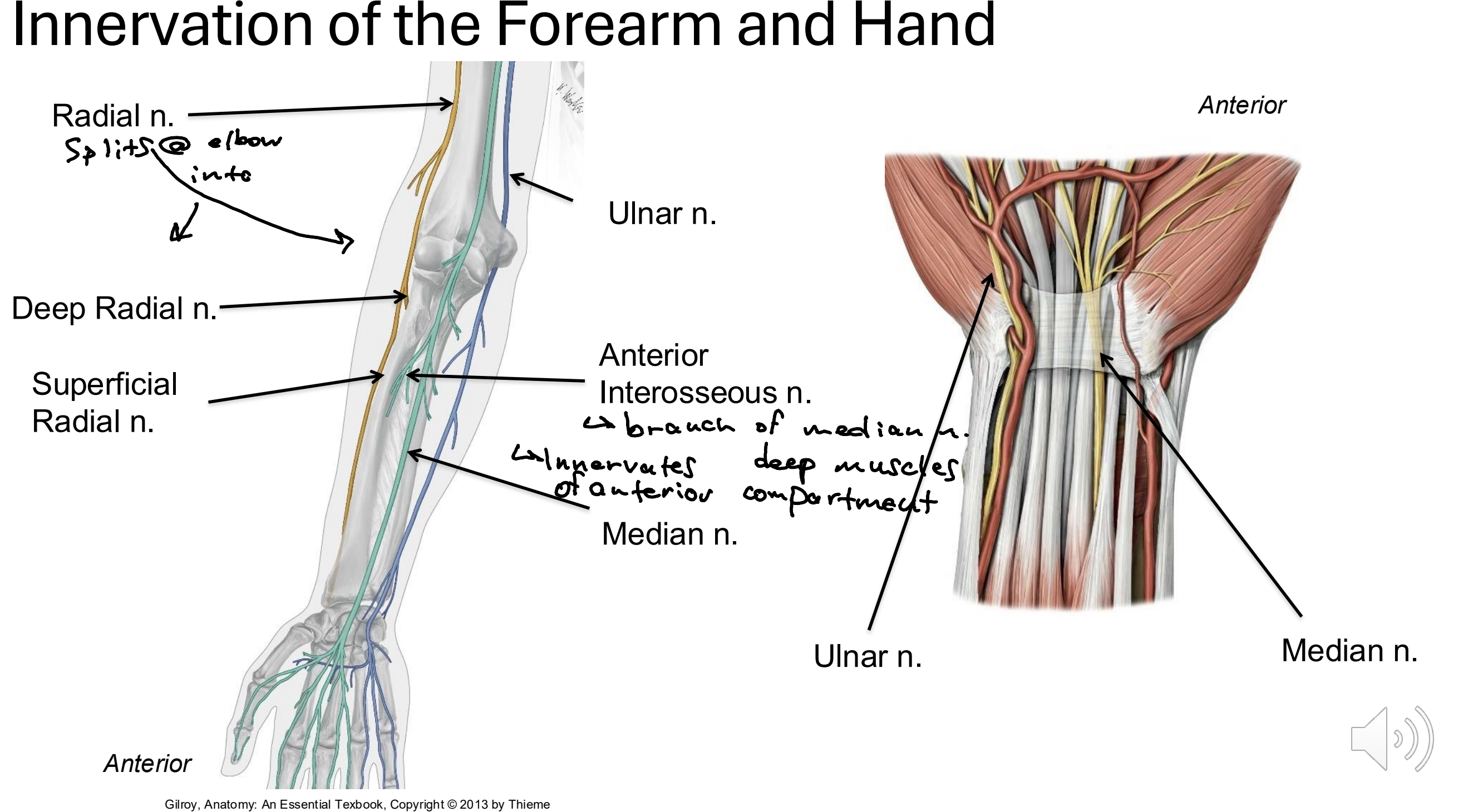
What is the anterior interosseus nerve?
A branch of the median nerve that innervates the deep muscles of the anterior compartment of the forearm

Which nerves are responsible for the cutaneous innervation of the hand (sensation)?

Which nerve and which muscles run through the carpel tunnel?
Median nerve
Flexor pollicis longus
Flexor digitorum superficialis and profundus
What ligament forms the anterior roof of the carpel tunnel? Hint: retina
Flexor retinaculum

What is carpel tunnel syndrome, and what are the symptoms?
Swelling in the confined space of the carpel tunnel, which puts pressure on median nerve.
Symptom include numbness, tingling, pain, weakness, difficulty gripping
Commonly affected digits: Thumb, index finger, half of ring finger
