(L20) IMED2004 - Stem Cells I
 Call Kai
 Learn
Learn Practice Test
 Spaced Repetition
 Match
Match Flashcards
 Knowt Play
1/66

There's no tags or description
Looks like no tags are added yet.
Name | Mastery | Learn | Test | Matching | Spaced | Call with Kai | Chat |
|---|
No analytics yet
Send a link to your students to track their progress
67 Terms
What reference materials were recommended for Stem Cells I?
- Developmental Biology 12E: Chapter 5, Stem Cells
- Online video "Tutorial" by Michael Barresi, an author of the textbook
- The tutorial link is in the lecture folder
.
Lecturer explanation:
The lecturer described these as optional supporting resources for this introductory stem-cell material.
What topics are covered across the three Stem Cells lectures?
Stem Cells I:
- Stem cells — definition
- Potency and lineage restriction
- Maintaining homeostasis — stem cells, proliferation, repair and cancer
.
Stem Cells II:
- Types, origins and niches
- Signalling, expansion and self-renewal
- Adult stem cell examples
.
Stem Cells III:
- Use of stem cells — research and clinical applications
- Cloning
- Approved therapies: skin grafts and haematopoietic stem cell transplants
- Therapies in development: regeneration and replacement
What are the eight learning outcomes for Stem Cells I?
1. Understand differentiation and lineage restriction as they apply to development.
2. Define the key properties of stem cells.
3. Define and distinguish different states of stem-cell potency and give examples from each lineage class.
4. Be familiar with developmental cues that may push cells toward a stem or differentiated state.
5. Understand the role of stem cells in maintenance of homeostasis.
6. Outline terminal differentiation and discuss the problems it causes for cell replacement.
7. Explain how different tissues use different strategies for stem-cell utilisation in repair.
8. Discuss biological problems with stem-cell maintenance throughout life.
Lecturer emphasis:
These were presented as the take-home messages for the lecture and correspond to the numbered cues on later slides.

What numerical facts illustrate the scale and specialisation of the human body?
Estimate shown:
- 37.2 trillion human cells
- Based on a 30-year-old male, 70 kg, 1.72 m, 1.85 m² surface area
- Plus approximately 100 trillion bacterial cells
- More than 200 different specialised human cell types
- All human body cells ultimately started from one cell
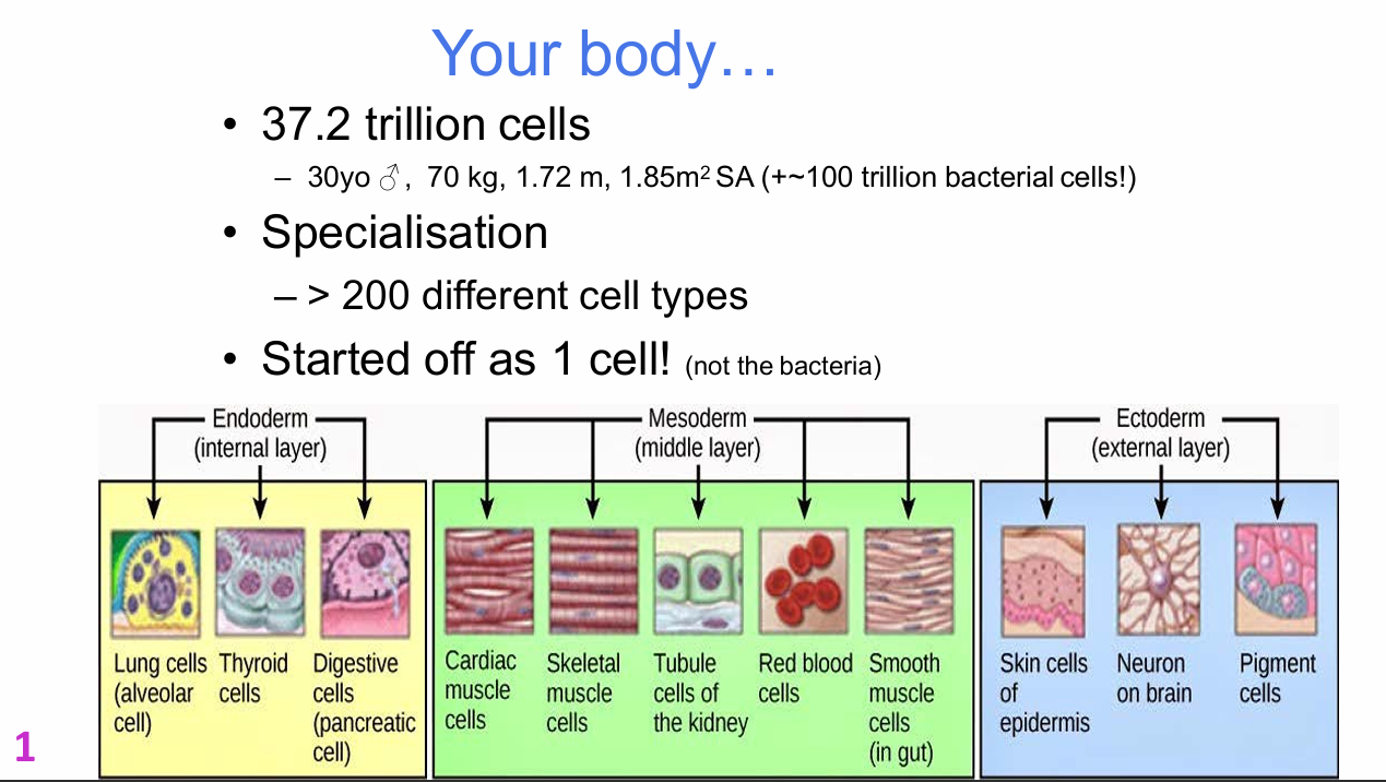

What germ-layer examples are shown for specialised cell types in the body?
Endoderm:
- Lung cells (alveolar cell)
- Thyroid cells
- Digestive cells (pancreatic cell)
.
Mesoderm:
- Cardiac muscle cells
- Skeletal muscle cells
- Kidney tubule cells
- Red blood cells
- Smooth muscle cells in gut
.
Ectoderm:
- Skin cells of epidermis
- Neurons in brain
- Pigment cells


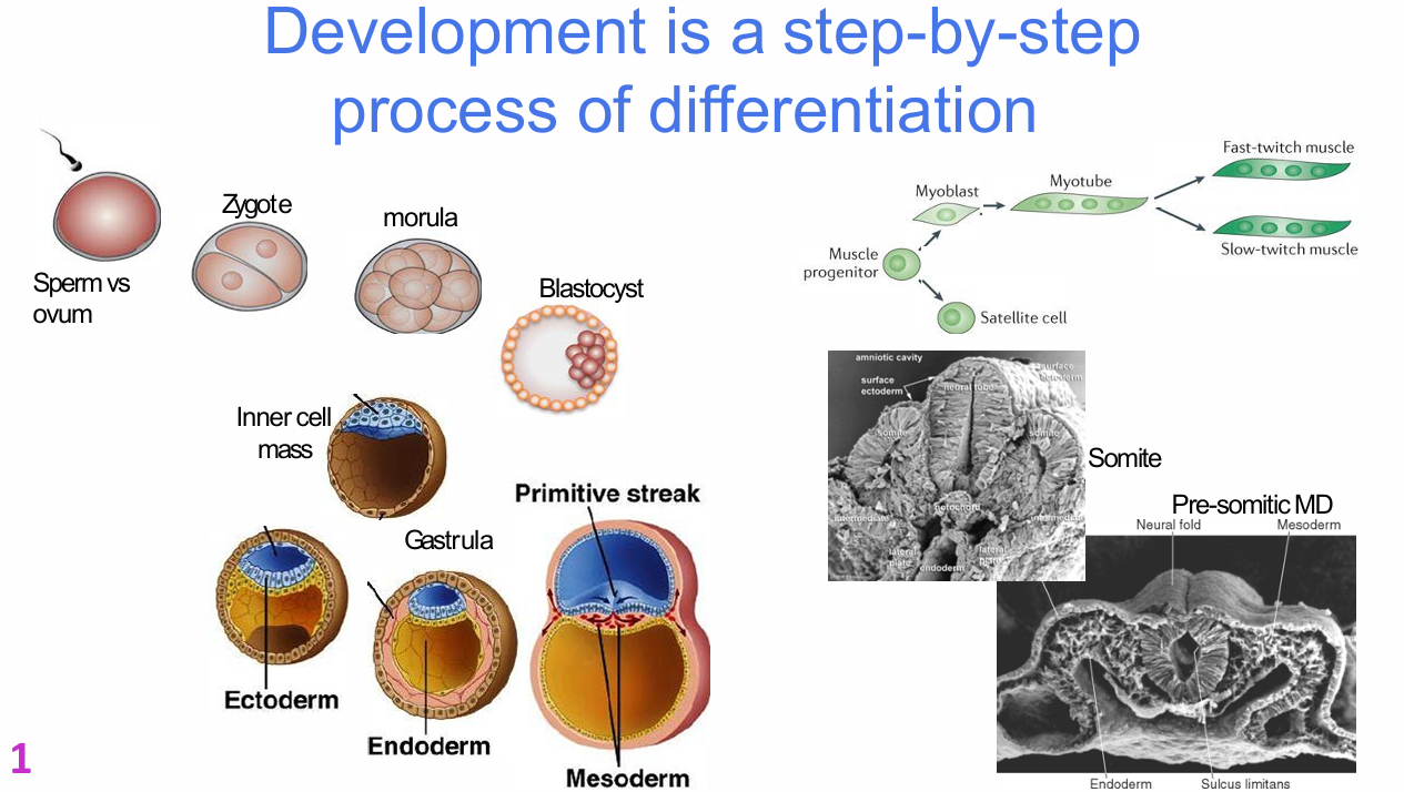
Why is development described as a step-by-step process of differentiation?
At each developmental stage, cells change:
- Gene expression
- Protein content
- Phenotype
- Developmental potential
.
The sequence shown progresses from:
sperm + ovum → zygote → morula → blastocyst → inner cell mass → gastrula → germ layers → increasingly specialised tissues.
Lecturer explanation:
The lecturer stressed that cells at later stages are not the same as their predecessors; each step represents a changed differentiation state.

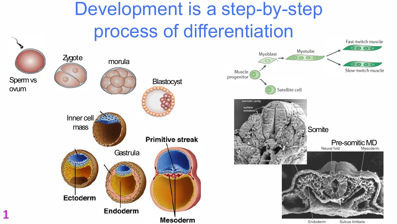
What developmental and muscle-lineage sequence is shown on the differentiation diagram?
Embryonic sequence:
- Sperm vs ovum
- Zygote
- Morula
- Blastocyst
- Inner cell mass
- Gastrula
- Ectoderm
- Endoderm
- Primitive streak
- Mesoderm
- Pre-somitic mesoderm
- Somite
.
Muscle lineage:
- Muscle progenitor
- Satellite cell
- Myoblast
- Myotube
- Fast-twitch muscle
- Slow-twitch muscle
What is differentiation?
Differentiation involves:
- Changes in gene expression
- Changes in phenotype
- Specification of a cell in a particular direction
- Lineage restriction
.
Differentiation is normally unidirectional.
Once a cell differentiates, it normally cannot return to a previous phenotype.

What is lineage restriction?
Lineage restriction is the progressive loss of alternative developmental possibilities as a cell differentiates toward a specific fate.
.
Lecturer explanation:
As a cell becomes specified for a particular lineage, it becomes less able to become something else.

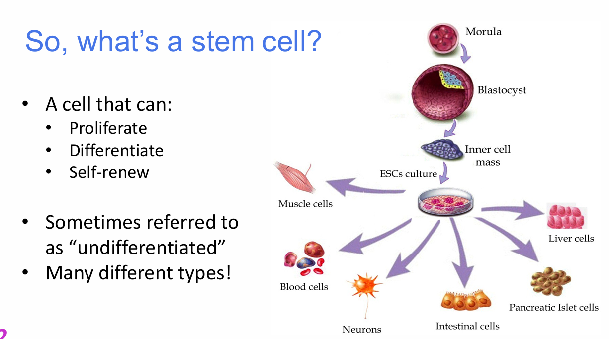
What three properties define a stem cell?
A stem cell must be able to:
1. Proliferate
2. Differentiate
3. Self-renew
.
Stem cells are sometimes referred to as "undifferentiated".
There are many different types of stem cells.


Why is self-renewal essential to the definition of a stem cell?
Self-renewal allows a stem cell to generate another stem cell and thereby maintain the stem-cell population.
Lecturer emphasis:
A cell is not considered a stem cell on differentiation capacity alone; it must also be able to self-renew.


What does unipotent mean?
Unipotent means a cell can generate only one cell type.
Slide wording:
- Cannot produce cells of any other type
.
Example shown:
- Eggs and sperm are presented as restricted examples in the potency spectrum.


What does totipotent mean?
Totipotent cells can produce cells of any type required to form a new individual, including extraembryonic tissues.
.
Examples shown:
- Very early embryo
- Zygote / morula-stage cells
.
Lecturer explanation:
Totipotency includes both embryonic tissues and extraembryonic tissues such as those required to support development.


What does pluripotent mean?
Pluripotent cells can produce all somatic cell types but not extraembryonic tissues (thats what the diagram shows, it cant make extraembryonic)
.
Classic example:
- Inner cell mass of the blastocyst
.
Lecturer explanation:
Pluripotent cells can generate derivatives of all three germ layers when given the appropriate differentiation signals.


What are the major ectoderm-derived tissues listed in the lecture?
Ectoderm:
- Central nervous system
- Retina and lens
- Cranial and sensory ganglia and nerves
- Pigment cells
- Head connective tissue
- Epidermis
- Hair
- Mammary glands

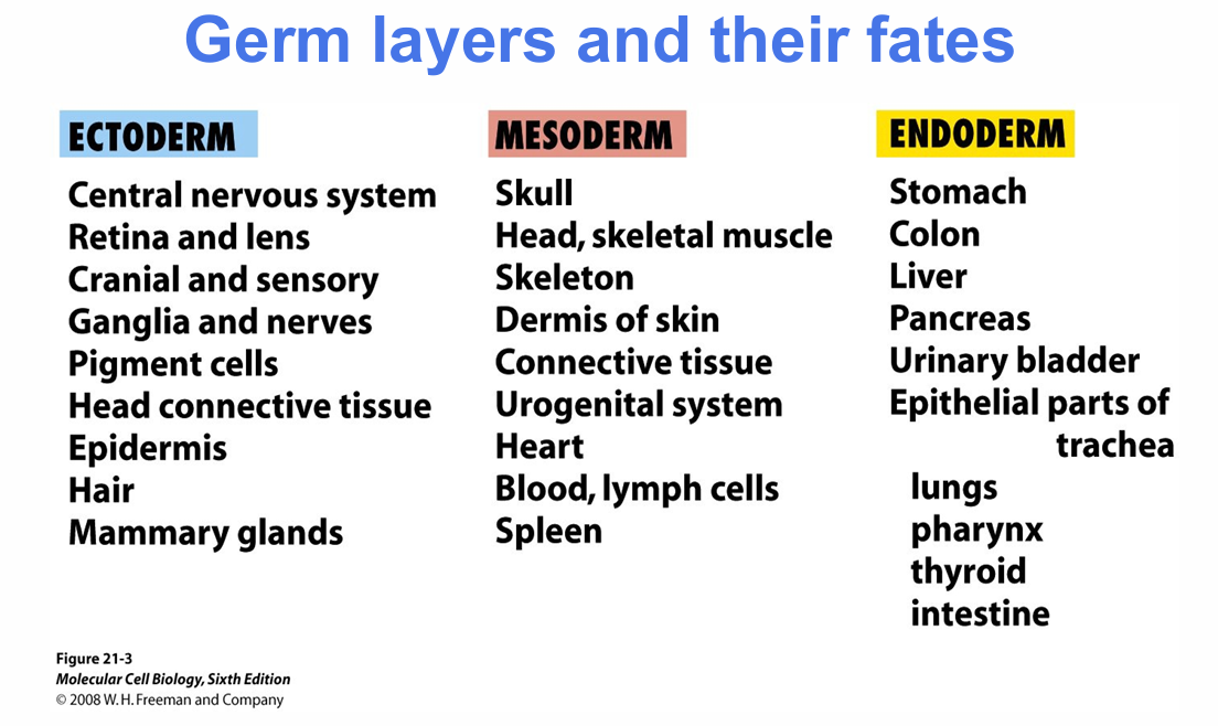
What are the major mesoderm-derived tissues listed in the lecture?
Mesoderm:
- Skull
- Head and skeletal muscle
- Skeleton
- Dermis of skin
- Connective tissue
- Urogenital system
- Heart
- Blood and lymph cells
- Spleen


What are the major endoderm-derived tissues listed in the lecture?
Endoderm:
- Stomach
- Colon
- Liver
- Pancreas
- Urinary bladder
- Epithelial parts of trachea
- Lungs
- Pharynx
- Thyroid
- Intestine


What should you identify in the germ-layers-and-fates diagram?
- Ectoderm and its characteristic derivatives
- Mesoderm and its characteristic derivatives
- Endoderm and its characteristic derivatives
- The principle that each germ layer gives rise to a restricted set of tissues

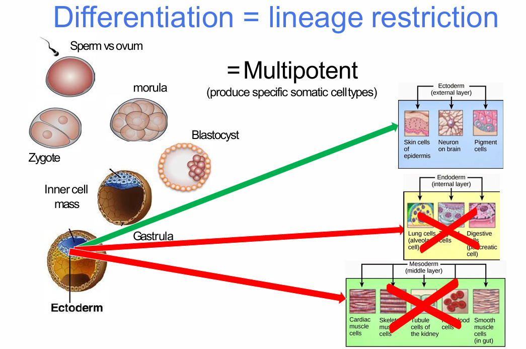
What does multipotent mean?
Multipotent cells can produce multiple specific somatic cell types, but only within a restricted set of lineages.
They cannot generate every somatic cell type.

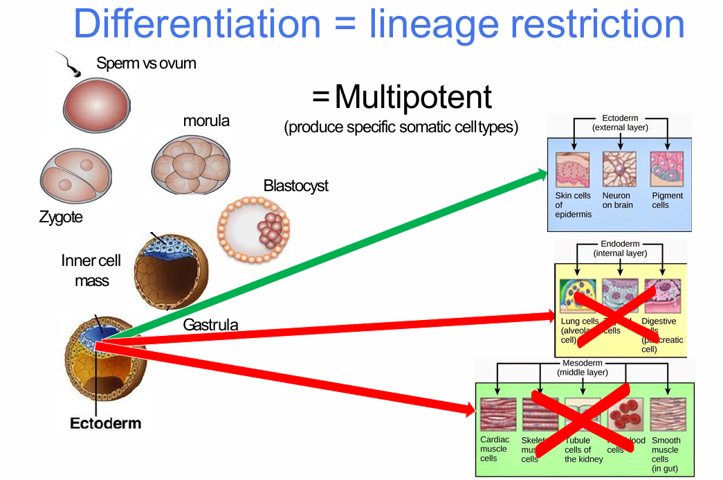
How does ectoderm illustrate multipotency?
Multipotent ectodermal cells can generate several ectoderm-derived fates, such as:
- Epidermal cells
- Neurons
- Pigment cells
They do not generate mesodermal or endodermal lineages.

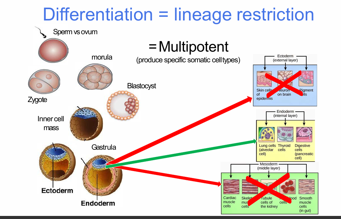
How does endoderm illustrate multipotency?
Multipotent endodermal cells can generate several endoderm-derived fates, including:
- Lung-derived cells
- Thyroid cells
- Digestive/pancreatic cells
They do not generate ectodermal or mesodermal lineages.


How does mesoderm illustrate multipotency?
Multipotent mesodermal cells can generate several mesoderm-derived fates, including:
- Cardiac muscle
- Skeletal muscle
- Kidney tubule cells
- Red blood cells
- Smooth muscle
They do not generate ectodermal or endodermal lineages.


What does bipotent mean?
Bipotent means a cell can generate two cell types.
The muscle lineage is used as an example of progressive restriction (muscle can make myoblast and satellite cell)


How does the muscle lineage illustrate progressive potency restriction?
Sequence shown:
- Muscle progenitor
- Satellite cell
- Myoblast
- Myotube
- Fast-twitch muscle
- Slow-twitch muscle
The pathway illustrates narrowing potency toward a single tissue lineage.


How are totipotent, pluripotent, multipotent and unipotent stem cells classified?
Totipotent:
- Each cell can develop into a new individual
- Example: cells from early embryos, approximately 1-3 days
.
Pluripotent:
- Can form any of >200 somatic cell types, but not extraembryonic tissues
- Example: inner mass cells of blastocyst, approximately 5-14 days
.
Multipotent:
- Can replace a number of other tissues
- Examples: foetal tissue, cord blood, adult stem cells
.
Unipotent:
- Generates one specific cell type only
- Example: some tissue-specific stem cells

What determines a stem cell's potency or plasticity?
The diversity of cell types that a stem cell can generate defines its potency.
.
Potency is also referred to as:
- Plasticity
Greater range of possible cell fates = greater potency/plasticity.
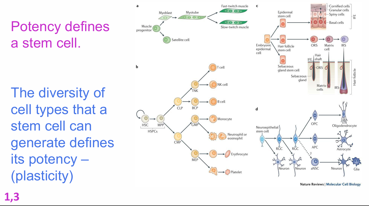
How do the example lineages on the potency slide differ in plasticity?
Examples shown include:
- Muscle lineage: relatively restricted
- Skin/epidermal lineages: several tissue-specific stem-cell branches
- Haematopoietic lineage: broad multipotent differentiation potential
- Neural lineage: generates multiple neural cell types

What three major factors can push a cell down one developmental pathway rather than another?
1. Signals
- Cells are exposed to different diffusible signals
- Examples: growth factors, cytokines
.
2. Transducers
- Transcription factors change which genes are switched on or off
.
3. Asymmetric cell division
- Cell contents are distributed asymmetrically between daughter cells during division
How do extracellular signals and transcription factors cooperate to influence cell fate?
Extracellular protein signals such as growth factors and cytokines activate intracellular pathways.
These pathways influence transcription factors, which alter gene expression.
Together, signalling environment and transcriptional state push cells toward particular differentiation pathways.
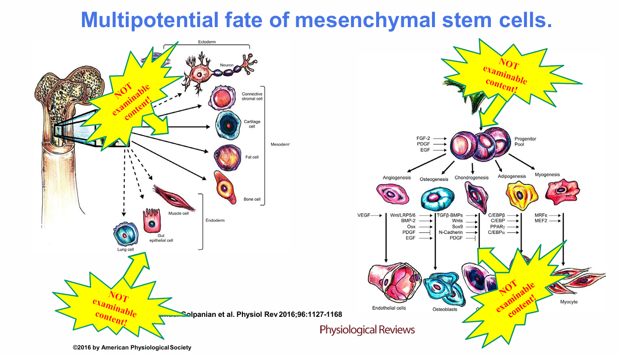
What does the mesenchymal stem-cell example illustrate about cell fate? (NOT ASSESSABLE)
Mesenchymal stem cells are presented as multipotent cells whose eventual lineage depends on the signalling context.
The figure shows that different combinations of growth factors and differentiation cues can direct cells into multiple fates.
Lecturer explanation:
The lecturer explicitly said the detailed signalling factors and pathways on this slide were for interest only and were not required for assessment.


What is asymmetric cell division?
Asymmetric cell division produces daughter cells with different cellular contents and/or developmental fates.
A common stem-cell outcome:
- One daughter retains stem-cell identity
- The other becomes a progenitor or committed cell


How do symmetric and asymmetric divisions contribute to stem-cell maintenance?
Symmetric stem-cell division:
- Can generate two equivalent stem cells
.
Asymmetric division:
- Generates one stem cell and one progenitor/committed cell
.
Symmetric progenitor division:
- Expands progenitor numbers
.
Terminal differentiation:
- Produces mature differentiated cells


What labels should you identify in the asymmetric-cell-division diagram?
- Symmetric cell division
- Asymmetric division
- Terminal differentiation
- Activated stem cell
- Dormant stem cell
- A = stem cell
- B = progenitor cell
- C = differentiated cell

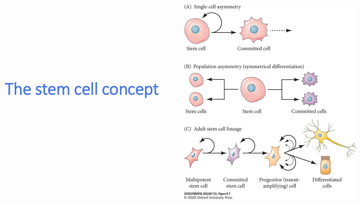
What is single-cell asymmetry in the stem-cell concept?
A stem cell divides so that:
- One daughter retains stem-cell identity
- The other becomes a committed cell
This preserves the stem-cell pool while generating differentiated descendants.


What is population asymmetry in the stem-cell concept?
A stem-cell population is maintained at the population level because:
- Some stem cells divide symmetrically to make more stem cells
- Others divide symmetrically to produce committed cells
The overall balance preserves the stem-cell population.
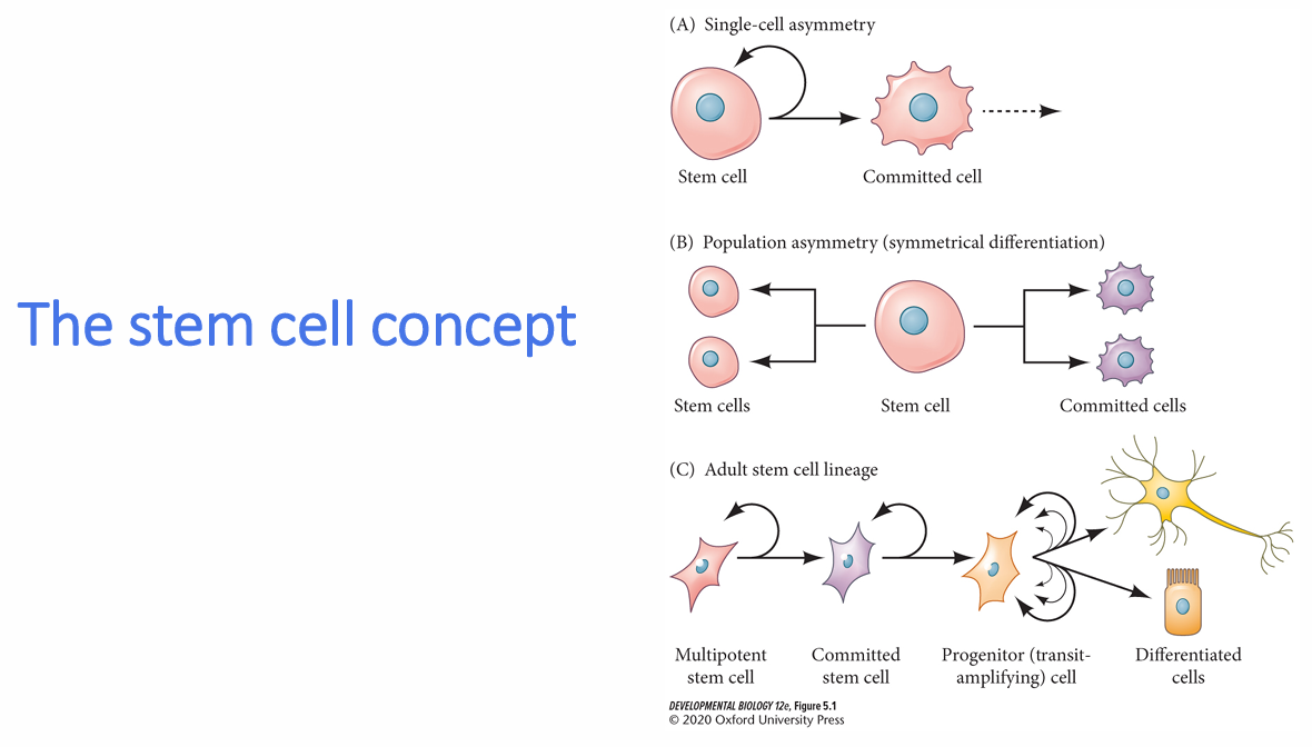
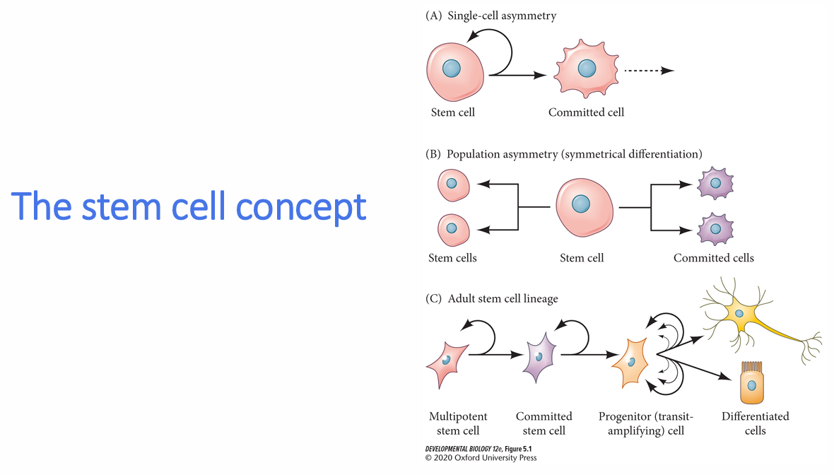
What is the adult stem-cell lineage model shown in the lecture?
Sequence:
1. Multipotent stem cell
2. Committed stem cell
3. Progenitor / transit-amplifying cell
4. Differentiated cells
The lineage includes self-renewal at earlier stages and rapid expansion at the progenitor stage.
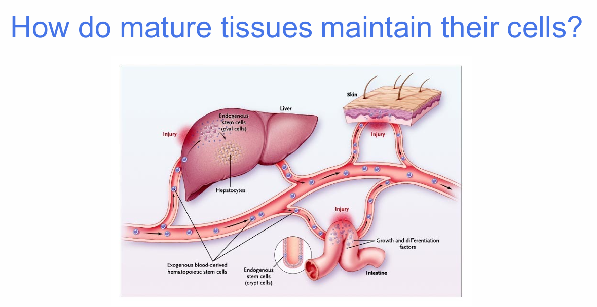
What determines how mature tissues maintain their cells?
Tissues differ in:
- Cell turnover rate
- Capacity of mature cells to re-enter the cell cycle
- Dependence on tissue-specific stem cells
- Degree of injury-induced regeneration
.
Examples considered:
- Liver
- Skin
- Intestine

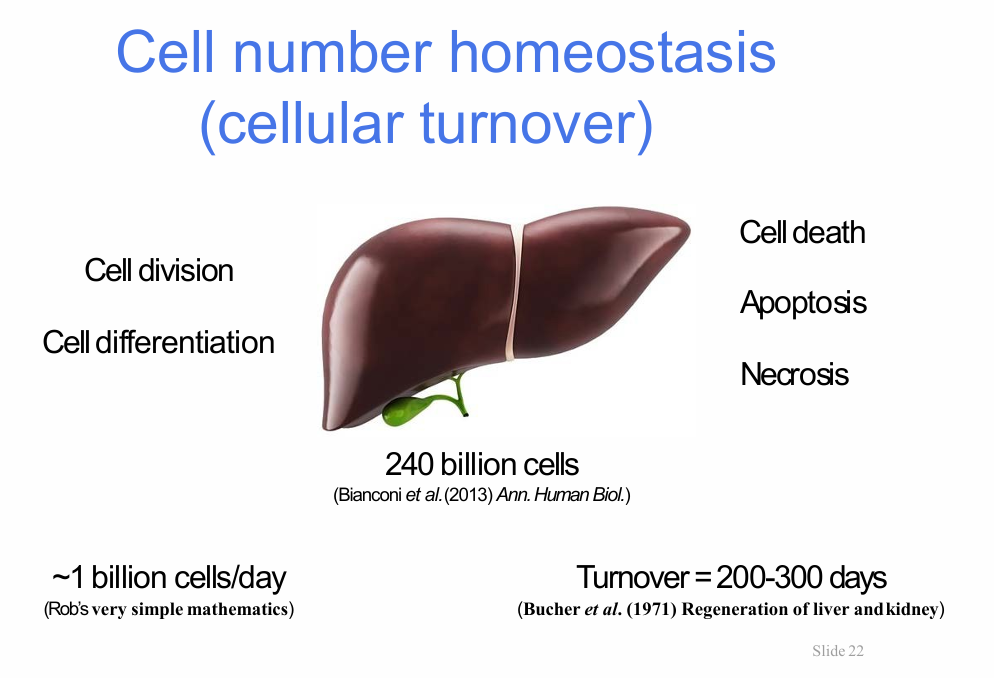
What is cell-number homeostasis?
Cell-number homeostasis is the balance between:
- Cell division
- Cell differentiation
- Cell death by apoptosis
- Cell death by necrosis
This balance maintains a relatively stable number of cells within a tissue.


What numerical example of liver cell turnover was given?
Liver example:
- Approximately 240 billion cells
- Turnover approximately 200-300 days
- Roughly 1 billion cells per day replaced
Lecturer explanation:
The lecturer used the liver to illustrate how continuous gain and loss of cells can still maintain a stable organ cell number.

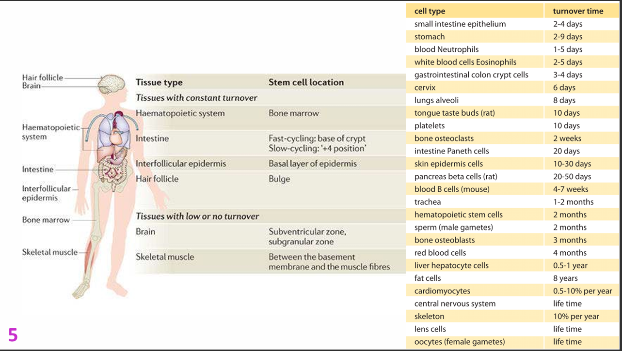
Where are stem cells located in tissues with constant turnover?
Haematopoietic system:
- Bone marrow (fastest regenerating organ)
.
Intestine:
- Fast-cycling base of crypt
- Slow-cycling "+4 position"
.
Interfollicular epidermis:
- Basal layer of epidermis
.
Hair follicle:
- Bulge

Where are stem cells located in tissues with low or no turnover?
Brain:
- Subventricular zone
- Subgranular zone
.
Skeletal muscle:
- Between basement membrane and muscle fibres


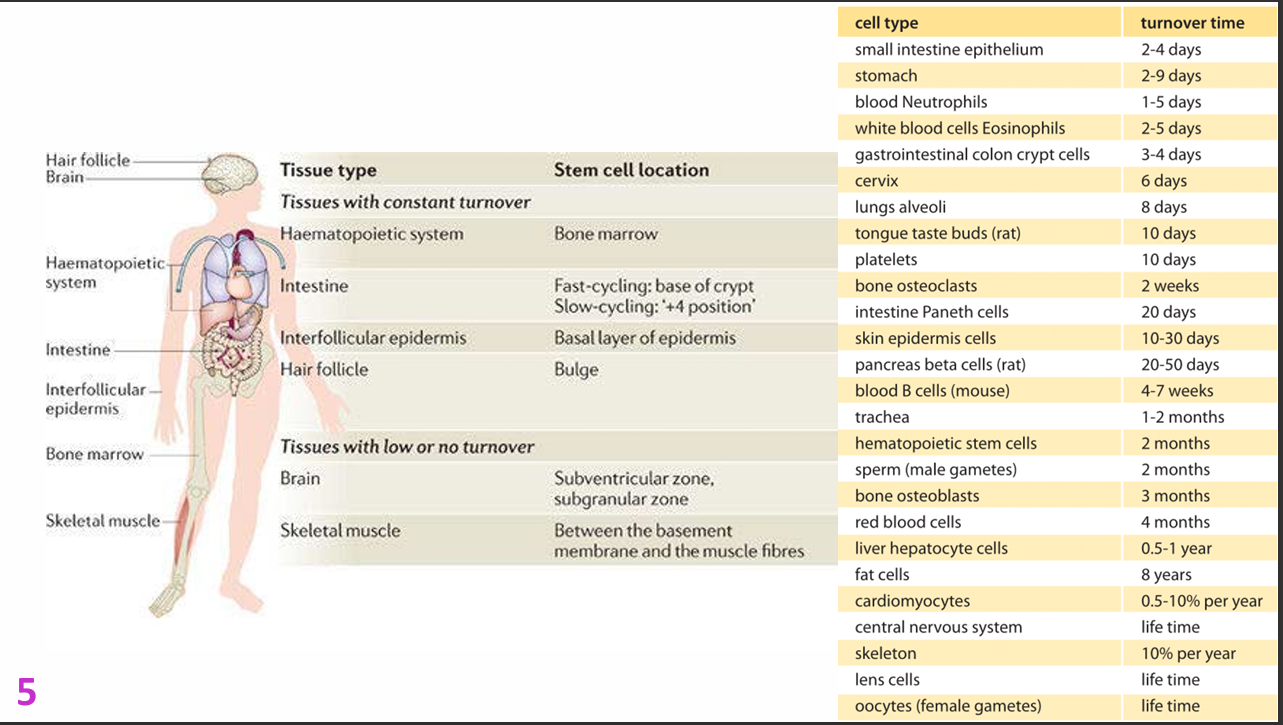
Which cell types have very short turnover times?
Examples:
- Small intestine epithelium: 2-4 days
- Stomach: 2-9 days
- Blood neutrophils: 1-5 days
- White blood cells/eosinophils: 2-5 days
- Gastrointestinal colon crypt cells: 3-4 days
- Cervix: 6 days
- Lung alveoli: 8 days
- Tongue taste buds (rat): 10 days
- Platelets: 10 days

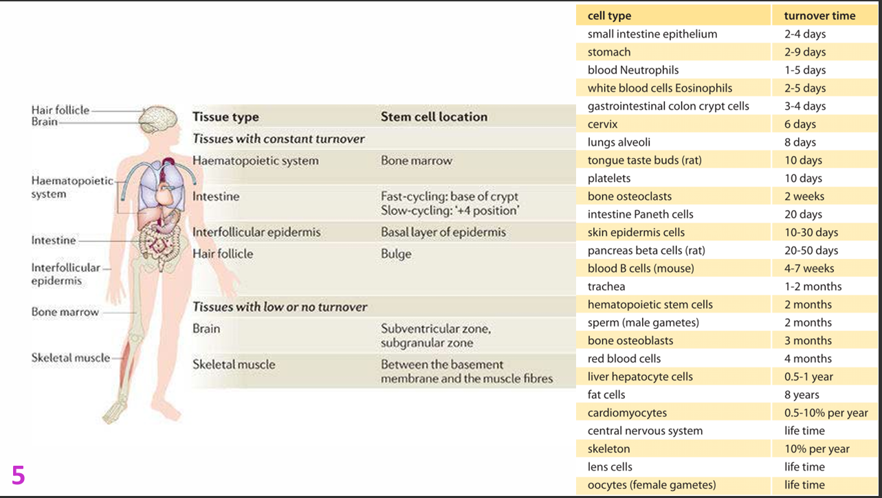
Which cell types have intermediate turnover times?
Examples:
- Bone osteoclasts: 2 weeks
- Intestinal Paneth cells: 20 days
- Skin epidermis cells: 10-30 days
- Pancreatic beta cells (rat): 20-50 days
- Blood B cells (mouse): 4-7 weeks
- Trachea: 1-2 months
- Haematopoietic stem cells: 2 months
- Sperm: 2 months
- Some osteoblasts: 3 months
- Red blood cells: 4 months
- Liver hepatocytes: 0.5-1 year


Which cell types have very long or lifetime turnover times?
Examples:
- Fat cells: ~8 years
- Cardiomyocytes: ~0.5-10% per year
- Central nervous system: lifetime
- Skeleton: ~10% per year
- Lens cells: lifetime
- Oocytes: lifetime


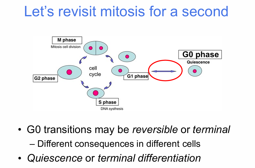
What is the significance of G0 in the cell cycle?
G0 is a non-dividing state that can have two different meanings:
Reversible G0:
- Quiescence
- Cell can re-enter the cell cycle
.
Terminal G0:
- Irreversible arrest
- Cell cannot re-enter mitosis
.
Different cell types have different consequences when they enter G0.


What cell-cycle stages are shown in the lecture?
- G1 phase
- S phase — DNA synthesis
- G2 phase
- M phase — mitosis/cell division
- G0 phase — quiescence
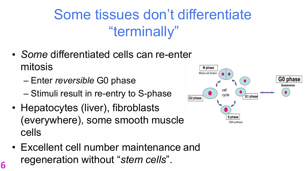
Which differentiated cells can re-enter mitosis from reversible G0?
Examples:
- Hepatocytes
- Fibroblasts
- Some smooth muscle cells
.
These cells can:
- Enter reversible G0
- Respond to appropriate stimuli
- Re-enter S phase
- Divide again
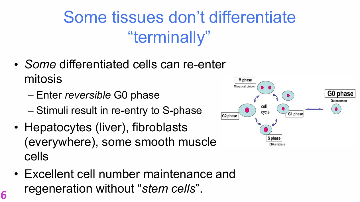

Why can some tissues maintain and regenerate themselves without relying heavily on stem cells?
If mature differentiated cells can re-enter the cell cycle, they can directly replace lost cells.
This provides excellent cell-number maintenance and regeneration without requiring extensive stem-cell recruitment.


Why does the liver have a remarkable regenerative capacity?
After major tissue loss:
- Remaining hepatocytes can re-enter S phase
- They proliferate
- They restore tissue mass and function
- They later return to quiescence
The liver has tissue-specific stem cells, but rarely needs to use them.
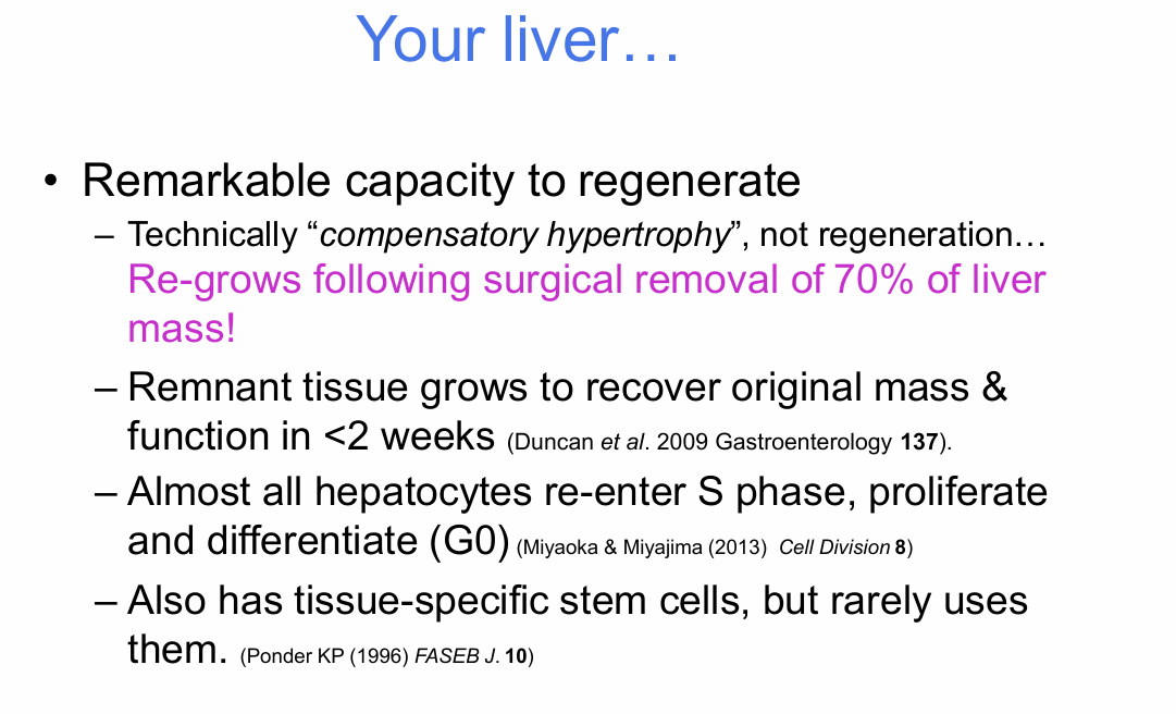
What quantitative details were given for liver regrowth?
- Surgical removal of about 70% of liver mass can be followed by regrowth of the remnant tissue.
- Original mass and function can be recovered in


Why are many mature cells terminally differentiated?
Possible reasons include physical or functional constraints.
Examples:
- Red blood cells are anucleate, so they cannot undergo mitosis.
- Skeletal muscle cells are multinucleate.
- Neurons and cardiac muscle rely on stable connectivity/function that division could disrupt.
- If gut epithelial cells divided while functioning as a barrier, tissue integrity could be compromised.

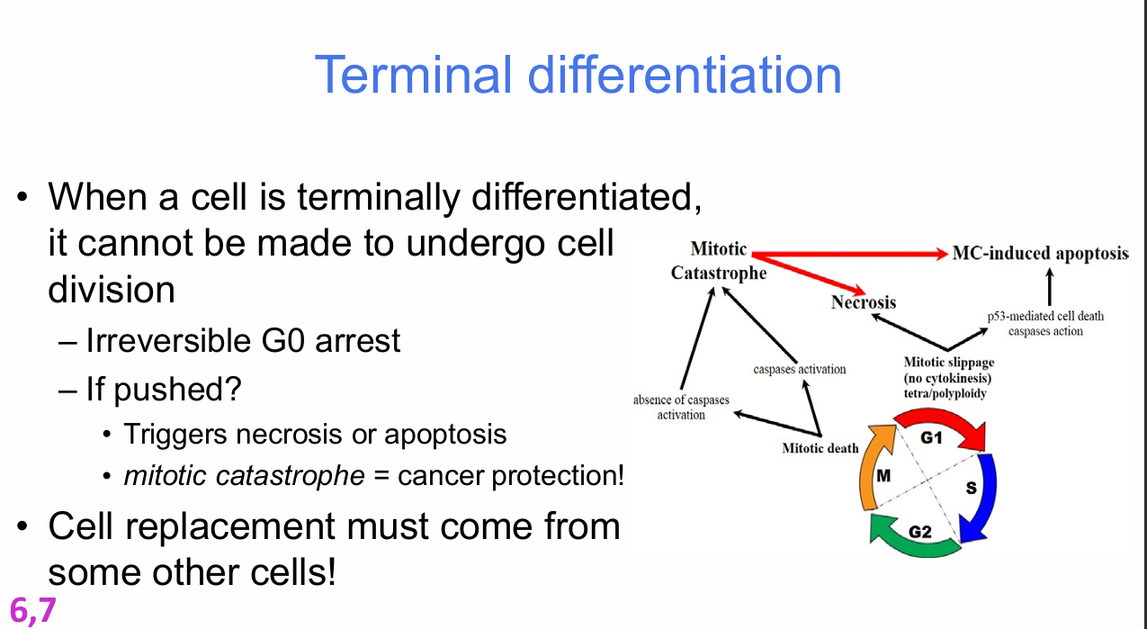
What is terminal differentiation?
Terminal differentiation means a mature cell is irreversibly arrested and cannot be made to undergo cell division.
Key feature:
- Irreversible G0 arrest
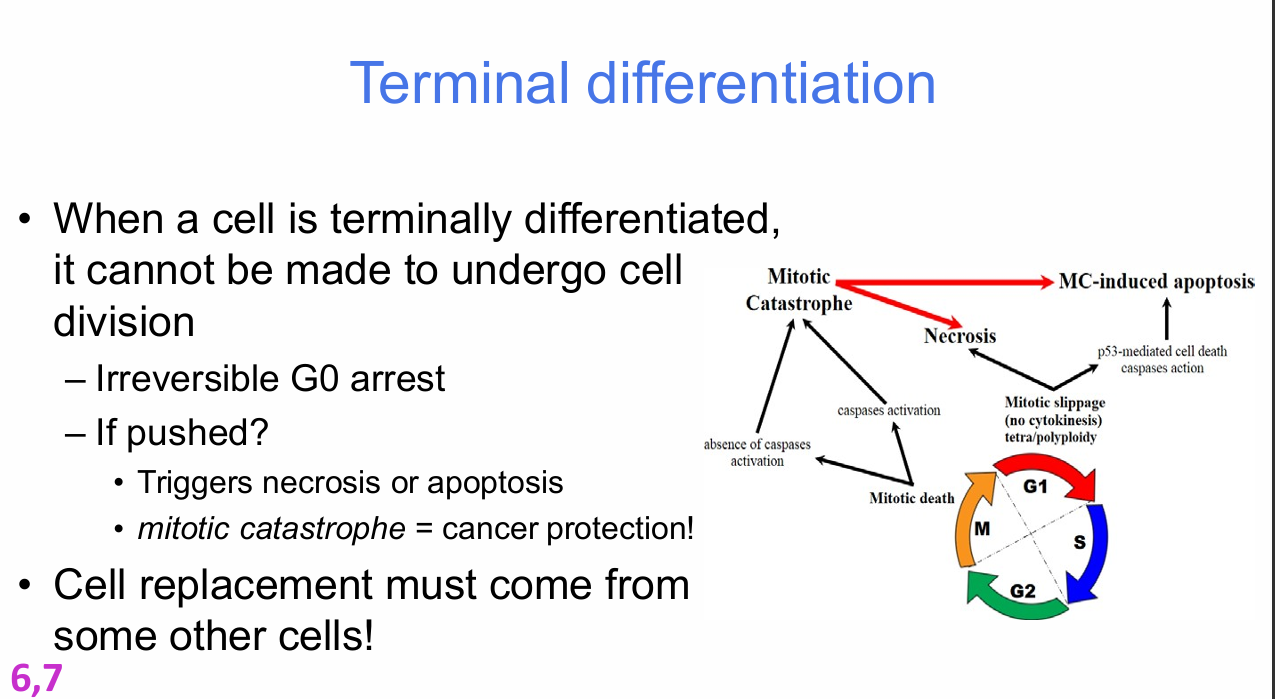

What happens if a terminally differentiated cell is pushed to divide?
Possible outcomes:
- Necrosis
- Apoptosis
- Mitotic catastrophe
Mitotic catastrophe acts as a form of cancer protection.
Consequence:
- Replacement of terminally differentiated cells must come from other cells.


What should you identify in the mitotic-catastrophe diagram?
- Mitotic catastrophe
- Mitotic death
- Necrosis
- MC-induced apoptosis
- Cell-cycle context
- Failure of normal division leading to cell death


How do tissue-specific stem cells maintain adult tissues?
Most adult tissues contain tissue-specific stem cells in reversible G0 (quiescence).
Their role:
- Maintain cellular homeostasis across the organism's lifespan
- Respond to tissue damage
- Activate from quiescence
- Proliferate extensively
- Generate cells that differentiate
- Regenerate damaged tissue

What is the sequence of stem-cell activation after tissue damage?
Tissue damage / external stimulus
→ activation of stem cells
→ extensive proliferation
→ differentiation
→ regeneration

What do you call an adult stem cell with a high rate of proliferation?
Correct answer:
A cancer cell / a cell behaving in a cancer-like uncontrolled proliferative state.
Why it is correct:
The lecturer's point was that adult stem-cell proliferation must be tightly controlled. If a stem cell proliferates at a persistently high, uncontrolled rate, this resembles cancerous behaviour.
.
Lecturer explanation:
Normal proliferation has an effective "off switch"; uncontrolled proliferation does not.

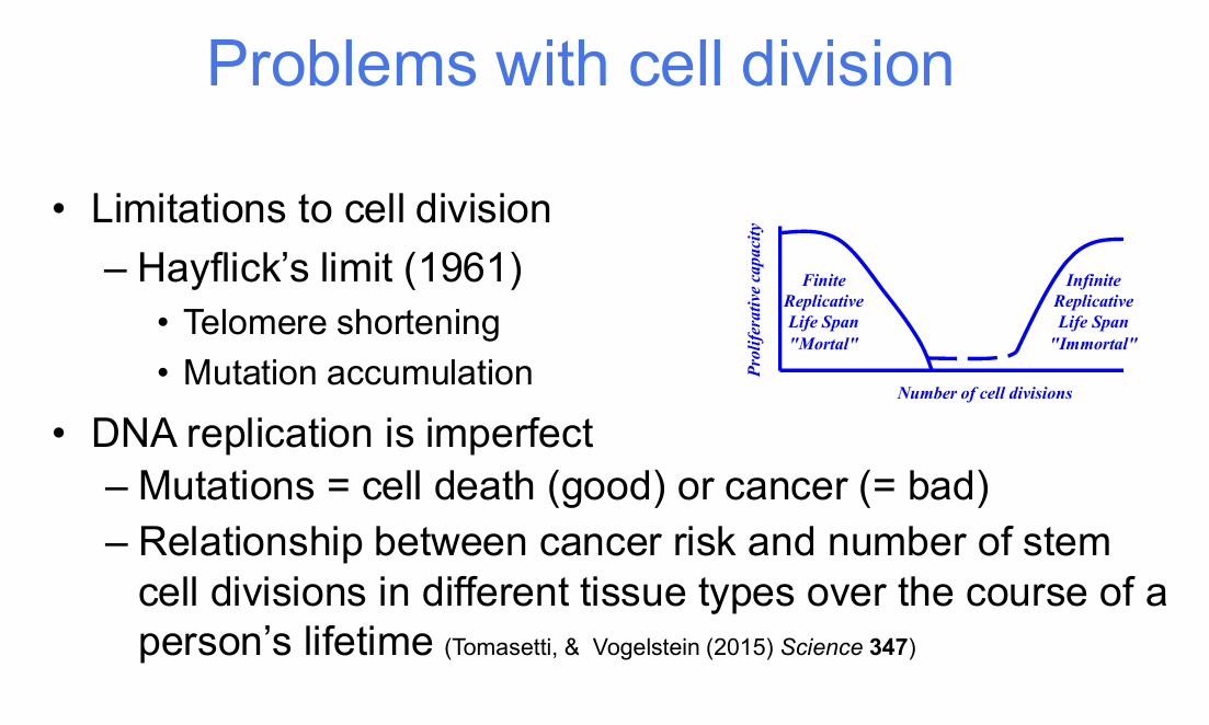
What are the major biological limitations to repeated cell division?
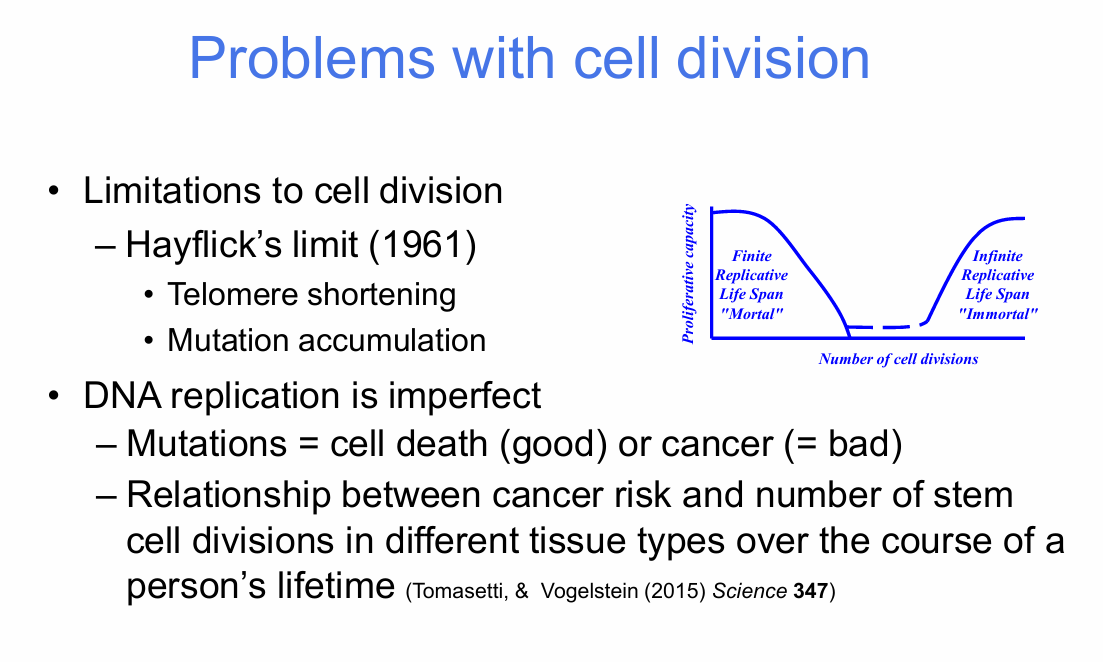
- Hayflick's limit (1961)
- Telomere shortening
- Mutation accumulation
- Imperfect DNA replication
Consequences of mutations:
- Cell death can remove damaged cells
- Some mutations can contribute to cancer

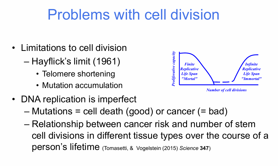
What is the Hayflick limit?
The Hayflick limit refers to the finite replicative lifespan of normal somatic cells.
The lecture links this limitation to:
- Progressive telomere shortening
- Eventual inability to continue dividing

How does mutation accumulation connect cell division to cancer risk?
DNA replication is imperfect.
More cell divisions:
→ more opportunities for replication errors
→ greater mutation accumulation
→ greater chance of oncogenic changes
The lecture highlights a relationship between lifetime stem-cell divisions and cancer risk in different tissues.


What do 'mortal' and 'immortal' mean in the proliferative-capacity diagram?
Mortal:
- Finite replicative lifespan
Immortal:
- Infinite replicative lifespan
The diagram contrasts limited normal proliferation with unlimited proliferative capacity.


What relationship is shown between lifetime stem-cell divisions and lifetime cancer risk?
The graph shows a positive association:
- Tissues with more lifetime stem-cell divisions generally have higher lifetime cancer risk.
Axes:
- X-axis: total stem-cell divisions
- Y-axis: lifetime risk
Lecturer explanation:
Highly proliferative tissues such as colorectal and skin-related tissues appear toward the higher-division/higher-risk end, whereas low-turnover tissues appear lower.

What does the rat small-intestine example illustrate about lifelong tissue maintenance?
Adult rat:
- Approximately 6 × 10^10 cells
.
Small intestine over a lifetime:
- Sheds >10^13 epithelial cells
- Requires large numbers of divisions to replace them
.
Slide values:
- 10^3 symmetric cell doublings from embryo to adult
- Followed by 10^13 asymmetric cell doublings during life
.
Key problem:
How can tissues be repaired while protecting the long-lived stem-cell pool?
How does the body protect true stem cells while still producing large numbers of replacement cells?
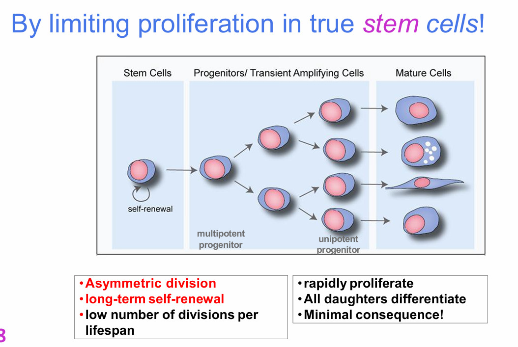
By limiting proliferation in true stem cells.
.
True stem cells:
- Undergo asymmetric division
- Maintain long-term self-renewal
- Divide relatively few times during the lifespan
.
Their progeny:
- Become multipotent progenitors
- Then unipotent progenitors / transit-amplifying cells
- Rapidly proliferate
- Ultimately differentiate into mature cells

Why are transit-amplifying cells useful for tissue maintenance?
Transit-amplifying/progenitor cells carry out most of the rapid expansion.
.
Advantages:
- True stem cells avoid repeated rounds of DNA replication.
- Mutation accumulation in the long-lived stem-cell pool is reduced.
- Daughter progenitor cells can proliferate rapidly.
- Their descendants then terminally differentiate.
.
Lecturer emphasis:
The expansion burden is shifted away from the true stem cells.

What should you identify in the stem-cell → transit-amplifying → mature-cell diagram?
Stem-cell compartment:
- Self-renewal
- Asymmetric division
- Long-term self-renewal
- Low number of divisions per lifespan
Progenitor / transit-amplifying compartment:
- Multipotent progenitor
- Unipotent progenitor
- Rapid proliferation
- All daughters differentiate
- Minimal long-term consequence
Mature-cell compartment:
- Terminally differentiated cells

What are the major take-home messages from Stem Cells I?
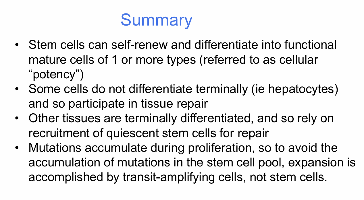
- Stem cells can self-renew and differentiate into functional mature cells of one or more types.
- The range of cell types they can generate is their cellular potency.
- Some differentiated cells, such as hepatocytes, are not terminally differentiated and can participate directly in tissue repair.
- Terminally differentiated tissues rely on recruitment of quiescent stem cells for repair.
- Mutations accumulate during proliferation.
- To limit mutation accumulation in the stem-cell pool, most expansion is performed by transit-amplifying cells rather than the true stem cells themselves.
