N245: chapter 27 - heart failure, circulatory shock, cardiac function
 Call Kai
 Learn
 Practice Test
 Spaced Repetition
 Match
 Flashcards
 Knowt Play
1/112

There's no tags or description
Looks like no tags are added yet.
113 Terms
Right and left coronary arteries
First branches of the ascending aorta --> supply oxygen to the myocardium

The left coronary artery divides into
1. Left anterior descending/interventricular artery
2. Left circumflex artery

The right coronary artery divides into
1. Right marginal artery
2. Posterior interventricular artery

At what phase during heart contractions does blood flow through the coronary arteries?
During ventricular relaxation (paradoxical because all other arteries receive the most blood during ventricular systole)

Coronary Heart Disease/Coronary Artery Disease
Abnormal narrowing or obstruction of coronary arteries --> causing impaired coronary blood flow

What are the dangers of Coronary Heart Disease
Angina, MI, arrhythmias, heart failure, death
The two broad categories of coronary heart disease are:
Chronic ischemic heart disease, acute coronary syndrome

Chronic Ischemic Heart Disease (Angina)
Definition: Chest pain that occurs due to a lack of oxygen delivery to the myocardium
S+S: Triggered by emotional or physical stress
Treatment: Vasodilators

Stable angina
Definition: A type of chronic ischemic heart disease; pain during exercise or exertion (when heart's O2 demand increases)
Treatment: vasodilators

Variant (vasospastic) angina
Definition: A type of chronic ischemic heart disease; chest pain not related to physical activity (possibly bc of nitric oxide deficiency?) --> occurs when coronary arteries spasm
Treatment: vasodilators

What does nitric oxide do?
Nitric oxide causes blood vessels to dilate

Silent myocardial ischemia
Definition: A type of chronic ischemic heart disease; clinical evidence of decreased blood flow/O2 to the heart without angina

Unstable angina
Definition: A type of chronic ischemic heart disease/acute coronary syndrome; chest pain that occurs while a person is at rest and not exerting himself --> will probs lead to MI
not relieved by vasodilators

How is angina diagnosed?
Using a stress test or echocardiograph
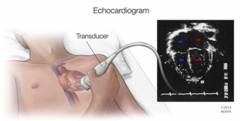
Where will angina manifest on the body?
Substernal pain that radiates to the jaw and left arm

How is angina managed?
1. Statins
2. Aspirin
3. Sublingual nitroglycerin
4. Calcium channel blockers
5. Beta-blockers
How will statins help angina?
By reducing cholesterol and heart rate
How will aspirin help angina?
By preventing blood clots and reducing pain
How will sublingual nitroglycerin help angina?
By dilating the blood vessels
How will calcium channel blockers help angina?
By preventing the contraction of blood vessels
How will beta-blockers help angina?
By blocking the release of adrenaline --> slowing the heart rate
Emergency Treatment for Angina
1) Stop activity
2) Patient seated in upright position
3) Administration of nitroglycerin and O2
When can you give a second dose of nitroglycerin?
Only when the pt has a history of angina

Acute coronary syndrome
A multitude of conditions; involve symptoms of sudden, reduced blood flow to the heart
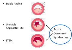
What are some possible causes of acute coronary syndrome?
Atherosclerosis, embolism, vasospasm (anything that quickly blocks the arteries)
Myocardial infarction
Death of cardiac muscle due to ischemia (lack of blood)

Signs and Symptoms of MI
Pressure/pain in chest
Sudden shortness of breath
Weakness, fatigue
Nausea, indigestion
Anxiety and fear

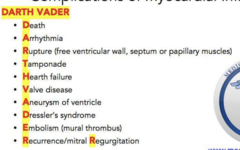
Complications of AMI
1. Sudden death
2. Cardiogenic shock
3. Cardiac tamponade
4. Aneurysm
5. Embolism
STEMI
ST elevation myocardial infarction

What is a STEMI?
A type of acute coronary syndrome; a heart attack that is characterized by elevated ST waves --> indicates a complete blockage
The most serious type of HA, called the "widow maker"

Treatment for STEMI
Angioplasty to break up clot/plaque
Percutaneous coronary intervention (PCI)
A balloon-tipped catheter is inserted into a coronary artery to force it open --> stents are put in place
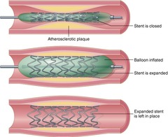
Angioplasty
Mechanically widening a narrowed or obstructed blood vessel

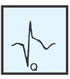
Abnormal Q waves
An ECG seen during a type of acute MI --> indicates necrosis of myocardium --> no contraction
NSTEMI
Non-ST elevation myocardial infarction

What is a NSTEMI?
A type of acute coronary syndrome; a heart attack that is characterized by ST depression and T wave inversion --> indicates a partial blockage

Treatment for NSTEMI
Administer anticoagulants (heparin) to break up the clot --> angioplasty if meds aren't effective

Serum Cardiac Markers
Proteins that are released into the blood from necrotic heart muscle after an MI

Myoglobin
Protein released by necrotic myocardium --> Elevated within 2 hrs of myocardial infarction

Creatine kinase MB (CK-MB)
Protein released by necrotic myocardium --> Elevated within 6-8 hours of myocardial infarction

Troponin I (TnI) and Troponin T (TnT)
Protein released by necrotic myocardium --> Rises within 3 hrs after MI, then decreases slowly over days

Coronary Artery Bypass Graft (CABG)
Open-heart surgery in which a blood vessel (usually a leg vein), is grafted to redirect blood around the blocked coronary artery

Pericarditis
Inflammation of the pericardium, usually secondary another condition

How is pericarditis classified
Pericarditis is classified by the cause or type of exudate
Acute pericarditis
Acute inflammation of the pericardium --> effusion may develop

Pericardial effusion
An accumulation of fluid in the pericardial cavity --> occurs as a result of pericarditis

Effects of pericardial effusion
1. Fluid around heart compresses heart wall
2. Heart cannot expand to fill
3. Blood backs up in systemic circulation
4. Decreased blood flow to lungs and tissues

Chronic pericarditis
Chronic inflammation of the pericardium
What are possible causes of chronic pericarditis?
Viral infections or autoimmune disorders
What are the effects of chronic pericarditis?
Adhesions form between the two membranes, fibrous tissue forms --> heart's movement is limited

Diastole
Relaxion of the heart

Systole
Contraction of the heart

CO
Cardiac output
CO equation
Stroke volume (amount of blood ejected per beat) x HR

Preload (EDV)
How much the heart expands when it fills with blood

Afterload
The amount of pressure that the heart needs to exert to eject the blood during ventricular contraction

Ejection Fraction
Percentage of blood pumped out to the body by the ventricles
Normal: 55-75%

Cardiac reserve
The heart's ability to increase CO during increased activity
Heart Failure
Condition in which the heart is unable to pump out enough blood to meet the body's metabolic demands; EF < 40%
Compensatory Mechanisms Heart Failure
1. Force of contraction increases
2. Myocardium increases
3. HR increases
4. Stimulates RAAS in kidney

How does RAAS affect heart failure?
Activation of RAAS compounds the problem --> aldosterone retains salt + water --> vasoconstriction and blood volume increase --> heart has to work even harder
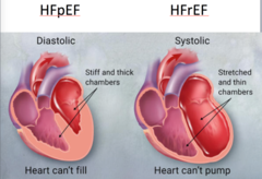
Systolic Heart Failure
A type of heart failure in which the heart cannot contract and eject blood --> decreased EF

Causes of Systolic Heart Failure
Inability to contract due to MI, increased afterload due to high BP, valve diseases

Diastolic Heart Failure
A type of heart failure in which ventricles can't relax and fill --> decreased SV and CO
Causes of Diastolic Heart Failure
Left ventricular hypertrophy, aortic stenosis (aortic valve won't open)

General consequences of heart failure
Less blood reaches the organs --> decreased cell function --> fatigue and lethargy --> acidosis
Left-sided congestive heart failure
When the left side of the heart fails, blood flows back into the lungs --> causing difficulty breathing, cough, and paroxysmal nocturnal dyspnea

Paroxysmal nocturnal dyspnea
Symptom of left-sided congestive heart failure; shortness of breath that occurs during sleep

Right-sided congestive heart failure
When the right side of the heart fails, blood flows back into the venous system --> high BP in jugular veins, edema of legs, spleen/liver enlargement

Generally, how do you treat heart failure?
Lower blood pressure, dilate blood vessels, if needed, restore contraction ability using an AED

What does Digoxin (Lanoxin) do for heart failure?
This medication helps the heart beat stronger with a more regular rhythm
it was derived from this pretty digitalis plant!

Cardiac anomalies
Structural defects of the heart that appear during embryonic development
Congenital Heart Defects
Structural abnormalities of the heart caused by improper development before birth (valvular or septal defects)

General S+S of Congenital heart disease
Pallor, tachycardia, dyspnea, squatting position, clubbed fingers

Ventricular Septal Defect
A hole between two ventricles --> allows blood to pass from the one ventricle to another

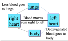
Right-to-Left Shunt (venous to arterial)
Blood from right ventricle moves to left ventricle BEFORE GOING TO LUNGS --> less blood goes to lung --> deoxygenated blood is delivered to tissues --> cyanosis
Left-to-Right Shunt arterial to venous
Oxygenated blood from left ventricle moves to right ventricle INSTEAD OF GOING THROUGH AORTA --> more blood goes to lung --> less blood goes to body

Valvular Defects
Defects of the heart valves, most commonly affect the aortic and pulmonary valves
Valve prolapse
One or more cusps in the valve will protrude in the wrong direction --> causing backflow

Valve Stenosis
A valve that cannot open all the way because it is narrower and stiffer --> less passes through the opening

Heart Murmur
An abnormal sound from the heart produced by defects in the valves

Regurgitant/Incompetent Valve
A valve that cannot close completely --> blood leaks backwards into previous chamber

Tetralogy of Fallot
A congenital heart condition caused by a combination of four heart defects
1. Pulmonary valve stenosis
2. Shunt in ventricular septum
3. Dextroposition of the aorta
4. Right ventricular hypertrophy

S+S of Tetralogy of Fallot
"Blue babies" and "tet spells"
The sudden development of cyanosis and fainting

Rheumatic fever
An inflammatory condition that usually occurs after an infection
Rheumatic heart disease
Heart disease caused by rheumatic fever
Acute stage of rheumatic fever
Inflammation of the heart (pericarditis, myocarditis, or endocarditis)

S+S of Rheumatic fever
Fever, inflammation of the heart, blood vessels, and joints, erythema marginatum

Mitral Valvulitis
Inflammation of the mitral valve; common symptom of rheumatic fever

Mitral Valve Stenosis
Narrowing of the mitral valve opening; caused by scarring from rheumatic fever

Chronic rheumatic heart disease
Valve scarring that arises as a consequence of rheumatic fever
How is rheumatic fever diagnosed?
Using a heart function test and/or electrocardiogram
How is rheumatic fever treated?
Antibacterial and anti-inflammatory agents
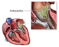
Infective endocarditis
Life-threatening inflammation of the endocardium due to a bacterial or fungal infection --> growths (vegetations) cause valve stenosis
Subacute infective endocarditis
Definition: Most commonly caused by streptococcus viridans (low virulence)
Symptoms:

Acute infective endocarditis
Definition: Most commonly caused by staphylococcus aureus (high virulence)
Symptoms: Sudden onset of fever, chills

Risk factors for infective endocarditis
Previous valve damage, bacteremia, compromised immune system
How is infective endocarditis diagnosed?
A blood culture is procured to determine the cause of infection
How is infective endocarditis treated?
Antibiotics for 4-6 weeks

Cardiomyopathies
Diseases of the heart muscles