A&P chapter 8
 Call Kai
 Learn
 Practice Test
 Spaced Repetition
 Match
 Flashcards
 Knowt Play
1/92

Earn XP
Description and Tags
The skeletal system pt 1 - AXIAL SKELETON
93 Terms
Axial skeleton
skull, vertebrae, sternum, ribs, & hyoid
number of bones
206
sesamoid bone
does NOT articulate with any other bone
bones that form within tendons
patella & hyoid bone
condyle on skull
articular surface on a bone (will form a joint with another bone)
fossae on scapula
shallow depression
trochanter
found on large bone like the femur
tubercle
found on small bones like the humerus
epicondyles are where on the femur and humerus?
above the condyles
Sinuses
frontal, sphenoid, ethmoid, and maxillary ( largest sinus in the skull). Sinuses lighten the skull & act as chambers that add resonance to the voice.
temporal
lateral
sphenoid
anterior to temporal bone
ethmoid
anteromedial
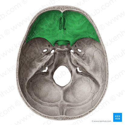
anterior cranial fossa
holds the frontal lobe of the brain (superior)

middle cranial fossa
holds the temporal lobes of the brain

posterior cranial fossa
contains the cerebellum (inferior)
frontal cranial bones
1 f
parietal cranial bone
2 p
temporal cranial bone
2 t
occipital cranial bone
1 o
sphenoid cranial bone
1 s
ethmoid cranial bone
1 e
sagittal suture
between parietal bones
coronal suture
at anterior margin (frontal bone)
lambdoid suture
at posterior margin (occipital bone)
squamous suture
at lateral border (temporal bone)
zygomatic process
on temporal bone
zygomatic arch
where 2 bones meet zygomatic bone + temporal bone
external acoustic meatus
ear canal
styloid process
pointed spine for muscle attachment
mandibular fossae
a smooth, concave depression on the inferolateral surface of the temporal bone of the skull
mastoid process
lump behind earlobe, filled with air sinuses
petrous part
seen in cranial floor (inside skull)
internal acoustic meatus
opening for nerve carrying signals for hearing & balance
jugular foramen
opening for passage of internal jugular vein
foramen magnum (occipital bone)
large opening for passage of spinal cord
occipital condyle
articulate with first vertebra of vertebral column (cervical → C1)
external occipital protuberance
attachment for nuchal ligament (bump of back of head)
diploe
found in skull
optic canal (sphenoid bone)
passage of optic nerve (CNII)
sella turcica (sphenoid bone)
houses pitutiary gland
pterygoid process
serves in muscle attachment (TMJ)
ethmoid bone
located between the eyes (anteromedial)
cribriform plate
horizontal plate forming roof of nasal cavity
cribriform foramen
CNI (cranial nerve) olfactory smell
facial bones
are anterior to the cranial cavity
maxillae
superior jaw → foundation of face. Form upper jaw & part of orbits,& meet each other at the median intermaxillary suture (between maxillary bones)
Intermaxillary fissure
failure to fuse → cleft palate
zygomatic bones
form angles of cheekbones
lacrimal bones
the smallest and most fragile bone of the facial skeleton in humans, and located at the front part of the medial (inner) wall of the eye socket (orbit).
vomer
inferior half of nasal septum
mandible
inferior jaw
body of mandible
teeth
ramus of mandible
superior
angle
body & ramus meet (corner of jaw)
condylar process
containing mandibular condyle for articulation with temporal bone (mandibular fossa) to form temporomandibular joint (TMJ)
coronoid process
anterior lip
hyoid bone
in the throat (sesamoid bone → axial skeleton)
mastoid fontanelle
posterolateral
sphenoid fontanelle
anterolateral
anterior fontanelle & posterior fontanelle
soft spot in the skull (only in babies)
cervical vertebrae
7 in the neck
thoracic vertebrae
12 in the chest. 12 PAIRS OF RIBS ARTICULATE WITH THORACIC VERTEBRAE
lumbar vertebrae
5 in the back
axis & atlas
articulates with occipital condyles
C7
vertebra prominent
kyphosis
PRIMARY curve - thoracic / sacral
lordosis
SECONDARY curve - cervical / lumbar
fetal position
kyphosis (primary curve)
tummy time in infants
2nd curve forms
scoliosis
muscle problem → happens when the bone grows faster than the muscle
hyperkyphosis
osteoporosis (hunch)
hyperlordosis
due to obesity (pelvis goes forward)
vertebrae body
weight-bearing portion (anterior)
spinous process
projects posteriorly
transverse processes
extend laterally
vertebra lamina
spinous process + transverse process
vertebra pedicle
body + transverse process
vertebral foramen
spinal cord goes though it
intervertebral disc
cartilaginous (fibrocartilage) pad located between bodies of adjacent vertebra

nucleus pulposus
inner gelatinous mass
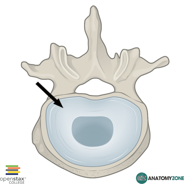
annulus fibrosus
outer ring of fibrocartilage (absorb shock )
herniated disc
a break in annulus fibrosis → puts painful pressure on spinal nerve or spinal cord
bulging disc
nucleus pulposus stays in disc
C1
also known as atlas. has NO BODY. Articulates (forms joint) with occipital condyles of skull (atlanto-occipital joint) and C2 (atlantoaxial joint)
atlanto-occipital joint
yes head motion
atlantoaxial joint
where atlas & axis articulate.
C2
also called the axis which allows for rotation of the head (no head motion). contain dens (odontoid process) → projects superiorly (surrogate body for C1) & held in place by transverse ligament
superior articular facet
surface that articulates with occipital condyle (atlanto-occipital joint→ yes head movement). Articulates with C1. Unique to C2
cervical vertebrae
small body, large foramen. Has transverse foramen (vertebral artery)
thoracic vertebrae
spinous process angles sharply downward. Larger body than cervical. Superior costal facet → where rib attach to thoracic vertebrae
lumbar vertebrae
large body, small foramen
sacrum & coccyx
ala (wings), sacral promontory → where L5 sits