(W3+4) Liver-Penny Ch. 2
 Call Kai
 Learn
 Practice Test
 Spaced Repetition
 Match
 Flashcards
 Knowt Play
1/59

There's no tags or description
Looks like no tags are added yet.
60 Terms

The patient in figure 2-55 presented with RUQ pain and elevated AFP. What is the most likely diagnosis?
a. Hepatic hematoma
b. Hemangioma
c. Hepatoma
d. Focal nodular hyperplasia
c. Hepatoma
The malignant mass associated w/ HCC is referred to as a hepatoma. The tumor marker for HCC is AFP. May appear as solitary, hypoechoic mass. (may yield target or bulls eye sign)

Which of the following benign liver masses is typically isoechoic and contains a central scar?
a. Hepatoblastoma
b. Cavernous hemangioma
c. Hamartoma
d. Focal nodular hyperplasia
d. Focal nodular hyperplasia
FNH- second most common benign liver tumor. Most commonly found in women. Asymptomatic with a central scar.
The covering of the liver is referred to as:
a. Glisson capsule
b. Gerota fascia
c. Morison pouch
d. Hepatic fascia
a. Glisson capsule
The left lobe of the liver can be separated from the right lobe by the:
a. Rt hepatic vein
b. Middle hepatic vein
C. Lt hepatic vein
d. Falciform ligament
b. Middle hepatic vein
The TIPS shunt is placed:
a. Between the main hepatic artery and main portal vein
b. Between a portal vein and a hepatic vein
c. Between the common hepatic duct and common bile duct
d. Between a portal vein and hepatic artery
b. Between a portal vein and a hepatic vein

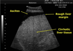
Which of the following is the likely diagnosis for the patient in Figure 2-56?
a. Hepatitis
b. Hepatic steatosis
c. Cirrhosis
d. Steatohepatitis
c. Cirrhosis
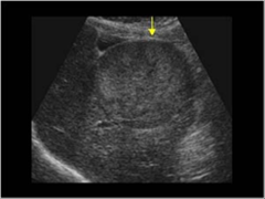
What is the arrow in Figure 2-56 most likely indicating?
a. Metastatic liver disease
b. Hemangioma
c. Amebic hepatic abscess
d. Hepatoma
d. Hepatoma

The right lobe of the liver is divided into segments by the:
a. middle lobar fissure
b. middle hepatic vein
c. right hepatic vein
d. left hepatic vein
c. right hepatic vein


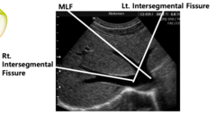
The right intersegmental fissure contains the:
a. right hepatic vein
b. middle hepatic vein
c. left portal vein
d. right portal vein
a. right hepatic vein
The main portal vein divides into:
a. middle, left, and right branches
b. left and right branches
c. anterior and posterior branches
d. medial and lateral branches
b. left and right branches

Which of the following is the patient suffering from in in figure 2-57?
a. Hepatopetal flow with hepatitis
b. Hepatofugal flow with hepatitis
c. Hepatopetal flow with portal hypertension
d. Hepatofugal flow with portal hypertension
d. Hepatofugal flow with portal hypertension


What is the normal velocity range of the structure being analyzed with the sample gate in Figure 2-57?
a. 20-40 cm per second
b. 90 to 100 cm per second
c. 0.5 to 0.8 cm per second
d. 1.2 to 1.3 cm per second
a. 20-40 cm per second


The ligamentum teres can be used to separate the:
a. Medial and lateral segments of the left lobe
b. Medial and posterior segments of the right lobe
c. Anterior and medial segments of the left lobe
d. Anterior and posterior segments of the right lobe
a. Medial and lateral segments of the left lobe


The main lobar fissure contains the:
a. right hepatic vein
b. middle hepatic vein
c. main portal vein
d. right portal vein
b. middle hepatic vein

All of the following are located within the porta hepatis except:
a. main portal vein
b. common bile duct
c. hepatic artery
d. middle hepatic vein
d. middle hepatic vein
Right-sided heart failure often leads to enlargement of the:
a. abdominal aorta
b. IVC and hepatic veins
c. IVC and portal veins
d. portal veins and spleen
b. IVC and hepatic veins

The mass noted by the arrow in Figure 2-58 was discovered in an asymptomatic 22-yr old female patient. What is the most likely diagnosis?
a. Focal nodular hyperplasia
b. Fibroma
c. Cavernous hemangioma
d. Lipoma
c. Cavernous hemangioma
Asymptomatic, benign, hyperechoic, Right lobe


Which of the following is typically transmitted through contaminated water found in places such as Mexico, Central America, South America, Asia, India, and Africa?
a. Amebic liver abscess
b. Hydated liver cyst
c. Candidiasis
d. Hepatoma
a. Amebic liver abscess
Comes from the parasite Entamoeba histolytica. Rt lobe, fluid filled-debris level.

The right portal vein divides into:
a. middle, left and right branches
b. left and right branches
c. anterior and posterior branches
d. medial and lateral branches
c. anterior and posterior branches
The diameter of the portal vein should not exceed:
a. 4 mm
b. 8 mm
c. 10 mm
d. 13 mm
d. 13 mm
The right lobe of the liver can be divided into:
a. medial and lateral segments
b. medial and posterior segments
c. anterior and medial segments
d. anterior and posterior segments
d. anterior and posterior segments
Which of the following is true about the portal veins?
a. Portal veins carry deoxygenated blood away from the liver
b. Portal veins have brighter walls than the hepatic veins
c. Portal veins should demonstrate hepatofugal flow
d. Portal veins increase in diameter as they approach the diaphragm
b. Portal veins have brighter walls than the hepatic veins
The left lobe of the liver can be divided into:
a. medial and lateral segments
b. medial and posterior segments
c. anterior and medial segments
d. anterior and posterior segments
a. medial and lateral segments

Normal flow within the hepatic artery should demonstrate a:
a. high-resistance waveform pattern, with a slow upstroke, and gradual deceleration with diastole
b. low-resistance waveform pattern, with a quick upstroke, and gradual deceleration with diastole
c. low-resistance waveform pattern, with a slow upstroke, and gradual acceleration with diastole
d. high-resistance waveform pattern, with a quick upstroke, and gradual deceleration with diastole
b. low-resistance waveform pattern, with a quick upstroke, and gradual deceleration with diastole

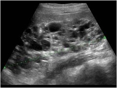
The patient in Figure 2-59 would be most likely suffering from:
a. Cirrhosis
b. Portal hypertension
c. ADPKD
d. Echinococcus granulosis
c. ADPKD

Budd-Chiaria syndrome leads to a reduction in the size of the:
a. hepatic arteries
b. portal veins
c. hepatic veins
d. common bile duct
c. hepatic veins
A tongue-like extension of the right lobe of the liver is termed:
a. papillary lobe
b. focal hepatomegaly
c. Riedel lobe
d. Morison lobe
c. Riedel lobe
The left portal vein divides into:
a. middle, left, and right branches
b. left and right branches
c. anterior and posterior branches
d. medial and lateral branches
d. medial and lateral branches
The left umbilical vein after birth becomes the:
a. falciform ligament
b. main lobar fissure
c. ligamentum teres
d. ligamentum venosum
c. ligamentum teres
Normal flow within the hepatic veins is said to be:
a. Biphasic
b. Irregular
c. High resistant
d. Triphasic
d. Triphasic
The inferior extension of the caudate lobe is referred to as:
a. papillary process
b. focal hepatomegaly
c. Riedel process
d. Morison lobe
a. papillary process

The patient in Figure 2-60 has other sonographic findings including splenomegaly and hepatofugal flow within the main portal vein. What are the three unlabeled arrows in this image identifying?
a. Irregular SMA
b. Esophageal varices
c. Splenic varices
d. Atrophic coronary artery
b. Esophageal varices
-Esophageal varices are dilated submucosal distal esophageal veins connecting the portal and systemic circulations.


What other sonographic findings would also most likely be evident in the patient with Figure 2-60? (splenomegaly, hepatofugal flow, ascites in the liver)
a. Periportal cuffing
b. Water lily sign
c. Subcapsular hematoma
d. Shrunken right lobe
d. Shrunken right lobe

The patient in Figure 2-60 would least likely be suffering from which of the following clinical findings? (splenomegaly, hepatofugal flow, ascites in the liver)
a. Hepatic encephalopathy (loss of brain function when a damaged liver doesn't remove toxins from the blood)
b. Narrowing of the hepatic veins
c. Caput medusa (cluster of swollen veins in your abdomen)
d. Tremors
b. Narrowing of the hepatic veins
Which of the following is the most common reason for a liver transplant?
a. hepatocellular carcinoma
b. Hepatitis C
c. Hepatitis B
d. Hepatic mesastasis
b. Hepatitis C
Clinical findings of fatty infiltration of the liver include:
a. elevated liver function labs
b. fever
c. fatigue
d. weight loss
a. elevated liver function labs
Shortly after birth, the ductus venosus collapses and becomes the:
a. falciform ligament
b. main lobar fissure
c. ligamentum teres
d. ligamentum venosum
d. ligamentum venosum
Sonographically, when the liver is difficult to penetrate and diffusely echogenic, this is indicative of:
a. portal vein thrombosis
b. metastatic liver disease
c. primary liver carcinoma
d. fatty liver disease
d. fatty liver disease
The most common cause of cirrhosis is:
a. portal hypertension
b. hepatitis
c. alcoholism
d. cholangitis
c. alcoholism
Clinical findings of hepatitis include all of the following except:
a. jaundice
b. fever
c. chills
d. pericholecystic fluid
d. pericholecystic fluid
What form of hepatic abnormality are immunocompromised patients more prone to develop?
a. hepatic adenoma
b. amebic abscess
c. hydatid liver abscess
d. candidiasis
d. candidiasis
All of the following are sequela of cirrhosis except:
a. portal vein thrombosis
b. hepatic artery contraction
c. portal hypertension
d. splenomegaly
b. hepatic artery contraction
Normal flow toward the liver in the portal veins is termed:
a. hepatopetal
b. hepatofugal
a. hepatopetal
Which of the following masses would be most worrisome for malignancy?
a. echogenic mass
b. cystic mass with posterior enhancement
c. isoechoic mass with a central scar
d. hyperechoic mass with a hypoechoic halo
d. hyperechoic mass with a hypoechoic halo
Which of the following statements is true of hepatic adenomas?
a. hepatic adenomas are more common in males
b. hepatic adenomas are also referred to as a stealth lesion
c. hepatic adenomas typically contain air owing to bacterial formation
d. hepatic adenomas can undergo malignant degeneration
d. hepatic adenomas can undergo malignant degeneration
Which of the following is the most common cancer found in the liver?
a. hepatocellular carcinoma
b. adenomcarcinoma
c. metastatic liver disease
d. hepatoblastoma
c. metastatic liver disease
Which hepatic mass is closely associated with oral contraceptive use?
a. hepatic adenoma
b. hepatic hypernephroma
c. hepatic hamartoma
d. hepatic hemangioma
a. hepatic adenoma
Which of the following is considered the most common benign childhood hepatic mass?
a. Hepatoblastoma
b. Hepatoma
c. Hematoma
d. Hemangioendothelioma
d. Hemangioendothelioma
All of the following are clinical findings of hepatocellular carcinoma except:
a. reduction in AFP
b. unexplained weight loss
c. fever
d. cirrhosis
a. reduction in AFP
The childhood syndrome Beckwith-Weidemann is associated with an increased risk for developing:
a. hepatoblastoma
b. cirrhosis
c. portal hypertension
d. hepatitis
a. hepatoblastoma
-malignant pediatric tumor, most common malignant tumor of childhood.
Which of the following is associated with E. granulosus?
a. Candidiasis
b. Amebic liver abscess
c. Hydatid liver cyst
d. Hepatocellular carcinoma
c. Hydatid liver cyst
Which of the following laboratory findings would be most likely associated with a decrease in albumin?
a. hepatic steatosis
b. hepatic steatohepatitis
c. cirrhosis
d. hepatitis
c. cirrhosis
Which of the following are the cells of the liver responsible for engulfing pathogens and damaged cells?
a. Kupffer
b. Morrison
c. Hepatocytes
d. Epstein
a. Kupffer
What liver pathology is associated with periportal cuffing?
a. cadidiasis
b. amebic hepatic abscess
c. steatosis
d. hepatitis
d. hepatitis
Which of the following is an inherited disease characterized by disproportionate absorption of dietary iron?
a. Wilson disease
b. Hemochromatosis
c. von Gierke syndrome
d. von Hippel-Lindau disease
b. Hemochromatosis
Wilson disease results from excessive copper accumulation, whereas hemochromatosis results from excessive iron.
What inherited disease is linked with the development of cysts in the liver and other organs?
a. von Gierke disease
b. Wilson disease
c. Epstein-Barr disease
d. von Hippel-Lindau disease
d. von Hippel-Lindau disease
Wilson disease will present sonographic similar to what disorder?
a. Hepatitis
b. Portal hypertension
c. Budd-Chiari syndrome
d. Focal fatty infiltration
a. Hepatitis
Wilson disease- accumulation of copper- eventually accumulation of copper in hepatocytes results their dysfunction manifesting as fatty liver disease and ultimately progressing to liver failure, hepatitis, and liver fibrosis.
Cavernous transformation of the portal vein is found in the presence of:
a. portal venous gas
b. portal vein thrombosis
c. autosomal dominant polycystic liver disease
d. multiple hydatid liver cysts
b. portal vein thrombosis
Which of the following hepatic lesions may occur following a recent bout of appendicitis, diverticulitis, or cholecystitis?
a. Focal nodular hyperplasia
b. Pyogenic hepatic abscess
c. Echinococcal abscess
d. Amebic liver disease
b. Pyogenic hepatic abscess
Can result from the spread of infection from inflammatory conditions.

The patient in Figure 2-61 is a 5 year old female patient who presented with increased abdominal girth. What is the most likely etiology of this mass?
a. Hematoma
b. Pyogenic liver abscess
c. Hepatoblastoma
d. Hepatoma
c. Hepatoblastoma
Aggressive tumors most often before the age of 5, may have palpable abdominal mass.