PHGY mod4
 Call Kai
 Learn
 Practice Test
 Spaced Repetition
 Match
 Flashcards
 Knowt Play
1/38

There's no tags or description
Looks like no tags are added yet.
39 Terms
What is intracellular signal transduction?
Signal must be relayed into cell and trigger a cascade of events resulting in a function
what are 2 types of communication?
Extracellular communication | Intracellular communication |
Occurs when a signal is received from outside the cell Because cells need to send signals to other cells | External changes cause a change within the cell |
Describe 2 ways extracellular communication occurs
cells in direct contact
cells in indirect contact
describe how cells in direct contact communicate
Use gap junctions to communicate (links cytoplasm together)
Made of “connexons” that dock together to form channels connecting the cells
Allows chemical signals to move between them
Not all types of molecules can pass through gap junctions
Only for passing of small particles (ions, etc)
Can pass chemical but also electrical signals (e.g. cardiac muscle cells coordinating contraction)
They have gates that open/close as a defence mechanism
Not freely open doors

describe how cells in indirect contact communicate
1. Autocrine
Substances are released by a cell and has an effect on the same cell
2. Paracrine
Substances are released by a cell and have an effect on nearby cells
3. Endocrine
Substances are released by a cell and have an effect on distant cells
4. Neurotransmitters
Substances are released by a nerve terminal into synapse
Synaptic secretions occur = nerve cell axon terminates on a target cell
When excitatory signal travels down axon to synapse, releases neurotransmitters into synapse → can bind to receptor in target cell or degraded by enzymes or taken back up nerve cell
Neurotransmitters in synapse is “transient”
*1-3 are primary types of secretions = only differ based on how far the recipient cell is
Can diffuse through EC space, or in bloodstream

What happens after an external stimuli interacts with sensors on PM?
Extracellular stimuli
Sensors
Information processing
Effectors (produces response)
purpose of intracellular communication? what are the 4 components
Signal
Can be membrane permeable/impermeable
Receptors
Interact with signal
Signalling proteins
Help conduct signal intracellularly
Second messengers
Non-protein molecules helping conduct signal intracellularly

describe molecules used in intracellular communication
Membrane-permeable signal | Its molecules bind to receptor proteins in cytosol |
Membrane-impermeable signal molecule | Binds to transmembrane cell surface receptor proteins, later activating second messengers |
Signalling proteins and second messengers | Amplify + process + distribute incoming signals from both classes of signal receptor proteins |
Cytoplasmic effectors | Some signals are sent to effector proteins in cytosol A fast, short-lived response to activating a signalling pathway |
Nuclear effectors | Some signalling pathways end at effectors in nucleus These effectors are transcription factors that control gene expression Results in slower, prolonged response to signalling pathway |
describe types of signal transduction
Linear | 1 receptor interacts with 1 signaling protein/second messenger |
Convergent | Several receptors share common signalling proteins or second messengers |
Divergent | A single receptor can interact with multiple signalling proteins/second messengers |
Multi-branched | A combination of convergence/divergence |
Describe types of signals
Signal (ligands) (keys)
Come from EC space
Must bind to a sensor (receptor)
Ligand = a substance forming a complex with a biomolecule for a biological purpose
Triggers signals
Binds to the target protein
3 types of ligands
1. Membrane-impermeable
The majority of ligands
Cannot penetrate membrane, so they bind to receptor proteins on cell surface (5 types)
2. Membrane-permeable
Steroids
Can interact with cytosolic receptors, not limited to membrane receptors
3. Physical signals
Includes pressure, temperature, light
describe types of receptors? how many classes are there?
Often found in PM, but can be found in cytoplasm
GPCR, ion channels, guanylate cyclase, protein kinase receptors, transmembrane scaffolds, nuclear receptors (only cytosolic one)
These 6 classes detect many environmental stimuli
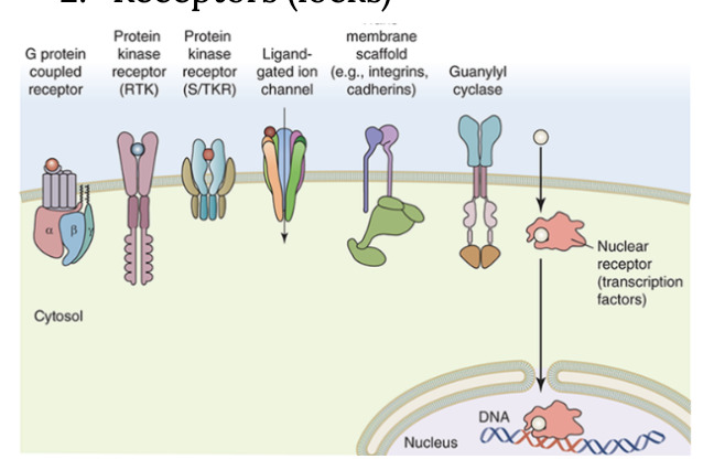
describe GPCR
A superfamily of receptors
Has hundreds of genes coding for receptors
Help detect smell, flight or fight
Structure:
7 transmembrane domains (H1-H7)
Has a heterotrimeric G-protein with alpha, beta, gamma subunits that interact with each other
Function:
When a ligand binds to GCPR, causes a conformational change in the receptor
This activates coupled G-protein subunits

describe ion channel receptors/ligand gated channels
A channel in the PM
Transmits signal information by allowing ions to move from one side of membrane to another
When specific ligands bind → conformational change for channel → pores open → ions flow through
These proteins are not enzymes
For voluntary muscle contraction
Common for neurotransmitters
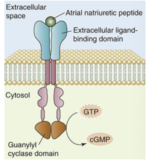
describe guanylate cyclase
A receptor
Bound to membrane or soluble in cytosol
Structure:
Has external ligand binding domain + transmembrane domain + internal catalytic domain
Soluble form: target for membrane-soluble ligands, mediating intracellular processes
Function:
When receptor is activated, catalytic domain of the membrane-bound guanylate cyclase converts GTP into cGMP
cGMP binds to other signalling proteins to initiate cellular processes
Important role in vision (convert light signal into electrical signal)
describe protein kinase and 2 classes
Not all of these are cell surface receptors, can also be cytosolic, affect enzyme activity, etc
In general: functions to phosphorylate other proteins with serine/threonine/tyrosine residues
Dysfunction can develop cancer
RTK (Receptor tyrosine kinase)
1. Before ligand binding, inactive receptors are separate polypeptides with inactive tyrosine kinase domains
2. After signalling molecule binds, this causes 2 subunits of the receptor to join together “dimerize”. Now, the kinase is active
3. Transautophosphorylation. Happens when the cytoplasmic tail of a subunit is brought close to the tyrosine kinase domain of the other subunit, and the opposite domain is phosphorylated on specific tyrosine amino acids.
4. The phosphotyrosine AAs are binding sites for more signalling proteins that pass the signal along the pathway
5. Ligand is released, and AAs are dephosphorylated by phosphoprotein phosphatases. Kinase resets to its inactive state (2 separate polypeptides)
S/TKR (serine/threonine kinase receptors)

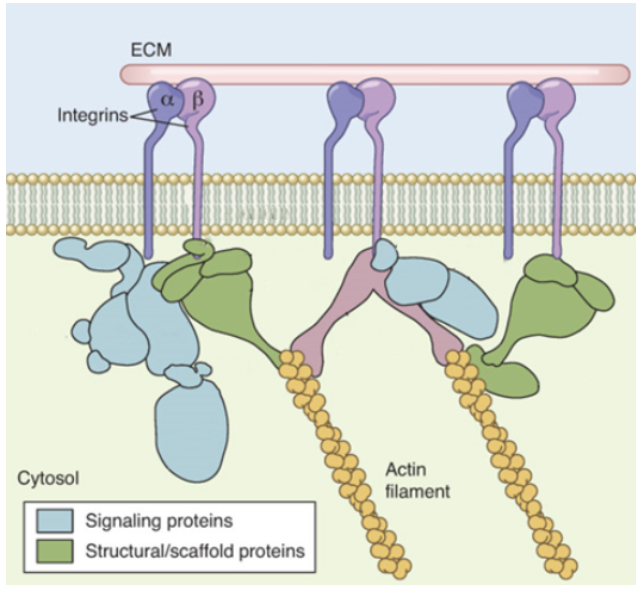
describe transmembrane scaffolds
Doesn’t always have a single, distinct function
Forms in large clusters of receptors and signalling proteins → can regulate signal transduction
Scaffold proteins = determine which signalling proteins can bind to a complex, associate with receptor, form signalling scaffold
Functions:
Brings signalling proteins together
Regulate signal transduction
Localize signalling proteins to specific cellular areas
Isolate specific signalling pathways
describe nuclear receptors
Found in cytosol
Some ligands (e.g. steroids) can directly cross PM and bind to these receptors
These receptors move through NPCs directly into nucleus
Inside nucleus, activated receptor complex can bind to DNA sequences (SRE, steroid response elements) to control gene expression
These receptors are also called transcription factors (they help regulate gene expression)
Can also respond to toxic substances

describe 6 classes of signalling proteins
G proteins, protein kinases, calcium binding proteins, adenylyl cyclase, lipid kinases, adaptor proteins
what are signalling proteins?
Purpose: to be the intermediary between receptor and effector
To transmit and amplify signal information
Can mobilize second messengers (non-protein molecules linking signalling proteins together to make more signalling pathways)
2 common features for all types:
1. Are highly mobile, can diffuse quickly through cytosol and may be able to move rapidly within PM (if membrane associated)
2. Can catalyze chemical reactions (enzymes) for signal amplification, or can bind to enzymes
describe G proteins
Bind GTP and propagate signals
2 classes:
Monomeric G-proteins | Heterotrimeric G-proteins |
Single polypeptides Has at least 2 different binding sites (one for GTP/GDP, one for target protein) + GTPase domain Not coupled to GPCR Is activated when GTP binds, then can bind to a target protein
| Has 3 different polypeptides Anchored to PM, activated by GPCR Alpha subunit = binds GTP/GDP and a target protein beta/gamma = attached together, stabilizes the inactive (GDP bound) form of alpha subunit |
how are G proteins activated?
4 steps
1. Binding
The heterotrimer with alpha and beta/gamma subunits is bound to GDP (inactive)
When a ligand binds to receptor, it changes conformation to interact with heterotrimeric G protein
2. Separate: Receptor protein causes exchange of GDP with GTP on alpha subunit
Heterotrimer separates into alpha and beta/gamma subunits
G proteins are active
3. Propagate
While separated alpha and beta/gamma subunits bind to downstream targets, propagating signal pathway
4. Cleave and reform. The alpha subunit cleaves GTP to form GDP. alpha and beta/gamma subunits bind to reform the heterotrimer. Returns back to inactive form
*Ga subunit catalyzes GTP hydrolysis. resulting Ga-GTP complex binds to Gby complex to reform heterotrimeric G complex

describe protein kinases
Enzymes that attach phosphate groups to tyrosine, serine, threonine amino acids
In addition to receptor protein kinases, there are also non-receptor protein kinases (most of them) = cytosolic
Cytosolic protein kinases = intermediaries, can activate other protein kinases/signalling proteins/phosphorylate effector proteins (enzymes) when activated
Usually, phosphorylating a target protein activates it, but some proteins are inactivated by it
Some protein kinases can enter nucleus but does not directly interact with DNA
Instead, they phosphorylate proteins that interact with DNA
describe calcium binding proteins
Ca2+ usually at low intracellular concentrations, so when levels increase, it can interact with other proteins
Calmodulin = a calcium-binding protein
When [Ca2+] rises, ca2+ binds to calmodulin inducing a conformational change (it wraps around binding site of target protein). Calmodulin can bind to its target protein
describe adenylyl cyclase
A nucleotide triphosphate is converted into another form
ATP is converted into cAMP, which perpetuates the signal
Adenylyl cyclase is not linked to membrane receptors
It binds to alpha subunit of heterotrimeric G-proteins
Adenylyl cyclase subunits
There are 2 types of heterotrimeric G-protein alpha subunits:
ɑs stimulates adenylyl cyclase
ɑi inhibits it
These 2 forms build different parts of heterotrimeric G-proteins and linked to different GPCR
describe lipid kinases
Phosphorylates phospholipids in cytoplasmic leaflet
Adds a phosphate to the polar head group
Results in a conformational change in phospholipid so it can bind to target protein in the membrane to pass the signal
describe adaptor proteins
Adaptors = proteins that are not receptors or enzymes
Have different binding domains that recognize phosphorylated AAs or other “activated” structures on signalling proteins
Forms part of the glue to hold signalling networks together
describe second messengers and key features
Refers to the nonprotein ions and molecules formed
Relay signalling information from signalling proteins to other cellular targets
First messengers = ligands that interact with receptors
Key features
Small
Diffuse rapidly in cytosol or membrane
Amplify signals so a few ligands can trigger larger responses
Doesn’t last long in cytosol
Second messengers (cAMP and cGMP) are degraded by phosphodiesterases
Ionic messengers like Ca2+ are moved into organelles
Other examples: DAG, IP3, NO
List the 4 steps in heterotrimeric G-protein signalling cascade
1. GPCRs
Over 800 different GPCRs that respond to stimuli (chemical or physical). Signal transduction pathway is initiated by binding of ligand to GPCR. enables the receptor protein to interact with heterotrimeric G protein
2. cAMP
Ligand-bound receptor stimulates replacement of GDP for GTP in Ga subunit
Causes heterotrimeric G-protein to dissociate from receptor and itself to leave a G(beta, gamma) subunit and an activated Gas-GTP
Gas-GTP can bind and activate signalling protein (adenylyl cyclase) to convert ATP into cAMP
3. PKA
cAMP can bind to PKA
Inactive PKA = tetrameric protein with 2 regulatory subunits + 2 catalytic subunits
When cAMP binds to regulatory subunits, the protein dissociates to release the active catalytic subunit
Catalytic subunit can phosphorylate cellular proteins
4. CREB
Active PKA catalytic domains can enter nucleus
Commonly targets CREB
CREB is phosphorylated by PKA and then binds CBP. These 2 proteins interact with DNA to initiate transcription
list 5 steps in phospholipid kinase signalling cascade
Ligand binds to GPCR, allowing receptor protein to interact with heterotrimeric G-protein. Ligand-bound receptor stimulates replacement of GDP for GTP in Ga. G-protein dissociates from receptor and itself to leave a G(beta, gamma) subunit and activated Ga-GTP
PLC
Ga-GTP binds the PLC (phospholipase C, a phospholipid kinase signalling protein)
PIP2/IP3
Activated PLC breaks down PIP2 to release 2 second messengers: DAG and IP3
Ca2+
IP3 diffuses in cytosol and activates its receptor on ER, opening a ligand-gated ca2+ channel
Ca2+ leaves ER and acts as a second messenger to activate ca2+ binding proteins
PKC
Membrane-bound diacylglycerol and cytosolic ca2+ bind to PKC, to activate it
PKC can phosphorylate targets
list 3 steps in protein kinase signalling cascade
1. FGF
Are proteins that stimulate growth of mammal cells
Binds to FGFRs receptors (a homodimeric receptor tyrosine kinase)
Binding of FGF to FGFR causes subunits to dimerize
FGFR then undergoes tyrosine transautophosphorylation to form phosphotyrosines on cytoplasmic side (can be bound by different proteins)
2. Grb2
An example of a phosphotyrosine binding protein
Is an adaptor protein
Binding to phosphotyrosine causes Grb2 to undergo conformational change to bind Sos
Sos is activated, binding to Ras (monomeric G-protein)
Replaces GDP with GTP, and Ras is now active
Ras binds to Raf (serine/threonine kinase)
Activated Raf can phosphorylate MEK (protein kinase) which phosphorylate another serine/threonine kinase (Erk)
3. Erk
Phosphorylated Erk forms a dimer that can phosphorylate other signalling proteins (cytosol/nucleus)
Erk enters nucleus to activate transcription factors
Initiate transcription
describe roles of lysosomes, proteasomes, peroxisomes
Lysosomes
Break down misfolded and damaged organelles/nucleic acids, etc
Proteasomes
Protein complexes that break down damaged and misfolded proteins in nucleus and cytosol
Peroxisomes
Handle dangerous free radicals (reactive oxygen species)
describe how cargo gets inside lysosome
M6P sugar tag is added onto cargo
Proteases are also directed towards lysosome using M6P (degrade proteins)
1. Vesicles deliver proteins to lysosome and are digested by proteases
2. Proteases are made in ER, tagged with M6P, delivered to lysosome by vesicles. Digest soluble and membrane proteins in lysosome
describe how lysosome digests
Responsible for breakdown of proteins from other organelles
Has high amounts of proteases
Also has enzymes to cleave and digest fats/sugars, can engulf organelles
Once broken down, are transported to cytosol for reuse
describe protein degradation by proteasome
Degrades intracellular proteins
Requires ubiquitination (form of post-translational modification of proteins)
Cytosolic proteins
Tagged with a polyubiquitin chain
Multiple ubiquitins needed for protein to be recognized by proteasome
Nuclear proteins
Proteasomes are also found in nucleus
Cell can degrade unwanted nuclear proteins without exporting it to cytosol (e.g. damaged histones)
Are polyubiquitinated in nucleus and degraded by nuclear proteasomes
describe peroxisomes
A place to keep and use these ROS safely using enzymes (catalase)
Are small, membrane bound organelles
Proteins = essential peroxisome proteins, made in cytosol, targeted to peroxisome by PTSs (peroxisomal targeting signals)
Decompose cargo like uric acid, AA, FA
list 2 types of cellular death
apoptosis
necrosis
describe apoptosis and 4 steps
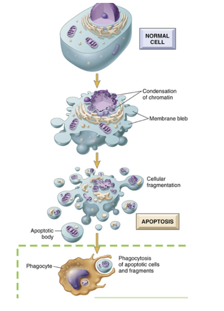
Regulated cell death
Energy consuming process
Protects body from damaged cells
Also used in development
4 steps:
1. Initiation
Using 2 different pathways, intrinsic and extrinsic
Cell initiates apoptosis
Intrinsic = originates in the outer membrane of mitochondria. Intracellular signals (Severe DNA damage, ROS, etc) turns on intrinsic pathway
External signals initiate apoptosis. Extrinsic pathway uses a PM receptor (death receptor). Neighbouring cells (immune cells) release death ligands, which binds to death receptors on damaged cell.
2. Membrane blebbing and enzyme activation
Cell shrinks and form blebs (first obvious sign of apoptosis)
Caspases are activated
Initiator caspases are activated by intrinsic/extrinsic pathway
Cleave and activate other caspases (executioner caspases)
3. Cell structure changes
Cell changes structure
DNA is fragmented, DNA repair halts. Nuclear membrane breaks down.
Cytoskeleton is disassembled
PM phosphoipid content changes with scramblases
PS is exposed on exoplasmic leaflet of PM
Organelles enclosed in apoptotic bodies
4. Engulfment
Phagocytes endocytose apoptotic bodies to dispose
Are disposed of by phagocytes’ lysosomes
Minimally disturbs cells
describe 3 steps in necrosis
A result of cellular injury
Cells will attempt to repair themselves
Show swelling in mito and ER
May return to normal
Occurs during severe damage
1. Damage
Cell is damaged beyond repair
Can be due to: toxins, extreme heat/radiation, freezing, ischemia (blood flow stopped), pathogens, mechanical trauma
2. Organelles begin to lose structures and swell
vacuoles/undefined bodies form
DNA may be degraded
3. destructed
Cell membrane and organelles lose structure
Can see holes
Cellular content spills, producing inflammatory signals
Mitochondria releases proteins, lysosomal contents exposed
Nearby cells are exposed to remains, and may be damaged
Unlike apoptosis, it’s difficult to clean up cellular remains
compare and contrast apoptosis and necrosis
Apoptosis | Necrosis |
Caused by DNA damage, withdrawal of growth factors/nutrients, detach from substrate, attack by cytotoxic lymphocytes | Trauma |
Key features:
| Key features:
|
End result
| Inflammatory response |