Exercise 11: Mammalian Anatomy & Physiology Study Guide
 Call Kai
 Learn
 Practice Test
 Spaced Repetition
 Match
 Flashcards
 Knowt Play
1/83

Earn XP
Description and Tags
Vocabulary flashcards covering mammalian anatomical cavities, external rat sex determination, superficial and deep musculature, human skeletal structures, internal organ systems, urogenital anatomy, and major circulatory vessels.
84 Terms
Thoracic Cavity
Body cavity superior to the diaphragm that contains the heart (pericardial cavity) and lungs (pleural cavities).
Abdominal (Peritoneal) Cavity
Body cavity inferior to the diaphragm containing the stomach, liver, intestines, kidneys, and internal reproductive organs.
Vaginal Orifice
Diaphragm
Sheet of skeletal muscle separating the thoracic and abdominal cavities that contracts to expand the thorax during breathing.

Urethral orifice

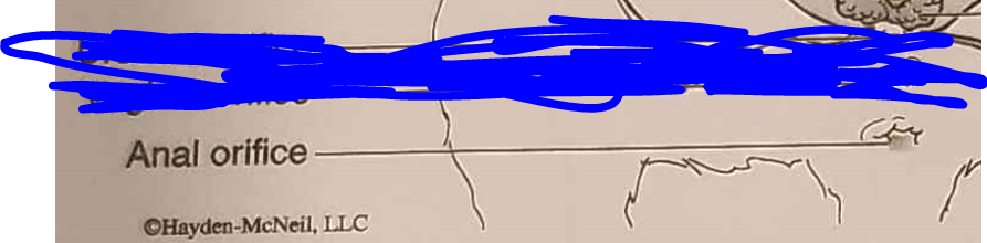
Anal orifice
Male Rat Sex Characteristics
Anatomical features including a prominent scrotal sac containing testes at the posterior end, a single urogenital orifice on the penis, and a larger anogenital distance.
Female Rat Sex Characteristics
Anatomical features including three distinct posterior openings (urethral orifice, vaginal orifice, and anal orifice), visible nipple rows on the ventral abdomen, and a shorter anogenital distance.
Masseter
Muscle located at the cheek / jaw (ventral and dorsal view) that elevates the mandible to close the jaw during chewing/mastication.

Biceps brachii
Muscle on the anterior upper forelimb (ventral view) that flexes the elbow and forelimb.

Triceps brachii
Muscle on the posterior upper forelimb (ventral and dorsal view) that extends the elbow and forelimb.
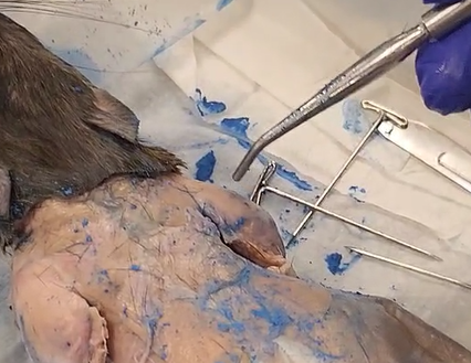
Pectoralis major / minor
Muscle located across the chest / ventral thorax that adducts and draws the forelimb toward the chest.

Latissimus dorsi
Muscle of the mid-back / dorsolateral trunk that pulls the forelimb dorsally and posteriorly.

External oblique
Superficial lateral abdominal muscle that compresses the abdomen and flexes the vertebral column.

Internal oblique
Muscle of the deep lateral abdominal wall that compresses the abdomen and aids in trunk rotation.
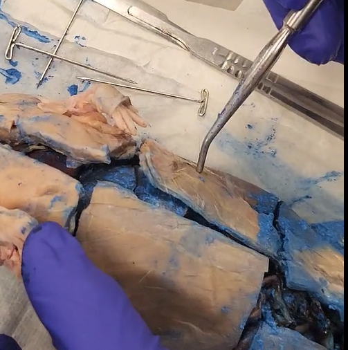
Rectus abdominis
Muscle along the ventral abdominal midline that compresses abdominal contents and flexes the lumbar spine.

Internal intercostals
Deep muscle layer located between ribs that depresses the ribs during forced expiration.
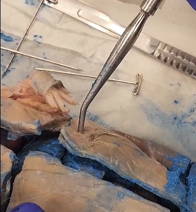
Gracilis
Muscle of the medial thigh (ventral view) that adducts the hindlimb.

Biceps femoris
Muscle of the lateral thigh (dorsal view) that flexes the leg at the knee and extends the thigh at the hip.

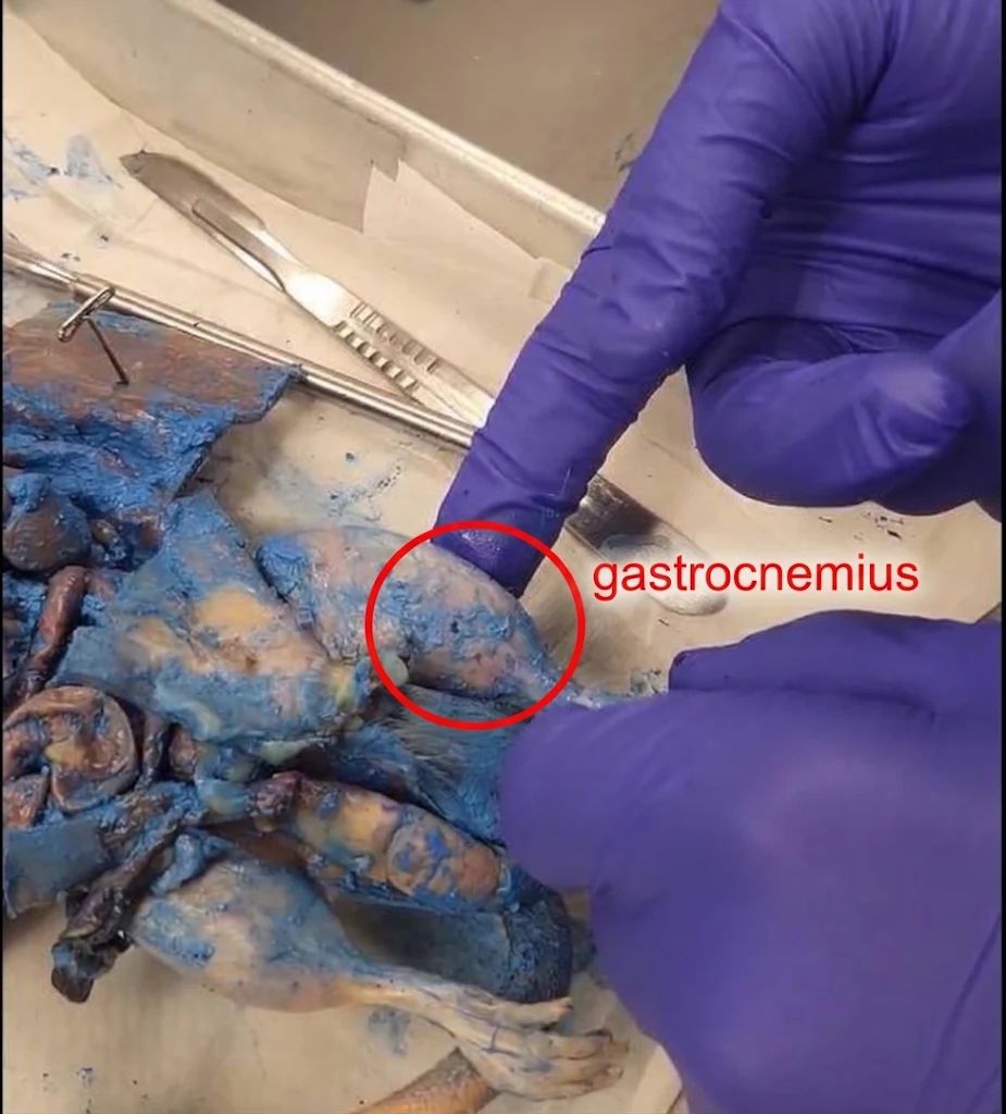
Gastrocnemius
Calf muscle on the posterior calf (dorsal view) that plantar flexes the hindfoot. It is on the underside of the biceps femoris
Cranium
Axial skeleton structure composed of bone that encloses and protects the brain.

Mandible
Axial skeleton bone forming the lower jawbone; it is the largest and strongest bone of the face and holds the lower teeth.

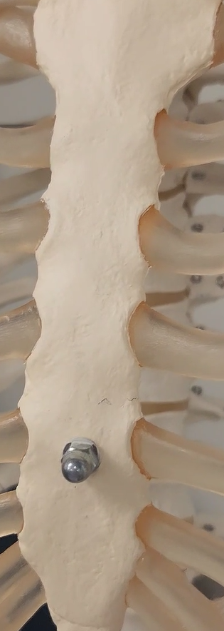
Sternum
Axial skeleton breastbone; a flat bone along the anterior chest midline that attaches ribs via costal cartilage.
Ribs
Axial skeleton structure consisting of 12 pairs of curved bones forming the thoracic cage to protect the heart and lungs.

Sacrum
Axial skeleton triangular bone composed of 5 fused vertebrae forming the posterior wall of the pelvis.
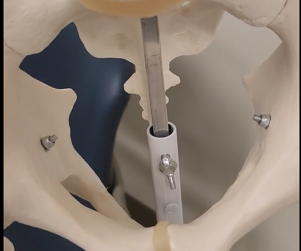
Coccyx
Axial skeleton tailbone; a small bone composed of 3–5 fused vertebrae located below the sacrum.
Clavicle
Appendicular skeleton bone (pectoral girdle); collarbone connecting the sternum to the scapula to anchor the upper limb to the trunk.

Scapula
Appendicular skeleton bone (pectoral girdle); shoulder blade, a flat triangular bone on the posterior thorax articulating with the humerus.
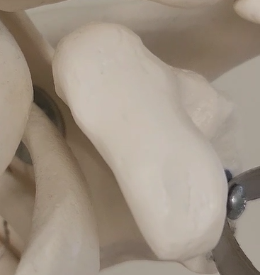
Humerus
Appendicular skeleton upper limb bone; a single long bone of the brachium (upper arm).

Radius
Appendicular skeleton upper limb bone; lateral bone of the forearm (thumb side) that allows forearm rotation. It is on the right side of this picture

Ulna
Appendicular skeleton upper limb bone; medial bone of the forearm (pinky side) forming the main hinge joint at the elbow. It is on the left side of this picture

Os coxa
Appendicular skeleton pelvic girdle bone formed by the ilium, ischium, and pubis; articulates with the sacrum and femur.

Carpals
Appendicular skeleton upper limb bones; a group of 8 short bones forming the wrist.
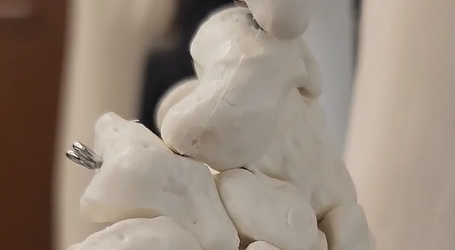
Metacarpals
Appendicular skeleton upper limb bones; five palm bones of the hand situated between the wrist and digits.


Phalanges (Hand)
Appendicular skeleton upper limb finger bones; 14 per hand arranged as proximal, middle, and distal, with the thumb having only 2.
Femur
Appendicular skeleton lower limb thigh bone; the longest, heaviest, and strongest bone in the human body. It is the top part of this image

Patella
Appendicular skeleton lower limb kneecap; a sesamoid bone protecting the knee joint.
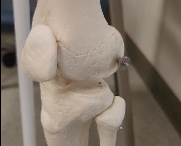
Tibia
Appendicular skeleton lower limb shin bone; the larger, weight-bearing medial bone of the lower leg.

Fibula
Appendicular skeleton lower limb bone; a slender lateral bone of the lower leg that serves primarily for muscle attachment.

Tarsals
Appendicular skeleton lower limb bones; a group of 7 short bones forming the ankle and heel.
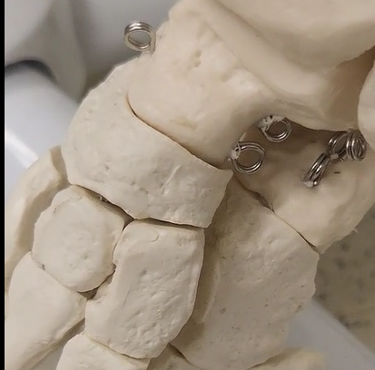
Metatarsals
Appendicular skeleton lower limb bones; five bones forming the arch and instep of the foot.

Phalanges (Foot)
Appendicular skeleton lower limb toe bones; 14 per foot arranged as proximal, middle, and distal, with the hallux/big toe having 2.

Esophagus
Muscular tube that transports swallowed food from the pharynx to the stomach.
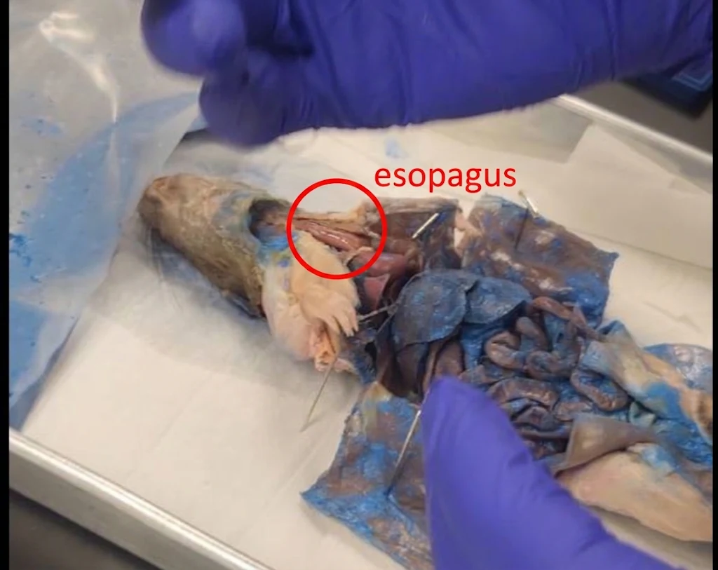
Stomach
Digestive organ that stores food, initiates protein digestion using HCl and pepsin, and churns contents into chyme.


Pyloric Sphincter
Smooth-muscle valve that controls the release of chyme from the stomach into the duodenum. it is underneath the liver

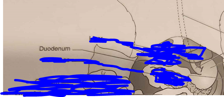
Duodenum
First C-shaped section of the small intestine that receives chyme, pancreatic enzymes, and bile for primary digestion.


Pancreas
Glandular organ that secretes digestive enzymes into the duodenum and hormones (insulin and glucagon) into the bloodstream.
Liver
Digestive organ that produces bile and processes nutrients.

Spleen
Lymphoid organ that filters blood, removes old red blood cells (RBCs), and stores lymphocytes.
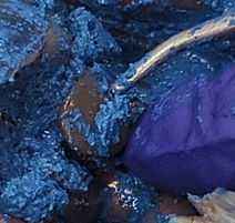
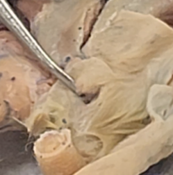
Mesentery
Double-layered membrane that supports intestinal loops and houses blood vessels. It is like the filmy stuff that is between the large and small intentine.

Caecum
Digestive organ pouch that ferments fiber.

Large Intestine
Digestive organ that reabsorbs water and salts from indigestible waste material.

Rectum
Terminal segment of the large intestine that stores feces before elimination.
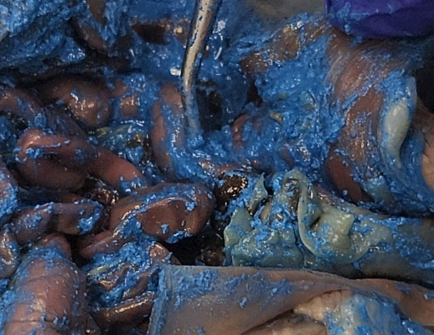
Anus
External sphincter-controlled muscular outlet for defecation.
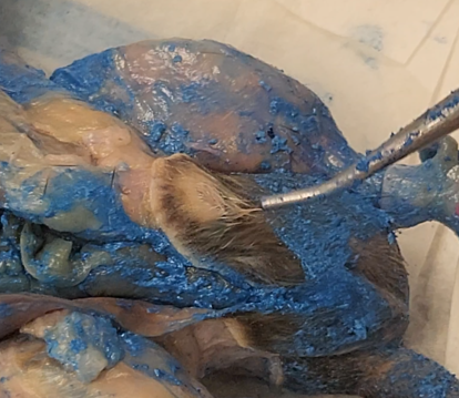
External Nares
Nostrils that serve as the entrance for air into the nasal cavity.
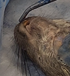
Larynx
Voice box; a cartilage structure housing the vocal cords and guarding the entrance to the trachea.

Trachea
Windpipe reinforced with C-shaped cartilage rings to maintain an open airway.

Lungs (Right & Left)
Primary organs of respiration; the right (multilobed) and left lungs contain alveoli for oxygen and carbon dioxide exchange.

Heart
Four-chambered muscular pump situated in the mediastinum that circulates blood throughout systemic and pulmonary circuits.

Atria (Right & Left)
Receiving chambers of the heart: the right atrium receives deoxygenated systemic blood, while the left atrium receives oxygenated pulmonary blood. They are on opposite sides of the heart. If you are looking at the rat from the right side, the left atrium is on the right side, and the right atrium is on the left side.
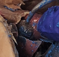
Mandibular (Salivary) Gland
Exocrine gland located beneath the jaw that secretes saliva containing amylase and mucus to lubricate food.

Thyroid Gland
Endocrine gland positioned anterior to the trachea that regulates metabolic rate via thyroid hormones (T3/T4).

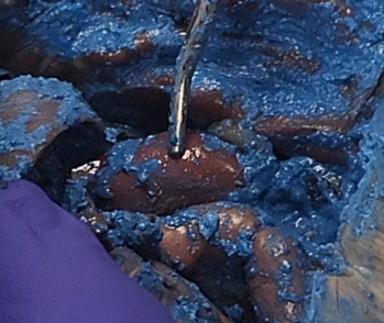
Thymus Gland
A bilobed lymphoid organ superior to the heart that serves as the site for T-lymphocyte maturation for immune defense. They are covered in this picture

Adrenal Gland
The adrenal gland sits atop each kidney and produces stress hormones, including cortisol and epinephrine. It is directly above the kidneys

Ureter
Excretory duct that drains urine from the kidney to the urinary bladder.

Urinary Bladder
Muscular sac that stores urine prior to excretion.

Ovary
Female gonad that produces ova and estrogen.

Oviduct
Female reproductive duct that transports ova from the ovary to the uterus.

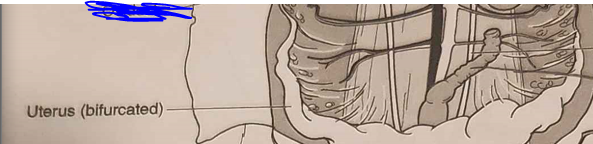
Bifurcated Uterus
Uterine structure in rats featuring two Y-shaped horns designed to accommodate multiple developing fetuses (litters).
Vagina
Muscular canal extending from the uterine body to the external environment.

Clitoris Gland
Small sensory erectile structure homeologous to the male penis.

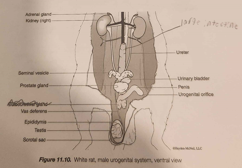
Testis
Male gonad producing sperm and testosterone; housed externally inside the scrotal sac for temperature regulation.

Epididymis
Male reproductive structure where maturing sperm are stored.

Vas Deferens
Male reproductive duct that conducts sperm from the epididymis to the urethra.

Seminal Vesicle & Prostate Gland
The small vesicle is located on the left, and the prostate gland is situated on the right when viewing the rat on its back. The small vesicle is on the left, and the prostate gland is on the right when looking at the rat on its back.

Common Carotid Arteries (Right & Left)
Arterial vessels that supply oxygenated blood to the head, neck, and brain. They should be above the heart, and they are not exposed in this picture.

Aortic Arch
Main arterial trunk exiting the left ventricle that supplies oxygenated blood to the systemic circulation. It is right above the heart

Jugular Vein
Venous vessel that drains deoxygenated blood from the head and neck back toward the heart. It is not exposed in the picture, but it is in the general area

Right Superior Vena Cava
Venous vessel returning deoxygenated blood from the upper body and forelimbs to the right atrium. It is located on the left side of the heart if looking down at a rat on its back. It is not exposed in the picture


Posterior (Inferior) Vena Cava
Venous vessel returning deoxygenated blood from the lower body and abdomen to the right atrium. It should be at the bottom of the heart, and it is not exposed.
Maxilla
The upper jawbone that holds the upper teeth and forms part of the orbits of the eyes.

Vertebreas
The individual bones that make up the spine, providing structural support and protecting the spinal cord.

Small Intestine
The part of the gastrointestinal tract where most of the digestion and absorption of nutrients occurs. It is located between the stomach and the large intestine.

Kidney
A vital organ that filters blood to produce urine, regulating fluid balance, electrolytes, and blood pressure.