Lecture 14: Fundamentals of Spine Pain
 Call Kai
 Learn
 Practice Test
 Spaced Repetition
 Match
 Flashcards
 Knowt Play
1/37

Earn XP
Description and Tags
Does not include surgeries
38 Terms
Low Back Pain Stats
2nd most common symptom-related reason to visit a physician
Lifetime prevalence 70-90%
Often resolves spontaneously, but high rate of recurrence
Biopsychosocial impact of pain
Physical impairment
Emotional burden
Decreased quality of life
Occupational impact
Cost of loss of productivity time among US workers from common pain conditions is > 61 billion dollars/year
Large burden financially & on resources
Patients with chronic pain use health care services up to 5X more frequently
Spine Anatomy Sections
ANTERIOR COMPARTMENT
VERTEBRAL BODIES
DISCS
NEURAXIAL COMPARTMENT
SPINAL CORD
CAUDA EQUINA
NERVE ROOTS
POSTERIOR COMPARTMENT
FACET JOINTS
MUSCLES

Differential Diagnosis of Back Pain
Mechanical
Non-mechanical
>90% of back pain is mechanical
Mechanical Causes of Back Pain
Lumbar strain
Herniated nucleus pulposus
Internal disc disruption
Compression fractures
Spondylolysis
Facet arthropathy
Spondylolisthesis
Sacroiliitis
Non-Mechanical Causes of Back Pain
Tumor
Infection
Inflammatory Arthritis
Visceral Diseases
Misc
Causes of Back Pain - Tumor
PRIMARY SPINAL TUMOR, METASTASES, MULTIPLE MYELOMA, LYMPHOMA
Causes of Back Pain - Infection
Vertebral osteomyelitis, diskitis, HIV, shingles, TB
Causes of Back Pain - Inflammatory Arthritis
Ankylosing spondylitis
Causes of Back Pain - Visceral Diseases
Nephrolithiasis, Prostatis, PID
Causes of Back Pain - Misc
Hemoglobinopathies
Psychosomatic D/O
Neuropathic Joints
More
Need of immediate spine imaging for mechanical low back pain?
Immediate routine lumbar spine imaging in patients with low back pain and without features indicating a serious underlying condition did not improve outcomes compared with usual clinical care without immediate imaging.
Routine lumbar imaging is associated with radiation exposure (radiography and CT) and increased healthcare costs
Red Flags for back pain
Unexplained fever
Unexplained weight loss
Neurologic deficit
Incontinence
Saddle anesthesia
Trauma
Age > 50
Night pain (laying down removes gravity)
History of IVDU
History of cancer
Immunosuppression
Prolonged use of corticosteroids
Facet Joint Pain
Posterior Compartment Pain
15-40% of non-radicular low back pain
40-60% of non-radicular neck pain
Can co-exist with other pain generators
H&P & imaging often unable to predict if pain originates from the facets
Lumbar Facet Joint Pain
With prolonged standing in lordotic posture, 16% of axial load assumed by facets
With lumbar spondylosis and degenerative disc disease → up to 70% of axial load can be transmitted to the facets
Facets are subject to microtrauma, capsular tears, synovial inflammation, degenerative arthritis, segmental instability, and post-surgical adjacent level stress
Low back pain with possible radiation into the buttocks, groin, and proximal leg
Dull, achy, deep, difficult to describe
Worse with twisting, bending, & standing
No neurologic deficit
Degenerative changes may be seen on radiologic studies
Neuraxial Compartment Pain
Typically causes radicular > axial pain
Causes:
Disc herniation into central canal or foraminal zone
Ligamentum flavum thickening, facet hypertrophy, and/or spondylosis
Can lead to:
Radiculopathy
Spinal stenosis
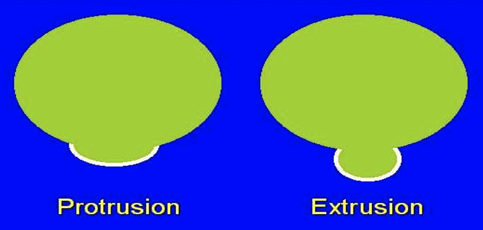
Protrusion vs Extrusion
PROTRUSION –DEPTH IN ANY PLANE < BASE OF HERNIATION IN SAME PLANE
EXTRUSION –DEPTH IN ANY PLANE > BASE OF HERNIATION IN SAME PLANE
Lumbar Disc Sequestration
Fragment of HNP is separated from main part of the disc

Nerve Root Pain
Mechanical Irritation:
Disc herniation or foraminal stenosis can compress the nerve root and/or DRG, causing abnormal neuronal discharges along the nerve root
Chemical Irritation:
Disc degeneration releases inflammatory mediators resulting in abnormal discharges from the DRG
Symptoms of Radiculopathy
Sharp, stabbing, shooting pain in affected extremity
Worse with coughing, sneezing, or straining
May have +SLR and neurologic deficit
May have loss of reflex, weakness, and/or gait impairment
Common Mechanisms Radiculopathy
lifting heavy object while bending and twisting at the waist
Repetitive spinal motions can be causative
L4 or L5 radiculopathy can cause
Foot drop

Etiology Spinal Stenosis
CONGENITALLY SMALL CANAL SIZE
DEGENERATIVE OSTEOPHYTES
SPONDYLOLISTHESIS
FACET JOINT HYPERTROPHY
THICKENING OF LIGAMENTUM FLAVUM
DISC HERNIATION
Spinal Stenosis
Hallmark symptom: Neurogenic claudication
Pain, fatigue, and/or paresthesias in lower extremities
Pain provoked by prolonged standing or walking
Relieved by rest or leaning forward on objects (shopping cart sign)
Anterior Compartment Pain: Discogenic pain
Disc degeneration can spread radially into the annulus fibrosis causing annular fissure
Nerve endings are only in the outer 1/3 of the annulus
Worsens with prolonged sitting, stooping forward, bending/twisting at the waist, valsalva maneuvers
Excessive axial compression may result in endplate edema and fracture
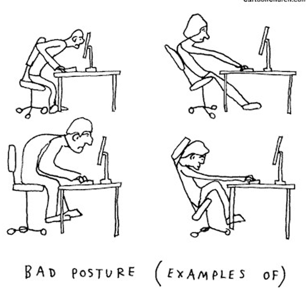
Posture and Back Pain

Anterior Compartment Pain: VERTEBRAL COMPRESSION FRACTURES
Most common fracture in osteoporotic patients
• ~ 700,000 fractures annually
• ~ 260,000 patients with 1st painful fracture diagnosed each year
• After 1st vertebral compression fracture, the risk of subsequent fracture increases 5-FOLD
Vertebral Compression Fractures
Acute > chronic back pain
Can be traumatic or related to minor activity
Sharp mechanical pain, difficulty finding a comfortable position
Pain typically improves with rest or lying down

Location of vertebral fractures
MOST COMMON LOCATIONS:
MIDTHORACIC REGION (T7–T8) THORACOLUMBAR JXN (T12–L1)
MIDTHORACIC REGION
THORACIC KYPHOSIS IS MOST PRONOUNCED & LOADING DURING FLEXION IS HEIGHTENED
THORACOLUMBAR JUNCTION
THE RELATIVELY RIGID T-SPINE CONNECTS TO MORE FREELY MOBILE LUMBAR SEGMENTS
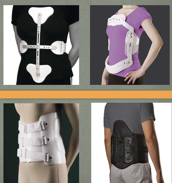
Bracing for Compression Fractures
CASH or Jewett Brace for thoracic compression fractures
Lumbosacral Orthosis (LSO) for lumbar compression fractures
Management of Spine Pain
REHABILITATION & MODALITIES
PHARMACOLOGIC THERAPY
PSYCHOLOGICAL THERAPY
INTERVENTIONAL PROCEDURES
Postural Training
Physical Therapy
Hamstring Stretches
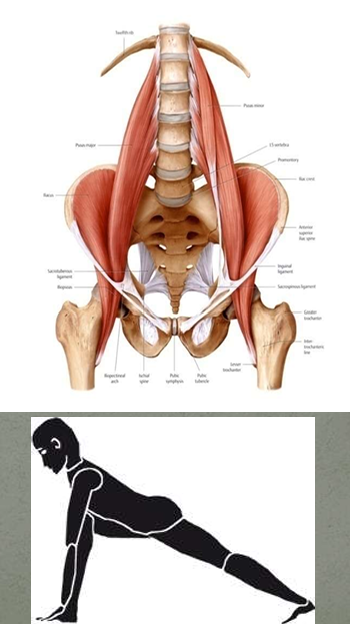
Psoas Stretches
Dynamic Lumbar Stabilization
McKenzie Exercises
Aquatic Exercises
Hamstring Stretches
defined connection between tight hamstrings and low back pain
Tight hamstrings → limited pelvic motion → increased strain on the lumbar spine

Psoas Stretches
Psoas: Primary hip flexor
Bilateral Psoas muscle action → efficient stabilization of lumbar spine in frontal plane
Range of exercises that aim to maintain the spine in neutral position, while progressing from:
static (lying) to dynamic (standing or jumping)
resisting gravity to resisting additional force
predictable to unpredictable movements
Muscular control of spine & core strengthening can reduce pain

McKenzie Exercises

Aquatic Exercise
Water-based exercises produced better improvement in disability and quality of life of the patients with CLBP than land-based exercise