powerpoint 7 - thorax, heart and lungs
 Call Kai
 Learn
 Practice Test
 Spaced Repetition
 Match
 Flashcards
 Knowt Play
1/112

There's no tags or description
Looks like no tags are added yet.
113 Terms

true ribs
ribs 1-7

false ribs
ribs 8-10

floating ribs
ribs 11-12

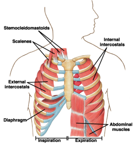
two main intrinsic muscles of the thoracic wall
external intercostal, internal intercostal

action of external intercostal muscles
inspiration
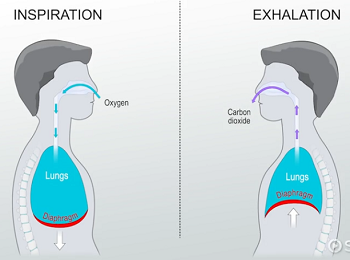
action of internal intercostal muscles
forced expiration
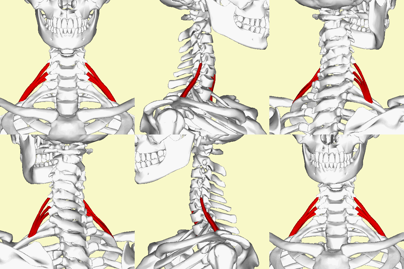
four main extrinsic muscles of thoracic wall
pectoralis major, pectoralis minor, serratus anterior, scalene

what scalene does not attach to the first rib
posterior scalene
what is the insertion, origin and innervation of the pectoralis major
anterior humerus, medial 1/3 of clavicle/sternum/ribcage, no innervation


horizontal adduction of humerus
what is the action of the pectoralis major
what is the insertion, origin and innervation of the pectoralis minor
coracoid process, ribs 3-5, no innervation
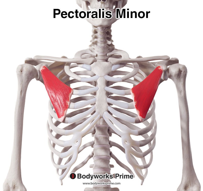
what is the action of the pectoralis minor
inspiration

what is the insertion, origin and innervation of the serratus anterior
medial border of scapula, ribs 1-8, long thoracic nerve

what is the primary action of the serratus anterior? secondary?
holds scapula to ribcage, protract the scapula and upward rotation

what is the insertion, origin and innervation of the anterior and middle scalenes
first rib, transverse process, no innervation

what is the insertion, origin and innervation of the posterior scalene
second rib, transverse process, no innervation

what is the primary muscle of forced inspiration? secondary?
diaphragm, external intercostals/scalenes/pectoralis minor

phrenic nerve C3-C5
what innervates the diaphragm
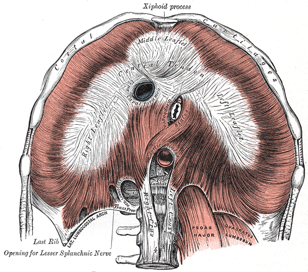
aortic aperture
what is the most inferior and posterior hole of the diaphragm

caval aperture
what is the most superior and anterior hole of the diaphragm

esophageal aperture
what is the most middle hole in the diaphragm

lungs
organ of respiration responsible for exchanging gases into/out of blood

left lung
which lung is smaller

cardiac notch
where is the heart located in the left lung

apex
what part of the heart sits within the cardiac notch

root
connects lung to mediastinum contains pulmonary vessels, nerves and bronchi

bronchopulmonary segments
discrete pyramidal units of lobes; divided by visceral pleura in the lungs

pleura
fibroserous membrane surrounding each lung and lining pulmonary cavities

parietal, visceral
layers of pleura superficial to deep (two)
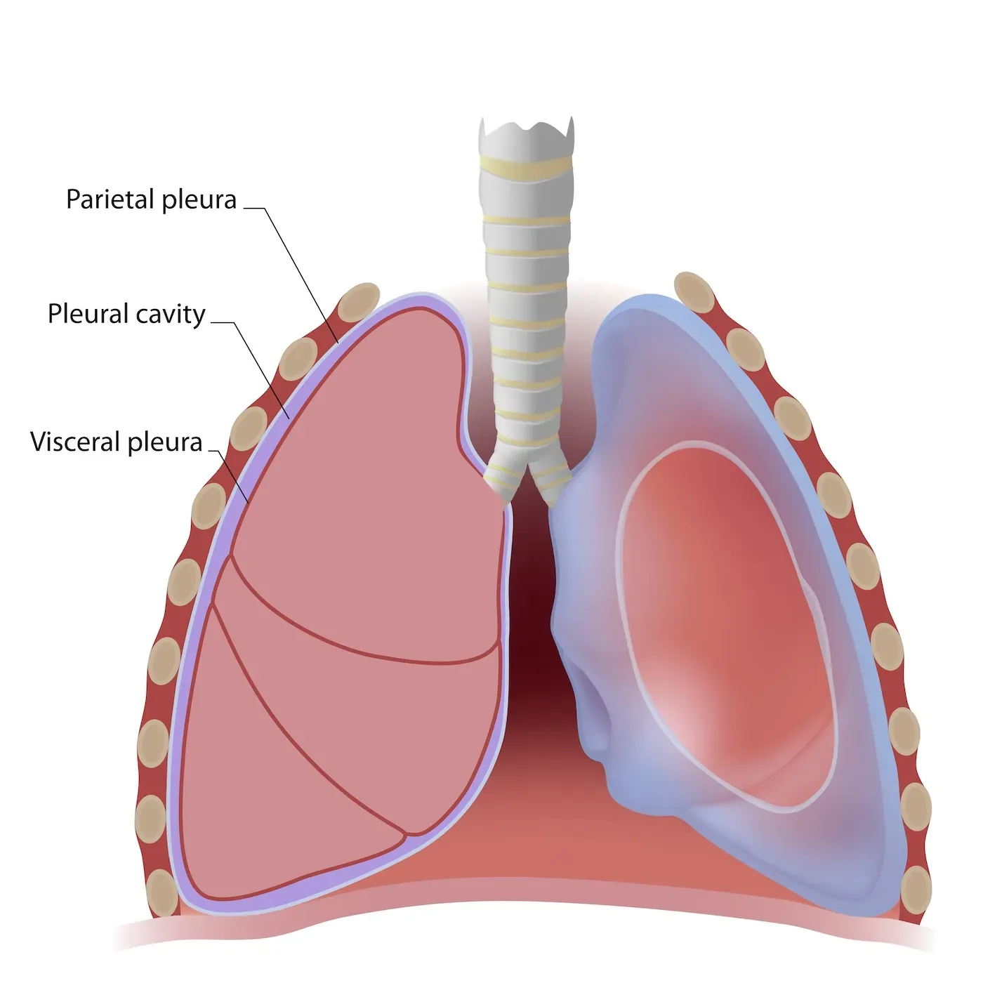
pleural cavity
potential space within pleural sac between visceral and parietal of the pleura; contains thin layer of serous fluid

pleuritis
inflammation of the pleura; creates friction and may produce adhesions between layers

nose route pathway of air up to common pathway
external nares
nasal cavity
nasal vestibule
internal nares
nasopharynx
common pathway
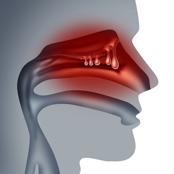

mouth route pathway of air up to common pathway
oral cavity
past tongue, hard palate, soft palate and uvula
common pathway
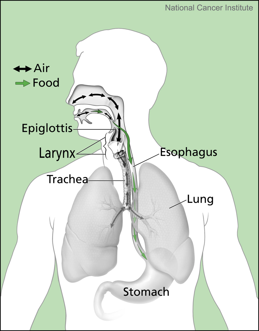
common pathway of air
oropharynx
laryngopharynx
trachea
tracheal bifurcation/carina
primary bronchi
secondary bronchi
tertiary bronchi
conducting bronchiole
terminal bronchiole
respiratory bronchiole
alveolar sac
alveolus
what does the hyoid bone articulate with
nothing
what is the epiglottis made of
elastic cartilage

what is a majority of the larynx made of
hyaline cartilage

what are the membranes of the larynx made of
dense regular connective tissue

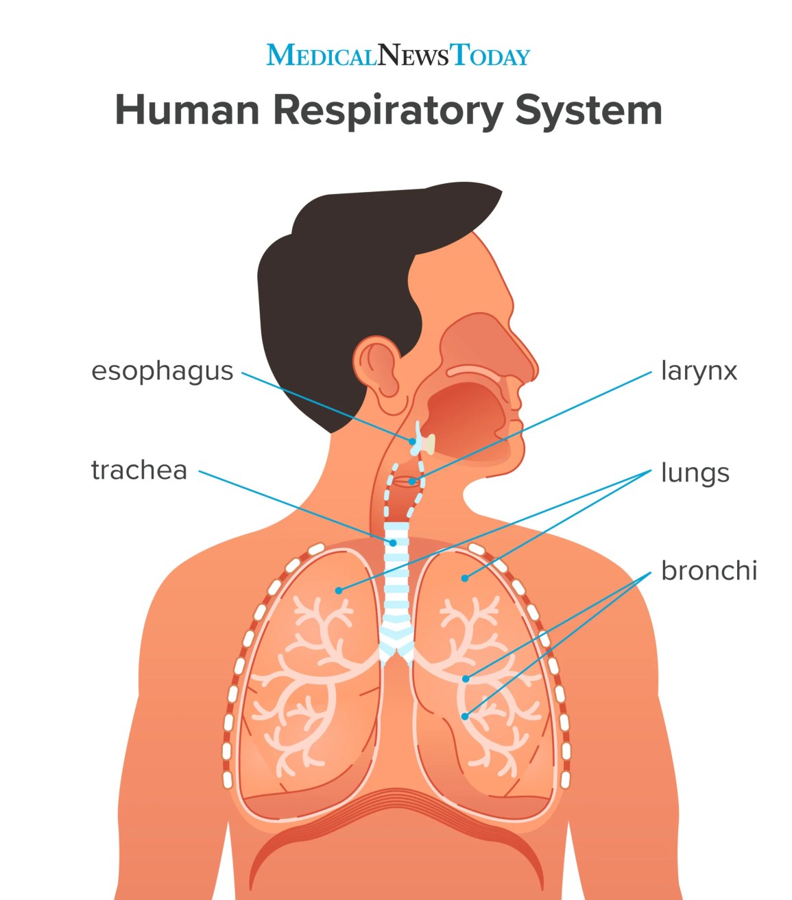
trachea
large membranous tube with cartilage rings

carina
internal split of the trachea

tracheal bifurcation
external split of the trachea

respiratory components
air conduction and gas exchange in location where alveoli are founds

what are the three respiratory components
bronchioles, alveolar sacs, alveoli

pneumocyte
type of cell in the alveoli responsible for gas exchange.

type I
pneumocyte that absorbs surfactant

type II
pneumocyte that produces and secretes surfactant

surfactant
a protein-phospholipid film that lowers the surface tension of alveoli

pulmonary artery
takes deoxygenated blood to lungs

pulmonary vein
takes oxygenated blood to heart

bronchial artery
supplies bronchial tree

bronchial vein
drains root of each lung
what is the cause of most respiratory system issues
diffusion distance

asthma
obstructed bronchioles
how is asthma treated
inhaler
emphysema
destroyed or damaged alveoli; reduces surface area for gas exchange
how is emphysema treated
slowing the damage of alveoli

choking
obstruction of upper airway due to an object
mediastinum
region between pulmonary cavities

heart, great vessels, pericardium, esophagus, trachea, thymus
what six items are in the mediastinum
layers surrounding the heart superficial to deep (include spaces)
fibrous pericardium, parietal layer of serous pericardium, pericardial cavity, visceral layer of serous pericardium


fibrous pericardium
tough inelastic connective tissue surrounding the heart
layers of the heart superficial to deep
epicardium, myocardium, endocardium

what does the myocardial layer of the heart contain
cardiomyocytes

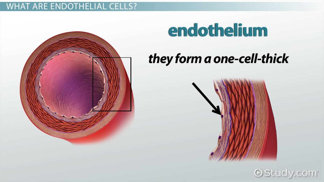
what is the endocardium made up
thin endothelium tissue

phrenic nerve C3-C5
what innervates the pericardium
heart
hollow, muscular organ that pumps blood through the circulatory system

auricles
extension/pouches of atria

pectinate
what muscle is found on auricles of the heart

trabeculae carneae
ridge like muscles found only in ventricles of the heart

chordae tendineae
cords connecting cusps of heart valves to papillary muscle

papillary muscles
muscles that contract when valves close to prevent prolapse of valve cusps

right atrioventricular valve, tricuspid valve
names for the valve that connects the right atrium and right ventricle

names for the valve that connects the right ventricle and pulmonary artery
pulmonary valve

left atrioventricular valve, bicuspid valve, mitral valve
names for the valve that connects the left atrium and left ventricle

left semilunar valve, aortic valve
names for the valve that connects the left ventricle and aorta

cardiac skeleton
dense fibrous connective tissue that separates chambers and provides anchoring points for muscle fibers/valves
which ventricle is thicker
left ventricle

blood circulation through the heart
superior/inferior vena cava
right atrium
tricuspid valve
right ventricle
pulmonary valve
pulmonary artery
arterioles
capillaries (at lungs)
venules
pulmonary vein
left atrium
bicuspid valve
left ventricle
aortic valve
base of aorta

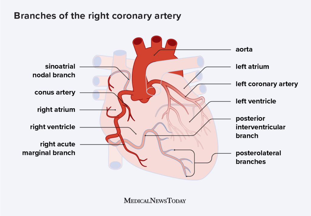
what arteries does the right coronary artery lead to
right marginal artery, posterior interventricular artery

what arteries does the left coronary artery lead to
anterior interventricular artery, left circumflex artery

what vein corresponds with the right marginal artery
small cardiac vein
what vein corresponds with the posterior interventricular artery
middle cardiac vein
what vein corresponds with the anterior interventricular and left circumflex arteries
great cardiac vein
where do all cardiac veins drain
coronary sinus
where does the coronary sinus drain
right atrium
what is the most selfish organ
heart
conduction system
coordinated electrical transmission leading to contraction of atria and ventricles
conduction system pathway
sinoatrial node
internodal bundles
atrioventricular node
bundle of his
right/left bundle branches
purkinje fibers
diastole
relaxation of the heart; 2/3 of cardiac cycle where ventricles elongate and filling occurs
systole
contraction of the heart; 1/3 of cardiac cycle where ventricles shorten and empty
initial diastole
atria and ventricles relaxed; AV and semilunar valves are closes
late diastole
atria fills, AV valve opens and blood passively fills ventricles
lub
sound produced by the closure of AV valves
dub
sound produced by the closure of semilunar valves

aortic valve stenosis
failure of valve to open fully; most common valve abnormality

heart murmur
one or both cusps prolapse; sounds caused by retrograde blood flow

pericarditis
inflammation of pericardium

cardiac tamponade
accumulation of fluid in pericardial cavity
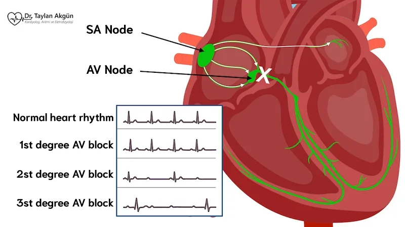
AV conduction system blocks
partial/complete blockage of conduction between atria and ventricles
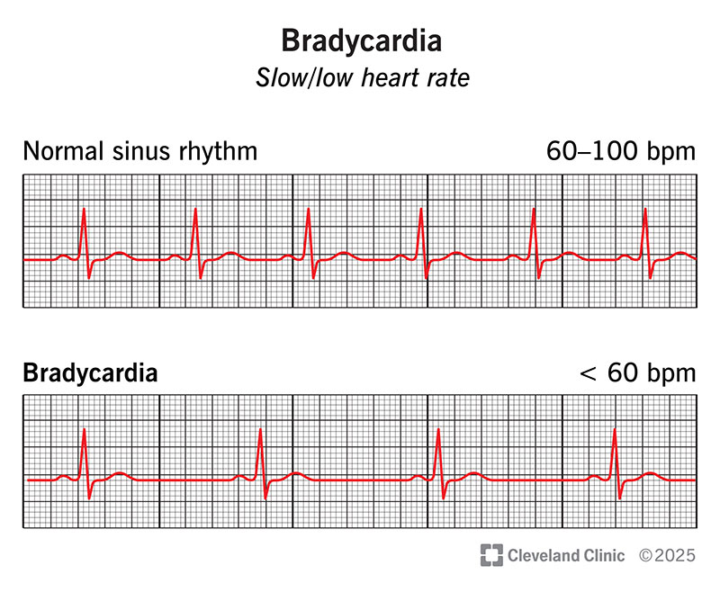
bradycardia
slow heartbeat