Body Tissues
 Call Kai
 Learn
Learn Practice Test
 Spaced Repetition
 Match
Match Flashcards
 Knowt Play
1/107

There's no tags or description
Looks like no tags are added yet.
Name | Mastery | Learn | Test | Matching | Spaced | Call with Kai | Chat |
|---|
No analytics yet
Send a link to your students to track their progress
108 Terms
Tissue
group of cells with a similar structure and common function
Epithelial
covering
connective
support
muscle
movement
nervous
control
Epithelium
sheet of cells that cover a body surface or line a cavity/forms boundaries between different environments. Skin separates body tissues from the environment and dinner linings of the bladder, stomach, and lungs.
Functions of epithelium
Protect, absorb, filtrate, excrete, secrete, and sensory reception
cellularity
Composed of tightly packed cells
Specialized contacts
Tight Junctions and desmosomes they are like snaps and hooks that hold adjacent cells tightly together and prevent molecules from passing between them
Polarity
each epithelial cell has 2 surfaces apical surface, and basal surface
Apical surface
The exposed surface (air,liquid,etc..)
Basal Surface
The attached surface
Basement Membrane
Composed of 2 layers (basal lamina and reticular lamina) not made of cells, boundary between epithelium and connective tissue, and reinforced epithelium by resisting stretching and tearing.
Basal Lamina
Adhesive sheet at the basal surface of epithelium. sticky and filters material from below.
Reticular lamina
Thin layer of collagen fibers at superficial surface of connective tissue
Innervated
Has nerves
Avascular
No blood vessels
How get O2 and Carbs?
Diffusion from blood vessels in the underlying connective tissue
Regeneration
Epithelium has a high regeneration capacity which means it makes more of themselves
Why does regeneration happen
You’re always losing epithelial cells from friction, damage, bacteria, and acid - but they’re replaced at the same rate you lose them
Microvilli
(some have) finger like extensions of the cell membrane function is to increase surface area = increased secretion or increased absorption
Cilia
(some have) small hair-like structures on outside of a cell to propel substances

Mobile cell
moves the cell (not in epithelium or humans)
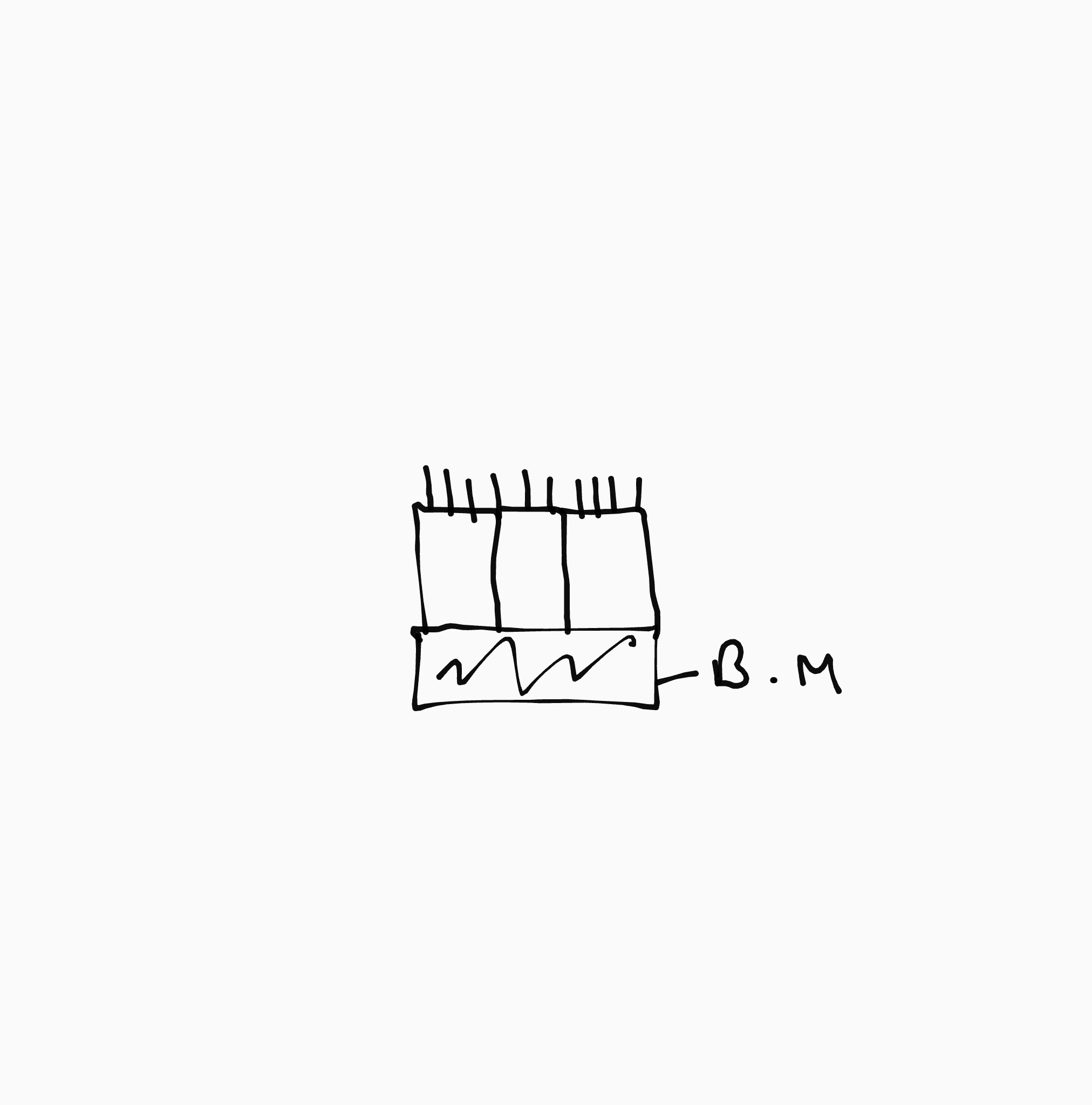
Stationary cell
Moves other stuff along the exterior of the cell (ex- egg in Fallopian tubes/mucus in trachea)
Classification of epithelium
Based on cell shape and arrangement
Simple
single layer of cells
Stratified
two or more layers of cells
squamous
Flat and scalelike
Cuboidal
Boxlike
Columnar
Tall and column shaped
When viewed from the top, all epithelial cells are…
6 sided (hexagon) and fit very tightly with their neighbors
Epithelial cells shape and order
Looks like honeycomb, and tight Junctions and desmosomes hold adjacent cells tight and do not allow stuff to pass between them
Why are hexagons stronger than boxes?
Fit tightly together with no gaps, attached to 6 other cells = stronger than 4, and joints are staggered and don’t line up=stronger
Connective tissue
Found everywhere in the body, and most abundant of the 4 tissue types
4 groups of CT in your body now
Connective tissue proper, cartilage, bone, blood
5 general functions of CT
Binding, support, protection, insulation, and transport of substances
All types of CT are derived from what during embryonic development
Mesenchyme
CT can be what regarding blood vessels?
Can be vascular/avascular/or anywhere in-between
CT is composed of 3 main elements
Ground substance (not alive), fibers (not alive), cells (alive)
Matrix
The nonliving parts of CT, composed of ground substance and fibers, bears weights, and withstands great and absorbs impact
Ground substance
Amorphous material that fills space between cells + contains fibers. and holds fluids
CT is a molecular sieve which means
Nutrients and other substances (O2,CO2) diffuse between the capillaries and the cells imbedded within it
Fibers
provide support (Collagen, Elastic, and Reticular)
Most abundant, made of fibrous protein (collagen) extremely strong - high tensile strength, and support for matrix
Collagen fibers
Made of a a fibrous protein elastin, allows stretching and recoils to original shape, more elastic fibers where CT expands and contracts EX - Blood vessels, and Lungs. Lots found in skin and elasticity decreases with age as the fibers wear out from use=wrinkles
Elastic fibers
Thin collagen-like fibers that branch out in between collagen fibers, branch out extensively like a spider web, and abundant where CT joins other tissues
Reticular Fibers
Blast
actively mitotic phase of a cell/secretes GS and fibers/Younger
Cyte
less active phase/a mature cell just doing its job/maintains the health of the matrix/if its area becomes injured, it reverts to a blast cell
Plasma cells
Produces antibodies (immune response)
Mast Cells
Detect foreign substances and initiates a local inflammatory response.
Macrophage
Phagocytizes foreign materials (bacteria,dust,junk) Engulfs dead cells+tissues and breaks them down (pac-man)
Mesenchyme
CT in the embryo that gives rise to the 4 other types of CT. None in your body right now!
Loose CT
cells are more spread out then in dense CT
Areolar CT
Located under the epithelium, and its function is to hold fluid in tissues = inflammation (swelling+bloating), and wraps and cushions organs
Adipose CT (fat)
Located under the skin/behind eyeballs/ and in breast and the function is to store reserved fuel/insulates against heat loss/some protection
Reticular CT
Location is lymphoid organs and function is it supports other cell types
Dense CT
Cells packed more tightly than loose CT
Tendons
attached muscle to bone
Ligaments
attach bone to bone
Dense regular CT examples
Tendons and ligaments both have great tensile strength
Dense irregular CT
Located dermis of skin/fibrous joint capsules and function is able to withstand tension exerted in many directions EX-Rotator cuff
Cartilage
Structurally between dense CT and bone, resists both tension and compression, tough but flexible, avascular/no nerves, heals slowly - avascular, less flexible as you age.
Perichondrium
well vasculized tissue that supplies oxygen and nutrients to cartilage cells
Hyaline cartilage
function is to support and reinforced and location is costal cartilage (skeleton)/external nose/ embryonic skeleton
Elastic cartilage
Function is maintains shape while allowing great flexibility and location is external ear
Fibrocartilage
Super strong and its function is great tensile strength/absorbs shock and pressure located in the inter vertebral discs / discs of knee joint
Bone
Hard-solid matrix with living cells throughout (absorbs calcium which makes it hard)
Blood
Liquid - filled matrix with living cells throughout
Fluid matrix in blood
Plasma
Nervous Tissue
No regeneration, parts are brain, nerves and the spinal cord. Composed of neurons and supporting neurons
Neurons
Generate and conduct never impulses
Supporting cells
Non conducting cells that support, protect, and insulate neurons
Muscle Tissue
Highly cellular and well vascularized
Skeletal Muscle
Function is voluntary movement, manipulation of environment, facial expressions and location is all skeletal muscles attached to bones or skin
Cardiac muscle
Cells contract pushing blood from the heart through the entire body and then back to the heart/involuntary. Location is the walls of the heart
Smooth muscle
Function is to propel substances or objects through internal passageways (food,urine,baby,etc) involuntary. location is the walls of hollow organs
Regeneration
Destroyed tissue is replaced by the SAME kind of tissue
Fibrosis
Destroyed tissue is replaced by SCAR tissue (fibrous CT)
3 steps of tissue repair
Inflammation - organization - regeneration or fibrosis
Inflamation
Injured cells release histamine and wound hormones that initiate tissue repair.
Capillaries dilate to do what?
brings WBC, plasma,clotting proteins + antibodies to the injured area which causes swelling.
Clotting proteins form
Blood clot
Blood clots happen to do what?
Stop the loss of blood, holds wounds ends together, and prevent penetration of bacteria or toxins
Scab
hardened outer portion of blood clot that is exposed to the air
Oragnization
New capillaries bud from nearby uninjured ones/must happen before all others can
Function of fibroblasts in organization process
Synthesize new collagen fibers to permanently connect the would ends together
What do macrophages do in the organization process?
digest the original clot and all the dead cells in the area
Blood clot becomes replaced by…?
granulation tissue (light pink), which releases a bacteria inhibiting substance
What kind of wounds is regeneration for
Minor to mild wounds
In regeneration how does surface epithelium regenerate?
epithelium cells migrate under the scab from the edges toward the center of the wound (how pieces of the scab fall off)
What kind of wounds is fibrosis for
more severe wounds
How does fibrosis work?
Epithelium regenerates just like regeneration, but the damaged tissue under the epithelium is replaced by fibrous CT which contracts and hardens as it matures
What does the fibrous CT in the process of fibrosis look like?
Visible under the epithelium as a scar
7 factors affecting tissue repair
type of tissue injured, type of injury,immediate care given, nutrition, blood supply,health, and age
What tissue heals the best
Epithelium
What tissue heals good
bone + fibrous CT
What tissue heals OK
Smooth muscle + dense regular CT
What tissue heals poor
Skeletal muscle + cartilage
What tissue heals the worst/not at all
Cardiac muscle + nervous tissue
In non-regenerating tissue and severe wounds, fibrous tissue replaces what
all lost tissue under the epithelium