The Visual System
 Call Kai
 Learn
 Practice Test
 Spaced Repetition
 Match
 Flashcards
 Knowt Play
1/63

There's no tags or description
Looks like no tags are added yet.
64 Terms
Provides sight for the ___ and __ of objects.
Provides __ movement control.
Provides information used in ___ and __ movement control.
Recognition and location
eye
postural * limb

Sight: Visual pathway from __ to __
Retina - cortex

Visual pathway begins with cells where that convert light into neural signals?
Retina
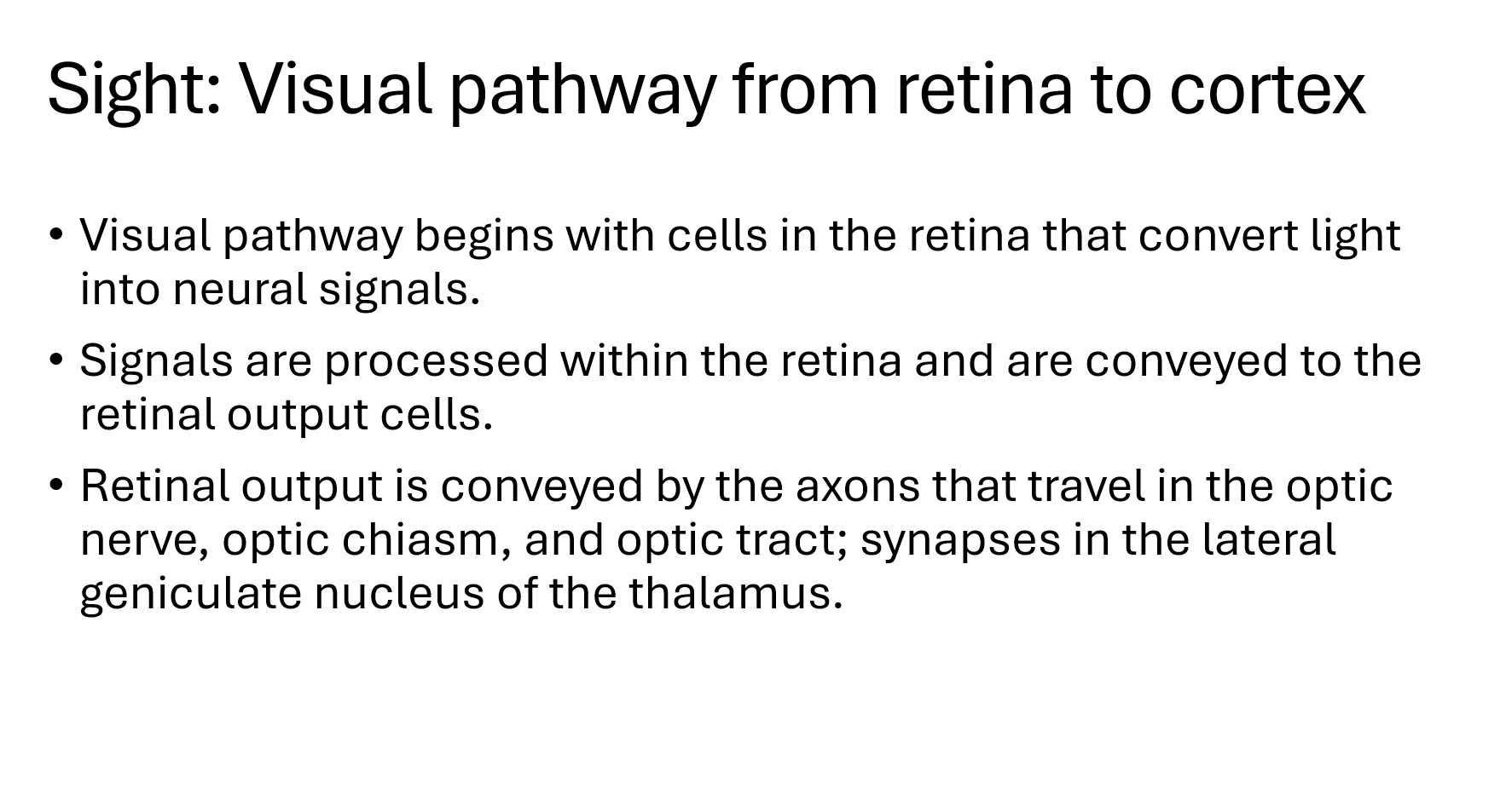
Signals are processed within the __ and are conveyed to the __
Retina - retina output cells
Retinal output is conveyed by the axons that travel in the?
optic nerve, optic chiasm, optic tract

Synapses in the?
LGN of the thalamus
Optic nerve is the bundle of axons that pass from __ to __
retina to optic chiasm
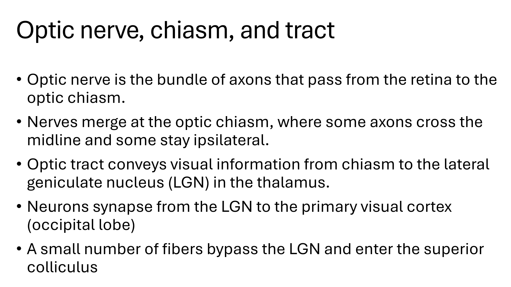
Nerves merge at the ___, where some axons cross the midline and some stay ipsilateral.
Optic chiasm
Neurons synapse from the __ to __
LGN to primary visual cortex (occipital lobe)
A small # fibers bypass the LGN and enter the ___
superior colliculus
Visual Pathways pathway

What area discriminates the shape, size, or texture of objects
info reaching the primary visual cortex stimulates neurons that do this
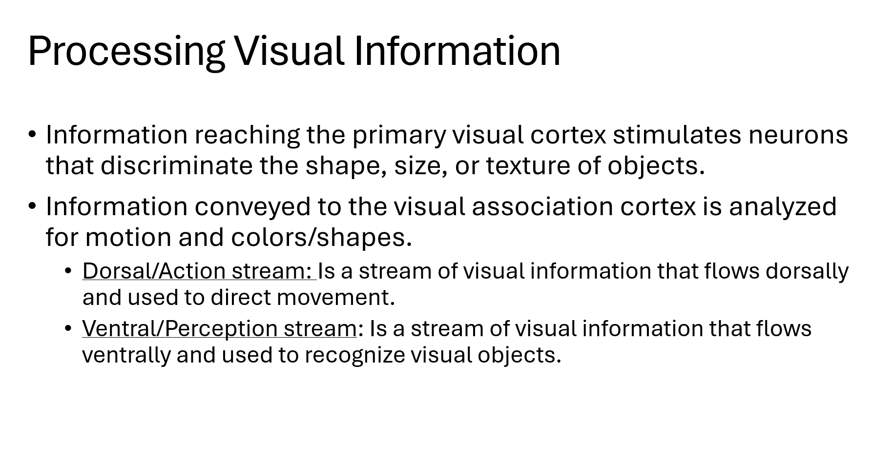
What area analyzes info for motion and color/shapes
Visual association cortex
What stream is used to direct movement?
Dorsal/action stream


What stream is used to recognize visual objects
Ventral/perception stream


A small number of fibers bypass the thalamus/LGN and go directly to the?
superior colliculus in the midbrain

Retina to Superior Colliculus
Function of
Tectum:
Pretectal area:
Tectum

Summary of Visual Pathways

Normal eye movements require synthesis of information concerning:
•Head movements (__ information)
•Visual objects (__ information)
•Eye movement and position (__ information)
•Selection of a visual target (__ and __ areas)

Eye movement system
Precise control of the ____ is vital for vision.
What part of the retina provides the best visual acuity?
fovea

To see one clear image, both eyes must aim at the same object so that the image lands on matching spots on each retina.
If the eyes are not aligned, vision may become blurry or double.
binocular vision: using both eyes together to see 1 image
Both eyes must point at the same object → image falls on corresponding retinal points → single clear image.
Fovea vs Peripheral Vision

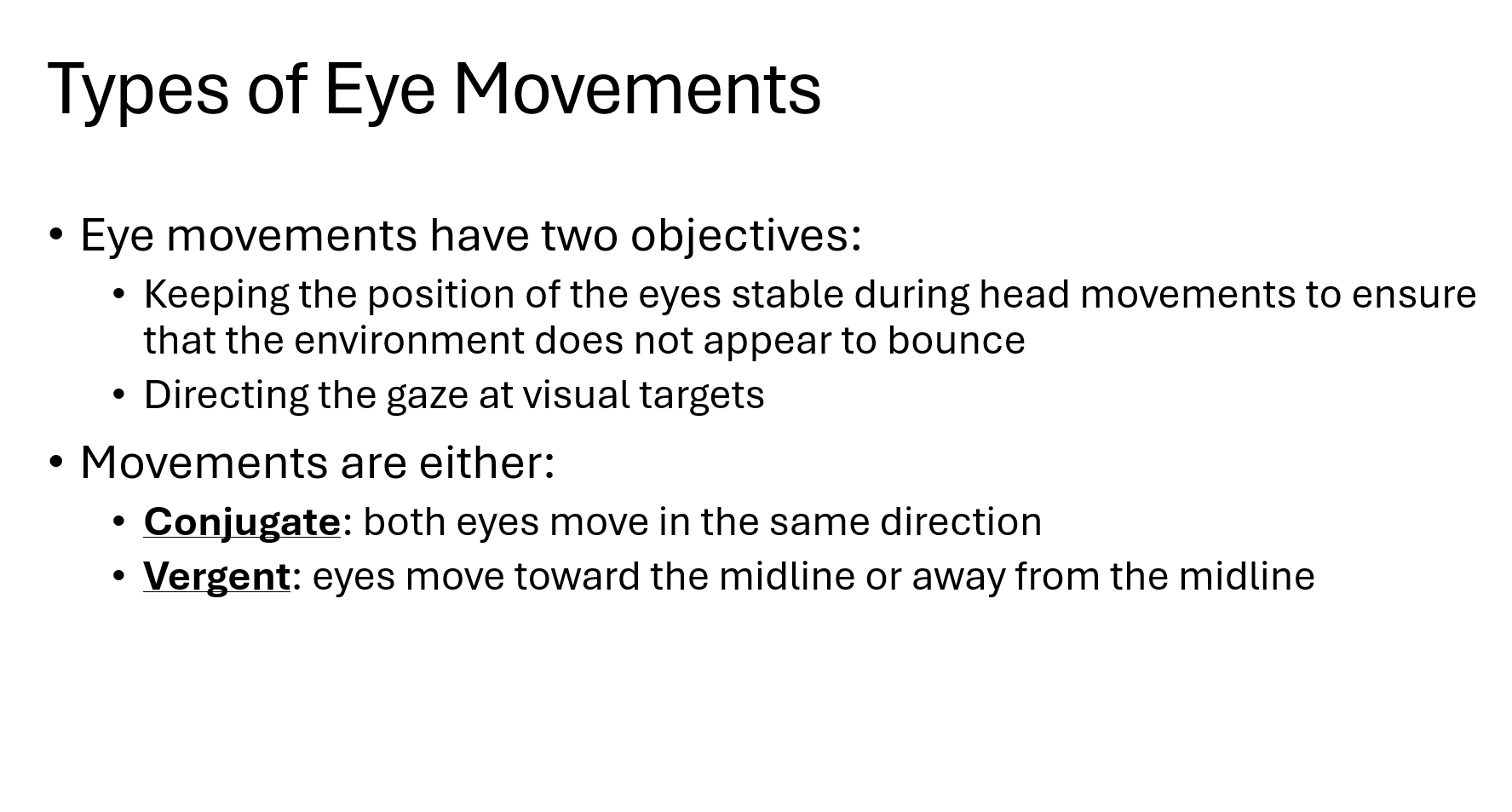
Eye movements have two objectives:

Eye movements are either
both eyes move in the same direction?
Conjugate
Eye movements are either
eyes move toward the midline or away from the midline?
Direction of gaze is accomplished by what 3 things?
Saccades, smooth pursuits, vergence movements

fast eye movements to switch gaze from one object to another
Saccades

eye movements that follow a moving object
smooth pursuits

movement of the eyes toward or away from midline to adjust for different distances between the eyes and the visual target

Vergence movements
What reflex stabilizes gaze during head movements?
Vestibulo-Ocular Reflex (VOR)

What is the main function of the Vestibulo-Ocular Reflex (VOR)?
To keep objects in focus during fast head movements via vestibular system
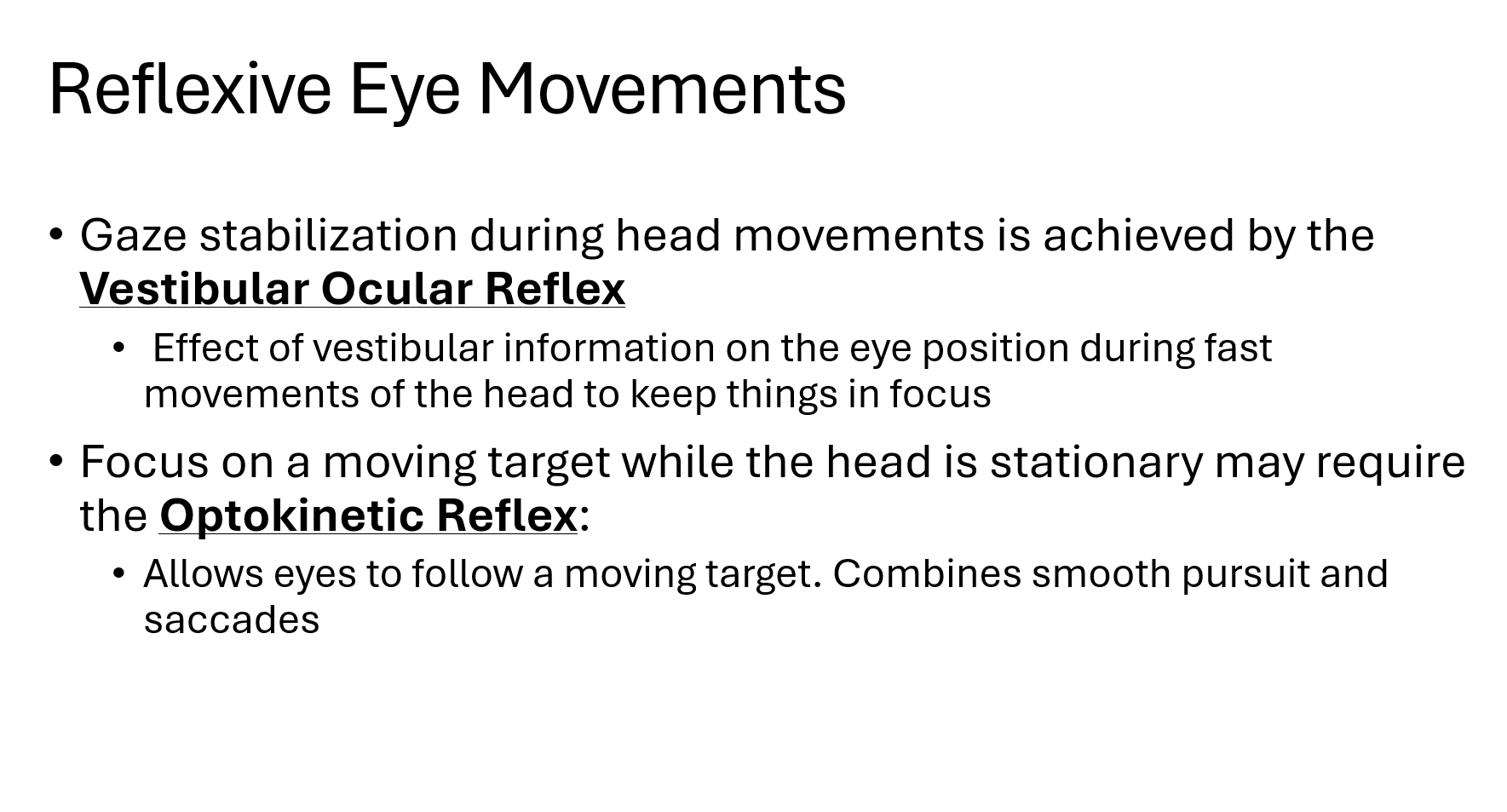
Focus on a moving target while the head is stationary may require the
Optokinetic Reflex (OKR)
What is the main function of the Optokinetic Reflex?
To allow the eyes to track a moving object.

What two types of eye movements are combined in the Optokinetic Reflex?
Smooth pursuit and saccades.

VOR vs. OKR?
VOR: Stabilizes gaze during head movement.
OKR: Tracks a moving object when the head is still.
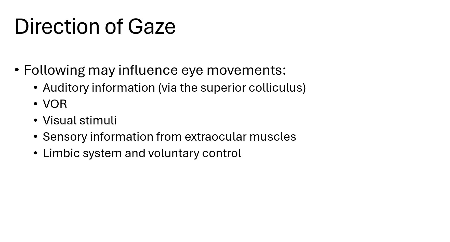
Following may influence eye movements:
quickly switch vision from one object to another.
Saccades
Saccades
What are the 2 ways saccades are generated?

Saccades
What 2 things control horizontal saccades?
used to follow a moving object.
Smooth pursuit
Smooth Pursuit
What is essential for the production of smooth pursuits movements?

Disorders of the Visual System

Cortical blindness occurs from damage to?
Loss of vision due to bilateral damage to the visual cortex.
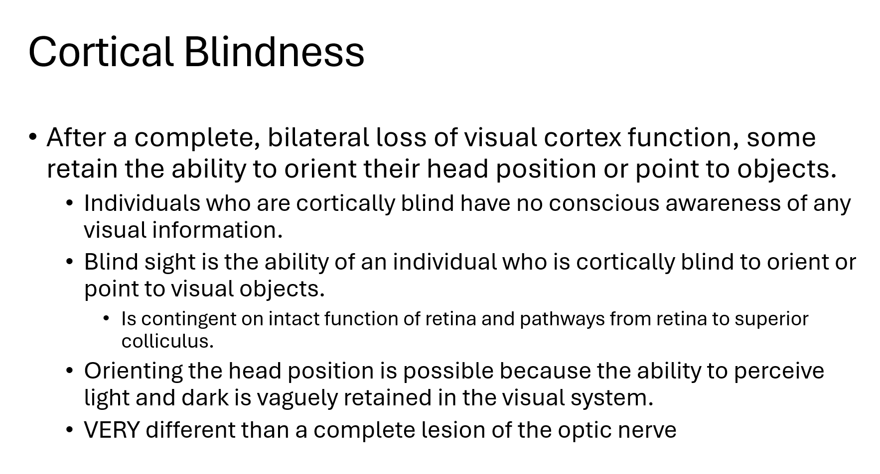
Can a person with cortical blindness consciously see objects?
No, they have no conscious visual awareness.

The ability of a cortically blind person to orient to or point to visual objects without conscious awareness of seeing them.
Blind sight

What pathways must remain intact for blindsight to occur?
The retina and pathways from the retina to the superior colliculus.
With blind sight, what is retained that allows them to orient head position
ability to perceive light and dark is vagally retained in the visual system
Abnormalities of eye movement occur with lesions involving:

MLF
Located:
Connects:
Responsible for:

Are deviations of the ability to align the eyes.
Tropia and Phoria

Is the deviation of one eye from a forward gaze when both eyes are open.
Tropia
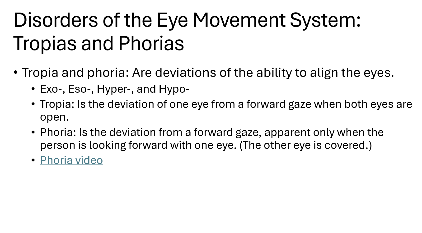
Is the deviation from a forward gaze, apparent only when the person is looking forward with one eye. (The other eye is covered.)

Naming Tropias

Cover test
Best to find?
Abnormal finding vs normal finding
Normal: eye does not move = properly aligned
Abnormal: uncovered eye moves to fixate on object
Any movement of the uncovered eye indicates a tropia.

Cover Test → Detects Tropia
Cover one eye.
Watch the other eye.
No movement = normal
Movement = tropia ✅
Alternate cover test (cross-cover test)
Best to find?
Abnormal finding vs normal finding
Cover one eye for at least 5 seconds.
Quickly move the cover to the other eye.
Repeat back and forth.
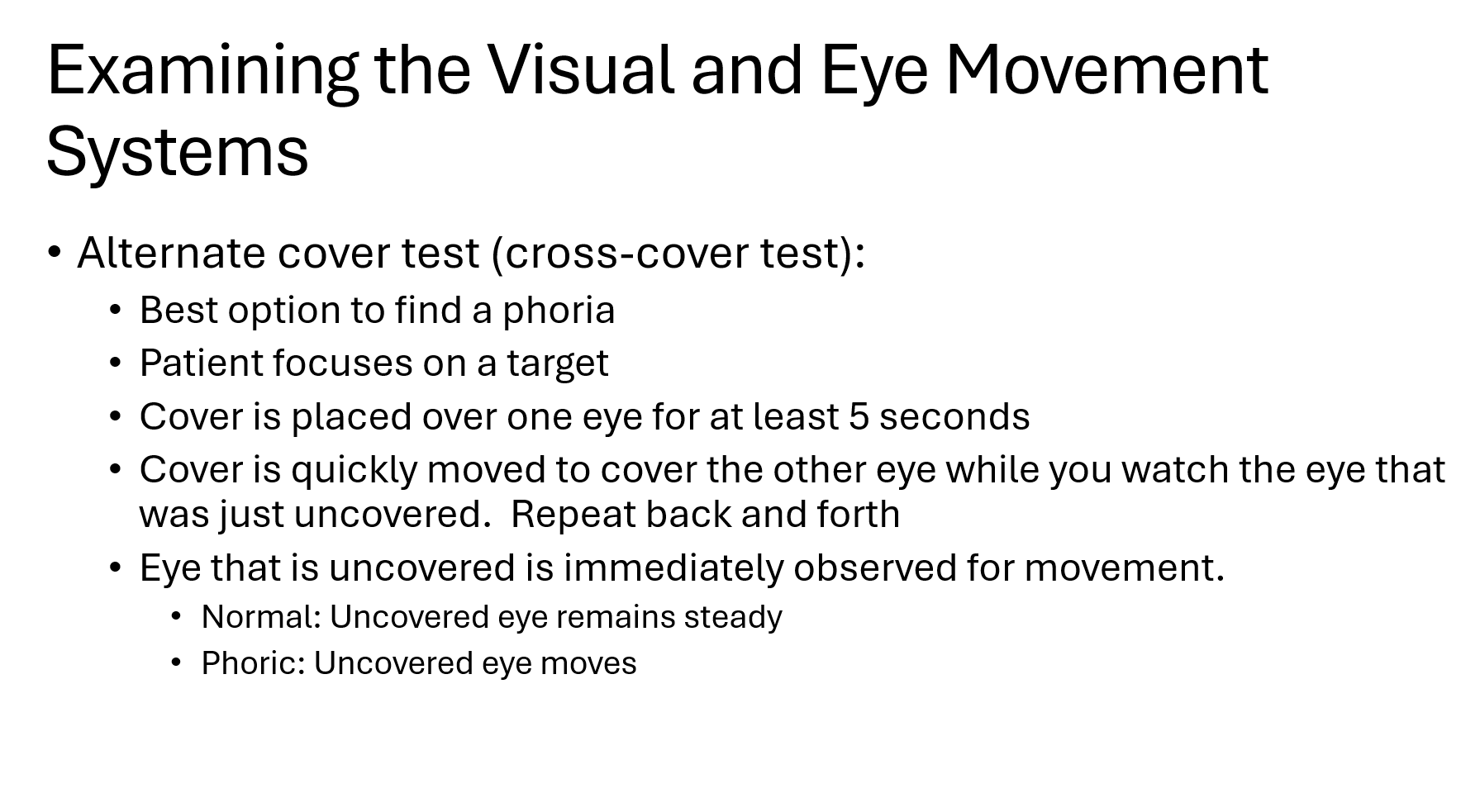
A phoria is hidden by binocular vision. When both eyes are open, the brain keeps the eyes aligned, so you can't see the deviation.
✅ Normal = No movement
Movement of the newly uncovered eye = Phoria
L visual field projects to?
R visual field projects to?
Left visual field projects to the right half of each retina
Right visual field projects to the left half of each retina
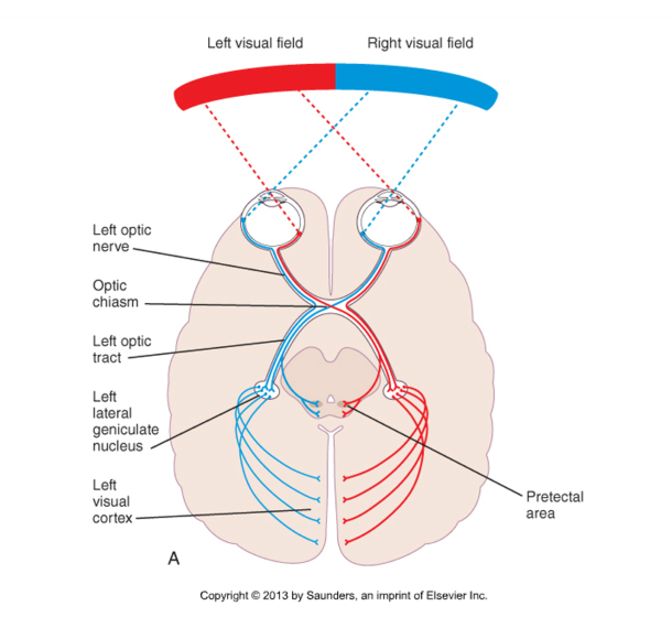
Do the nasal or temporal retinal fibers cross? where do they cross at?
Nasal = Crosses
Temporal = Stays
Nasal retina
Closest to the nose
Fibers cross at the optic chiasm
Temporal retina
Closest to the temples/ears
Fibers do NOT cross
Stay on the same side (ipsilateral)
The left visual field is detected by? (which part of retinas)
At the optic chiasm:
The left visual field is detected by:
Left eye nasal retina
Right eye temporal retina
At the optic chiasm:
Left nasal retina fibers cross
Right temporal retina fibers stay on the right
Both end up in the:
➡ Right optic tract
➡ Right visual cortex
Right brain sees LEFT visual field
The right visual field is detected by:
At the optic chiasm:
The right visual field is detected by:
Right eye nasal retina
Left eye temporal retina
At the optic chiasm:
Right nasal retinal fibers cross
Left temporal retinal fibers stay left
Both end up in the:
➡ Left optic tract
➡ Left visual cortex
Therefore:
Left brain sees RIGHT visual field
Left Optic Nerve Lesion
Damage occurs before the optic chiasm.
All visual information from the left eye is lost.
Result:
✅ Complete left eye blindness
Optic Chiasm Lesion
Only the crossing fibers are affected.
The crossing fibers come from the:
Left nasal retina
Right nasal retina
Remember:
Nasal retina sees the temporal visual field.
Result:
✅ Loss of both temporal visual fields
Right Optic Tract Lesion
Right Optic Tract Lesion
The right optic tract carries:
✅ Left visual field from BOTH eyes
Damage causes loss of:
Left temporal visual field (left eye)
Left nasal visual field (right eye)
Result:
✅ Left homonymous hemianopia
("Same side visual field loss in both eyes")
Left Optic Tract Lesion
Carries:
✅ Right visual field from both eyes
Result:
✅ Right homonymous hemianopia
Loss of:
Right temporal field (right eye)
Right nasal field (left eye)
Right Occipital Lobe Lesion
The right visual cortex processes:
✅ Left visual field
Result:
✅ Left homonymous hemianopia
Same deficit as a right optic tract lesion.