chpater 9 joints
 Call Kai
 Learn
 Practice Test
 Spaced Repetition
 Match
 Flashcards
 Knowt Play
1/63

There's no tags or description
Looks like no tags are added yet.
64 Terms
What is a joint
An articulation
contact point between two points
briefly list the three types of joints based on how they are joined
Fibrous joints
cartilaginous joints
Synovial joints
What are fibrous joints
united by dense irregular connective tissues
Movement ranges from immovable to slightly movable
depends on the length of the collagen fibres
two subs types
sutures, syndesmoses,
Ex: teeth, …..
Sutures
fibrous joint of thin layer of dense irregular connective tissues
Slightly movable in children
Only occur between cranial bones
irregular interlocking edges
add strength—> decreases chance of fracturing
Synostosis
when fibrous joint (ex: suture, forearm interosseous) loses its connective tissues and is replaced by bone through aging/development
Not moveable
Syndesmosis
fibrous joint
Greater distance between articulating surfaces + more dense connective tissues than in sutures
3 subtypes
interosseous ligament, interosseous membrane, gomphosis
Interosseous ligament
permit limited movement
Ex: anterior tibiofibular ligament
connects tibia and fibula at the distal tibiofibular joint
Interosseous
broad sheet of dense fibrous connective tissue
Binds long bones
Allows slight movement
Ex: between radius and ulna
Tibia and fibula
Gomphosis (dentoalveolar)
cone shaped peg fits into cavity w/ dense irregular tissue
Roots of teeth
Permits minute shock absorbing movements
Immovable joint
Small amount of dense irregular tissue
Cartilaginous joints
characterized by presence of solid piece of hyaline cartilage/ fibrous cartilage uniting bones
Range from immovable to slightly moveable
Two subtypes:
synchondroses and symphyses
Synchondrosis
Connecting material is a solid piece of cartilage
Little to no movement
Articulating bones connected by hyaline/fibrous cartilage
Cartilage replaced by bone
Ex: joint between first rib and manubrium of sternum, epiphyseal plates
Epiphyseal cartilage
Type of synchondrosis
hyaline cartilage growth centers during endochondral bone formation
Not joints associated with movement
Ex: epiphyseal growth plate
Symphysis
ends of bones covered with hyaline cartilage
Flat disc of fibrous cartilage connects bones
Ex: b/w Manubrium and sternum, pubic symphasis, invertebral discs
What are two types of joints that can form synostoses
synostosis
Synchondrosis
Synovial joints
most joints in body
Slightly moveable to freely moveable
Characterized by 2 layered articular capsule uniting bones
surrounds a lubricated space (articular cavity)
What is an articular cavity?
surrounded by an articular capsule
Permits wide range of moments from slightly-freely moveable
ex: synovial joints in carpals—> limited movement
ex: synovial joints in shoulder joints —> move freely
What is articular cartilage
surfaces of bones within synovial joint
Reduces friction b/w bones in joint during movement—> helps absorb shock + reduce friction between bones in joint
Covers surfaces of bones w/ smooth slipper surface
doesn’t bind them tgt
Articular capsule
sleevelike articular capsule/joint capsule
Of Fibrous layer
outer and inner layer
Dense irregularconnective tissue
Attaches to periosteum of articulating joints
Surrounds synovial joint
Encloses articular cavity
Unites articulating bones
What is a ligament
fibrous membrane arranged as parallel bundles of fibrous connective tissue
Adapted to resist strains
Hold bones close tgt in synovial joint
synovial membrane
of areolar connective tissue w/ collagen and elastic fibres
Includes accumulations of adipose tissue (articular fat pads)

Synovial fluid
secreted by synovial membrane
Hyaluronic acid secreted by synovial cells
Thin film over surfaces in articular capsule
Reduces friction
absorbing shock
Lubricating joint
Supplying nutrients/removing waste from articularcartilage
Phagocytize cells to remove microbes and cell debris
Where are synovial cells locsted
interstitial fluid
Synovial membrane
accessory ligaments
Extracapsular ligaments: lie outside articular capsule
Ex: Fibular collateral ligaments of knee joint
intracapsular ligaments: occur within articular capsule
ex: anterior/posterior crucifer ligaments of knee joint
Articular discs (menisci)
Crescent shaped pads of fibrous cartilage lie between articular surfaces of bones
Menisci of knee joint
For shock absorption
Better fit between articulating surfaces
Divides synovial cavity into two spaces
separate movements occur in each space
Labrum
in ball socket joints of shoulde and hip
Ring like fibrous cartilage
Increases contact between socket and surface of head

Nerve supply in synovial joints
same as those supplying skeletal muscles moving joint
many nerve endings distributed to articular capsule and associated ligaments
Some nerve endings convey pain from joint to spinal chord
Some respond to degree of movement/stretch at joint
Artery supply in synovial joints
Arteries penetrate ligaments and articular capsule to deliver O2 + nutrients
deliver to chondrocytes in articular cartilage
CO2 + waste moves from articular capsules—> synovial fluid —> veins
Waste from other joint structures directly pass to veins
Bursae
saclike structures
Resemble articular processes
walls of dense irregular tissue w/ synovial membrane
Alleviate friction in some joints
Ex: shoulder/knee
between skin, bones, muscle, bones ligaments and bones
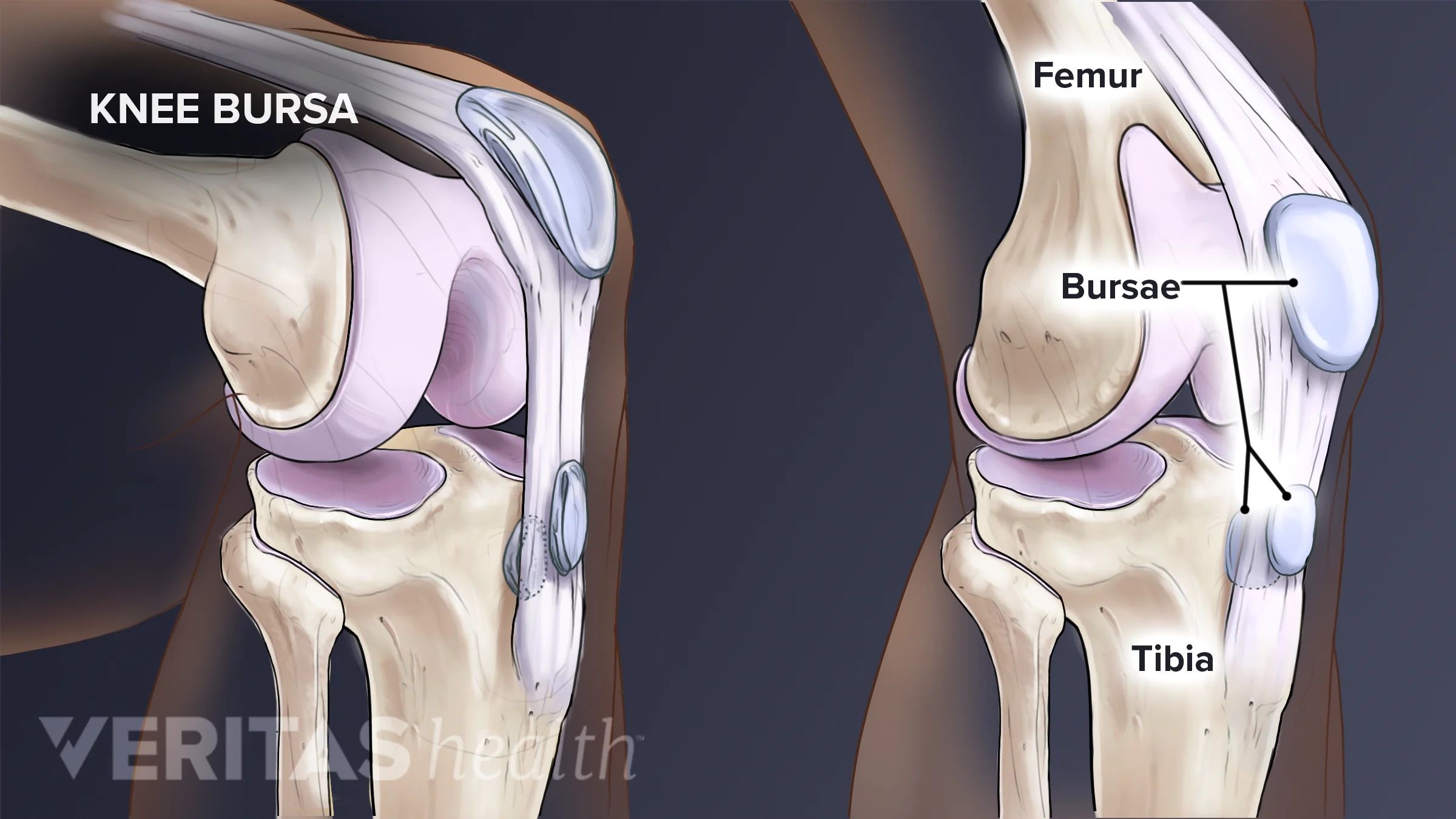
Tendon/synovial sheaths
Tube Like bursae
reduce friction around joints
wrap around tendons experiencing lots of friction
ex: wrist, ankle (many tendons in a confined space)

Briefly list the different synovial joints (6)
plane
Hinge
Pivot
Ellipsoid
Saddle
Ball and socket
Triaxal definition
back and forth
Side to side
Rotation
hip joint, shoulder joint
Biaxial
Freely movable
allows movement around two perpendicular axes
Flexion/extension and abduction/adduction
Ex: metacarpophalangeal joints (knuckles), carpometacarpal (joint), radiocarpal (wrist) joint
Uniaxial
Movement strictly around single axis
Hinge joints
ex: elbow, knee, finger/toe
pivot joints
ex: atlantoaxial joint, proximalradioulnar (forearm to turn up & down)
Plane
Articulated surfaces flat or curved
Many are Biaxial: back-forth, side to side
Some are triaxial: back-forth, side to side, rotation
Ex: intercarpal, intertarsal, sterrnocostal, vertebrocostal joints
Hinge
convex surface fits into concave surface
Uniaxial: Flexion-Extension
Ex: elbow, ankle, interphalangeal joints
Pivot
Rounded/pointed surface fits into ring
formed partly by bone and partly by ligament
Uniaxial: Rotation
Ex: Atlanto-axial and radioulnar joint
Ellipsoid
oval shaped projection fits into oval shaped depression
Biaxial: flexion-extension, abduction-adduction
Ex: radiocarpal and metacarpophalangeal joints
Saddle
Articular surface of one bone Is saddled shaped
Other bone “sits” in saddle
Biaxial: Flexion-extension, abduction, adduction
Ex: carpometacarpal joint b/w trapezium and metacarpal of thumb
ball and socket
Ball like surface fits into cup like depression
Triaxial: Flexion-extension, abduction, adduction, rotation
Ex: glenohumeral and hip joints
Temporomandibular joint (TMJ)
structure, function and what limits movement
mandibular fossa and mandibular condyle
Fibrocartilage instead of hyaline
Articular disc
superior cavity: gliding
Inferior: hinging
capsule and synovial fluid
extracapsular ligaments
Articular disc, ligaments bones, surrounding muscles limit movement
Glenohumeral joint (shoulder)
Structure and Function
ball and socket
Large round head into Glenoid cavity
Glenoid labrum
Bursar
Joint capsule and ligaments
Rotator cuffs (muscles and tendons) compressing numeral head into socket
Triaxial
Elbow Joint
Structure, function and what limits movement
ulnohumeral, radiohumeral and proximal radioulnar joint
hyaline cartilage
Joint capsule and membrane
Medial and lateral collateral ligaments Ligaments, annular ligament
Biceps, brachialis, triceps
Pronation, supination (pivot)
Flexion and extension (hinge action)
Olecranon and coronoid proces, soft tissue, ligaments, joint capsule limits movements
Coxal (hip) joint
Structure, function
acetabulum, head of femur
articular cartilage
Iliofemoral, pubofemoral and ischiofemoral ligament
Ligamentum teres
Lots of muscles
Weight bearing
Multiaxial
Tibiofemoral joint (knee)
Structures, functions and what limits movement
femur and tibia duh
Menisci
Hyaline cartilage
Lateral and medial collateral ligaments
Cruciate ligaments
Weight bear king shock absorption
Flexion and extension
Ligaments, tendons, joint capsules and bones limit movement
Alanto-occipital
Between superior articular facets of atlas and occipital condyle of occipital bone
Synovial (Ellipsoid)
Flexion and extension of head: slight lateral Flexion of head to either side
Alanto-axial
between dens of axis and anterior arch of atlas
Synovial (pivot)
Rotation of head
Intervertebral
between vertebral bodies
synovial (pivot)
Between vertebral arches
synovial ( planar between vertebral arches
flexion, Extension, lateral flexion, and Rotation of vertebral column
sternocostal
between sternum and first seven pairs of ribs
Cartilaginous (synchondrosis
sternum & first pair of ribs
Synovial (plane)
sternum and ribs 2-7
slight gliding
between sternum & ribs 2-7
Lumbosacral
b/w fifth lumbar vertebra & base of sacrum
cartilaginous (symphasis)
b/w inferior & superior articular facets
synovial (planar)
Flexion, extension, lateral Flexion, Rotation of vertebral column
Sternoclavicular
b/w sternal end of clavicle, manubrium and first costal cartilage
Synovial (plane, pivot)
Gliding, w/ limited movements most direction
Acromioclavicular
b/w acromion of scapula & acromial end of clavicle
Synovial (plane)
Gliding and rotation of scapula on clavical
Radioulnar
Proximal b/w Head of Radius & radial notch of ulna
Distal b/w ulnar notch of radius & head of ulna
Synovial (pivot)
Rotation of forearm
Wrist (radiocarpal)
b/w distal end of radius and scaphoid, lunate, triquetrum
Synovial (Ellipsoid)
Flexion, extension, abduction, adduction, circumduction + slight hyper extension of wrist
Intercarpal
b/w proximal and distal row of carpal bones and midcarpal joints
Hamate, scaphoid and lunate (midcarpal) joint
Rest are Synovial (plane)
Gliding, Flexion, Extension, abduction, adduction
Slight rotation at midcarpal joints
Carpometacarpal
Thumb joint b/w trapezium of carpus & first metacarpal
synovial (saddle)
Flexion, Extension, abduction, adduction, circumduction
remaining digits b/w carpus & 2nd-5th metacarpals
synovial (plane)
Gliding
Metacarpophalangeal & metatarsophalangeal
b/w heads of metacarpals (or metatarsals) and bases of proximal phalangeals
Synovial (ellipsoid)
Flexion, Extension, abduction, adduction and circumduction of phalanges
Interphalangeal
b/w heads of phalanges and bases of proximal phalanges
Synovial (plane)
Flexion & Extension of phalanges
Sacroiliac
b/w sacrum & ilia of hip bones
Synovial (plane)
Slight gliding
more in pregnancy
Pubic symphasis
b/w anterior surfaces of hip bones
Cartilaginous (symphysis)
Slight movements
even more during pregnancy
Tibiofibular
proximal joint b/w lateral condyle of tibia & head of fibula
synovial (plane)
Slight gliding
Distal joint b/w distal end of fibula and Fibular notch of tibia
fibrous (syndesmosis) at distal joint
Slight rotation of fibula during dorsiflexion
Ankle (Talocrural)
b/w distal end of tibia and medial malleolus & talus
B/w lateral malleolus of fibula and talus
Synovial (hinge)
Dorsiflexion and plantar Flexion
Intertarsal
synovial (plane)
subtalar & calcaneocuboid joints
synovial (saddle)
At talocalcaneonavicular joint
inversion and eversion
Tarsometatarsal
B/w 3 cuneiforms of tarsus & bases of 5 metatarsal bones
Synovial (plane)
Slight gliding