NESC/BIOL 4030 Lab Exam 1 Structures Review
 Call Kai
 Learn
 Practice Test
 Spaced Repetition
 Match
 Flashcards
 Knowt Play
1/32

There's no tags or description
Looks like no tags are added yet.
Name | Mastery | Learn | Test | Matching | Spaced | Call with Kai | Chat |
|---|
No analytics yet
Send a link to your students to track their progress
33 Terms


Dorsal view (of brain covered with dura mater):
Left vs. right sides
(Rostral vs. caudal axis)


Ventral view:
Tissue surrounding olfactory bulbs
Tissue surrounding optic nerves
Optic chiasm
Pituitary gland
Capillary beds
*Notice which side is right and which is left!
(Rostral vs. caudal axis)


Lateral view:
Trigeminal nerve (V)
(Dorsal vs. ventral axis)
(Rostral vs. caudal axis)


Dorsal view:
Sulcus vs. gyrus
Longitudinal fissure
Transverse fissure
Cerebellum
(Rostral vs. caudal axis)
(Left vs. right side)

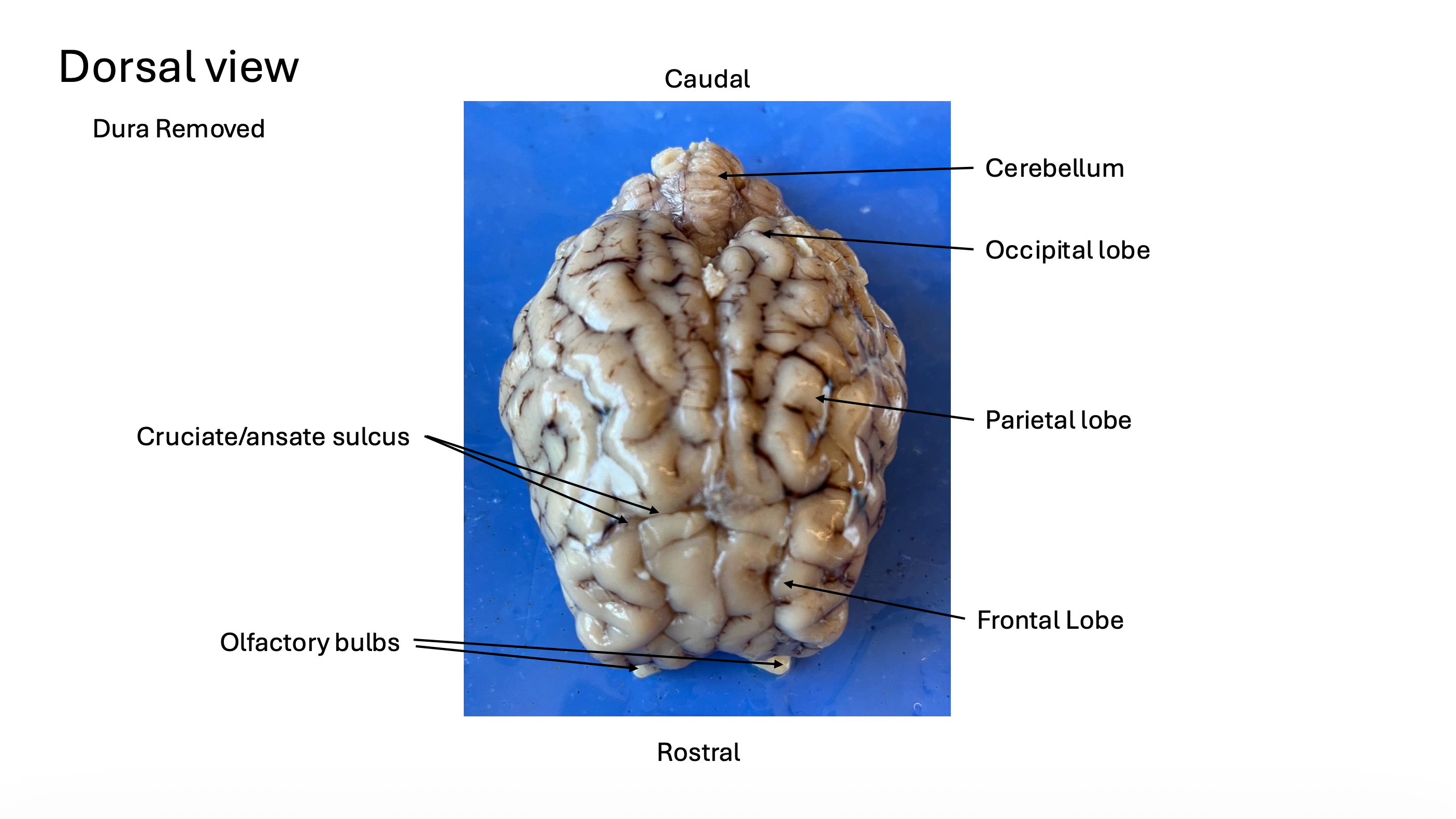
Dorsal view:
Cerebellum
Occipital lobe
Parietal lobe
Cruciate/ansate sulcus
Frontal lobe
Olfactory bulbs
(Rostral vs. caudal axis)


Lateral view:
Spinal cord
Medulla
Pons
Pyriform lobe
Rhinal fissure
Optic nerve
Olfactory bulb
Insula
(Dorsal vs. ventral axis)
(Rostral vs. caudal axis)


Caudal view (cerebrum and cerebellum pulled apart):
Superior colliculi
Inferior colliculi
Corpora quadrigemina (part of midbrain tectum)


Caudal view (separating superior colliculi from cerebrum to reveal pineal body):
Pineal body
Superior colliculus
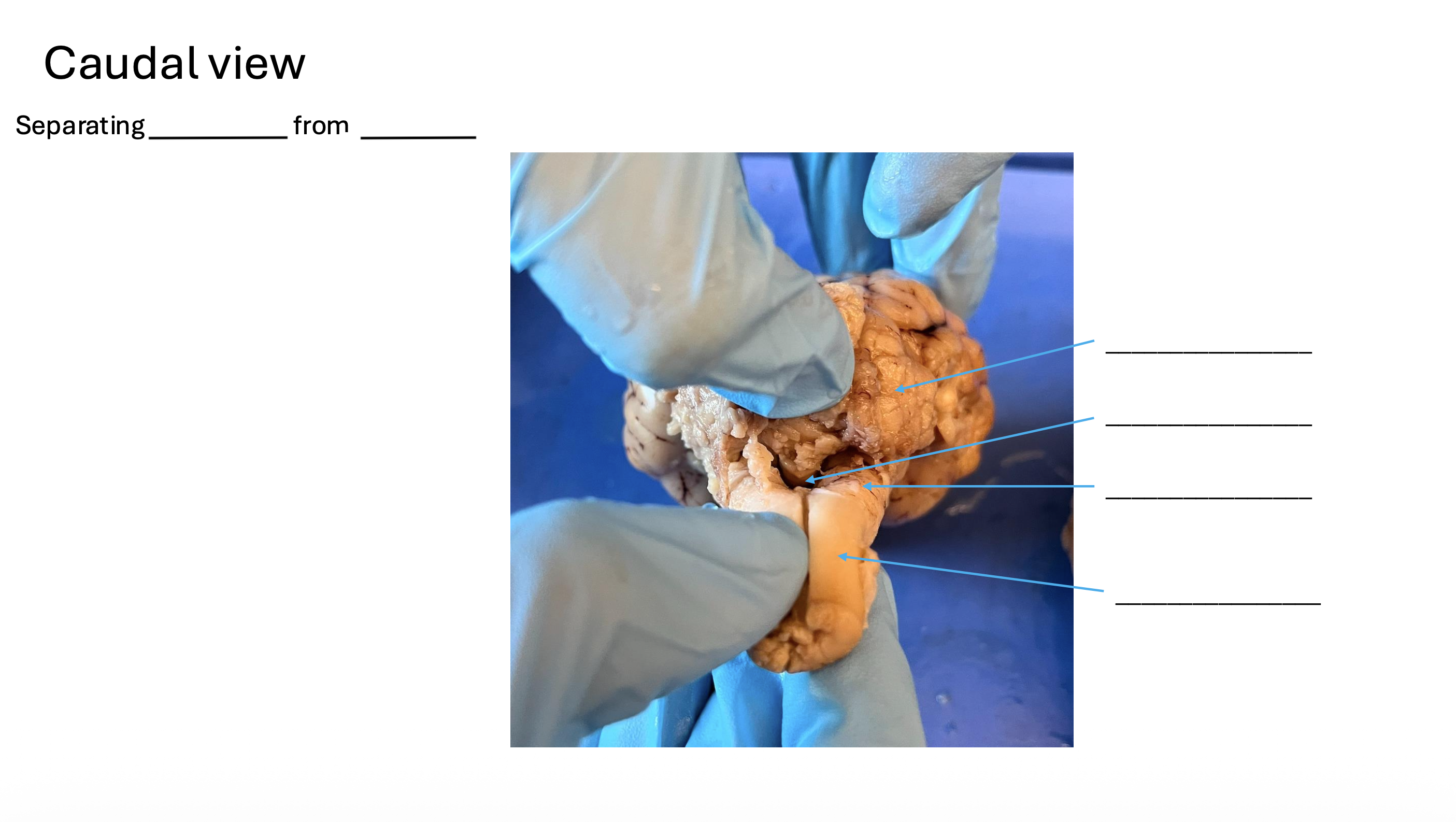

Caudal view (separating cerebellum from medulla to reveal 4th ventricle):
Cerebellum
4th ventricle
Medulla
Spinal cord


Ventral view:
Olfactory bulb (I)
Optic nerve (II)
Optic chiasm
Optic tract
Oculomotor nerve (III)
Trigeminal nerve (V)
Abducens nerve (VI)
(Rostral vs. caudal axis)


Ventral view:
Olfactory bulb (I)
Olfactory tract
Pyriform area


Ventral view:
Infundibulum (removed)
Mammillary body
Cerebral peduncles (part of midbrain)
Pons
Trapezoid body
Medulla
Pyramids (part of medulla)
Spinal cord


Midsagittal view:
Cingulate gyrus
Hippocampal formation
Fornix
Mammillary body
Optic chiasm


Midsagittal view:
Arbor vitae of cerebellum
Superior colliculus
Inferior colliculus
Spinal cord
Medulla
Pons
Midbrain


Midsagittal view:
Corpus callosum
Pineal body
Thalamus (massa intermedia)
Hypothalamus


Midsagittal view:
Lateral ventricle (septum pellucidum)
Lateral ventricle (space)
Cerebral aqueduct
4th ventricle
3rd ventricle


Coronal section 1:
Cingulate gyrus
Corona radiata
Corpus callosum
Lateral ventricle
Head of the caudate nucleus


Coronal section 2:
Cingulate gyrus
Corona radiata
Corpus callosum
Lateral ventricle
Head of the caudate nucleus
Septum pellucidum
Optic chiasm


Coronal section 3:
Cingulate gyrus
Corona radiata
Corpus callosum
Lateral ventricle
Head of the caudate nucleus
Putamen
Anterior commissure
Internal capsule
Globus pallidus
Optic chiasm


Coronal section 4:
Cingulate gyrus
Corona radiata
Corpus callosum
Lateral ventricle
Tail of caudate nucleus
Fornix
Putamen
3rd ventricle
Optic chiasm


Coronal section 5:
Cingulate gyrus
Corpus callosum
Lateral ventricle
Fornix
Thalamus
3rd ventricle
Amygdala
Hypothalamus


Coronal section 6:
Cingulate gyrus
Corpus callosum
Lateral ventricle
Fornix
Hippocampus
3rd ventricle
Thalamus


Coronal section 7:
Cingulate gyrus
Corpus callosum
Lateral ventricle
Hippocampus
Thalamus
Mammillary body


Coronal sections 8 & 9:
Superior colliculus
Cerebral peduncle
Cerebral aqueduct


Ventral view:
Removal of the dura


Midsagittal view:
Left vs. right hemisphere
(Dorsal vs. ventral axis)
Coronal sections of the sheep brain (i.e., Where is dorsal vs. ventral? Where is left vs. right hemisphere?)

Arranged as brain is section from rostral end to caudal end
Laid out dorsal to ventral (right sight-up)
Right hemisphere is on left, left hemisphere is on right
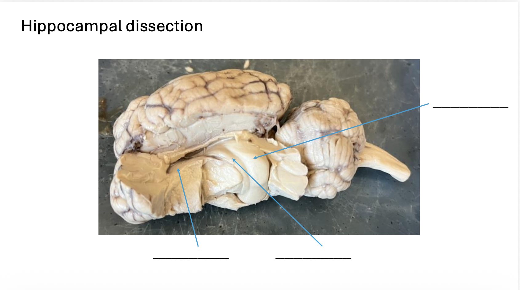
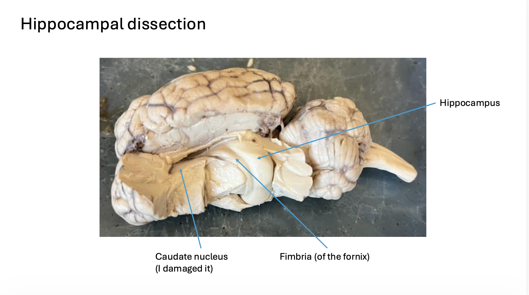
Hippocampal dissection:
Caudate nucleus (damaged in the image)
Fimbria (of the fornix)
Hippocampus
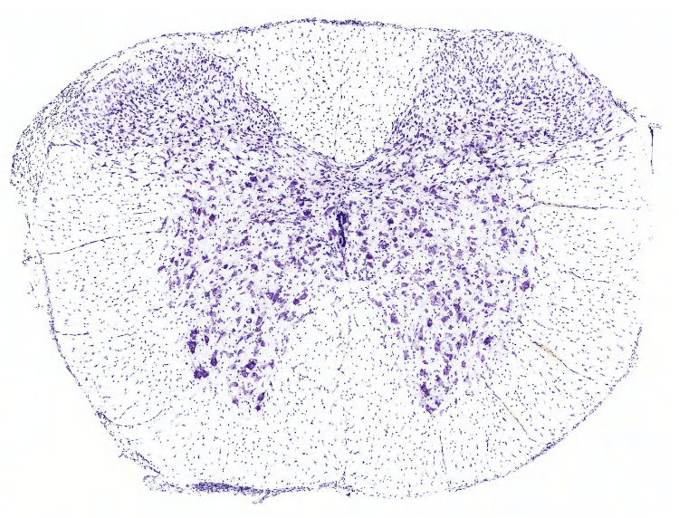
Cervical spinal cord (C3)
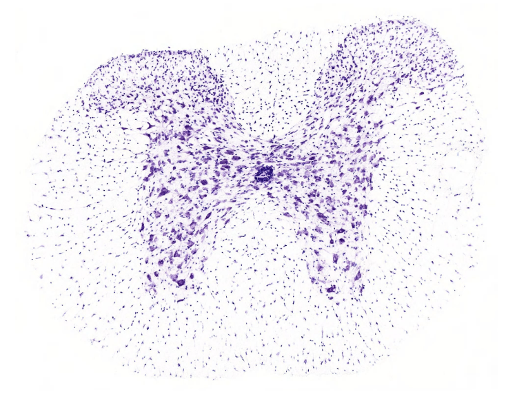
Thoracic spinal cord (T6)
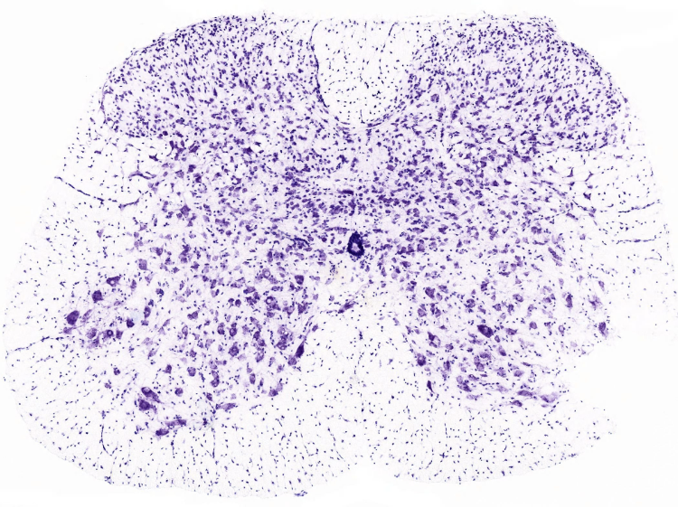
Lumbar spinal cord (L6)
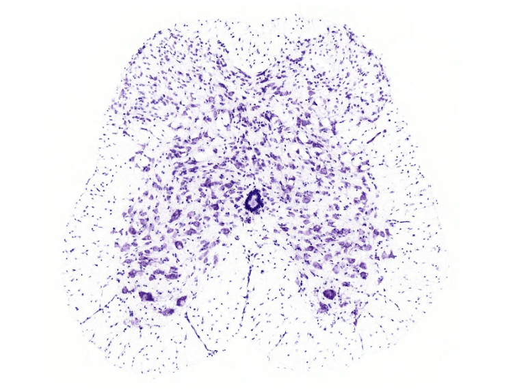
Sacral spinal cord(S4)
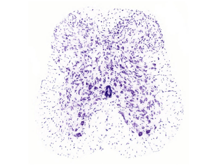
Coccygeal spinal cord