45-48. bovine lameness + deep digital sepsis
 Call Kai
 Learn
 Practice Test
 Spaced Repetition
 Match
 Flashcards
 Knowt Play
1/44

There's no tags or description
Looks like no tags are added yet.
45 Terms
what are characteristics of cows that increase the likelihood of becoming lame?
older cows (cows in their 2nd, 3rd, or more lactations)
cows with low BCS
cows with overgrown hooves
how often should lameness be evaluated?
“as frequently as possible”
every 2-4 wks
prior to hoof trimming
all cows at the end of lactation (at the start of the dry period)
what are challenges of locomotion scoring?
practical: time-consuming, subjective, requires training, scorers after disagree, needs a flat, non-slip, well-lit surface
biological: cows mask pain; a lag between pain and a visible lesion; many cows with lesions don’t look lame
result: lameness underestimated by producers
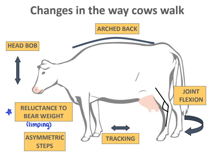
indicators of lameness
head carriage — head bob or uneven head movement; usually most obvious with forelimb lameness
arched back — convex arch between withers and tails
asymmetric steps or “cadence” — arrhythmic hoof placement
tracking — rear feet falling short of front feet imprint, which is often impacted by flooring surface and walking speed
weight transfer between limbs — sink of dew claws on the contralateral limb to the lame limb
joint flexion — stiffness in movement and abduction of the limb away from the mid-line
locomotion score targets / goals
lame (mild): <10%
failed to prevent
severe lameness: ≤1%
failure to prevent & treat
steps in lameness exam
locmotion evaluation
physical limb examination
proper restraint, thorough inspection, evaluate pain reaction
other diagnostic tools
IV regional anesthesia
radiographic imaging
U/S
what is the hoof wall vs. sole growth rate? how long does it take to replace each?
wall: ~5mm/month
wall is 75 mm long → ~15 months to replace
sole: ~3mm/month
sole is 7 mm thick → ~1.5-2 months to replace
what are the main goals of functional hoof trimming?
restore a more upright claw angle (aim ~52 degrees)
dorsal hoof wall — 75mm / 3”
sole thickness — 7mm / 0.25”
balance weight distribution between inner & outer claw
avoid doing harm!

common hoof trimming regimes
in general, dairy cattle should have their hooves inspected and, if necessary, trimmed every 4-6 months
routine lactation cycle trimming
at dry off / move from far-off to prefresh
early / mid-lactation ~80-120 days in milk
heifers prior to first calving
interventional trimming
lame
observed signs of overgrowth / claw rotation
what is digital dermatitis?
polymicrobial infection of the skin that commonly occurs in the interdigital cleft of the hoof
digital dermatitis — pathogenesis
damage to skin (microlacerations) + softened by water & chemicals → bacterial invasion
particularly spirochetes → treponema spp.
bacteria invade and damage the deeper layers of the epidermis / dermis → local inflammation
chronic inflammation → hyperkeratosis & proliferative lesions
digital dermatitis — acute, active lesions
stages M1, M2, M4.1
infectious, painful
should be treated ASAP
early topical treatment
easier to control
digital dermatitis — chronic / recurring lesions
stages M4 & M4.1
reservoir of disease
encysted treponema → deep skin
difficult to treat
long term consequences
key drivers of outbreaks
digital dermatitis — prevention
effective footbath
hygiene: clean, dry feet and alleys, manure management
biosecurity
what makes an effective footbath?
correct dimensions
frequency
4x/wk → adapt to minimize usage
product
ensure evidence of efficacy
<5% copper sulfate; <4% formalin
maintain pH 3.5-5
renewal
at ~200 cow passes OR depth ≤ 3” (8cm) OR pH > 5
consistent routine
digital dermatitis — biosecurity
monitor all groups of animals
youngstock, dry cows, close-up group
footbathing or spray feet
in-feed organic trace materials and other additives
start preventive practices (footbath) in the group that is 60-90 days prior to where DD is detected
time from initial infection to clinical signs
challenges of treating digital dermatitis
evidence of effectiveness
multiple treatments required
milk residues
digital dermatitis — treatment
topical tetracycline (~2g) — off label use!
milk residue
salicylic acid (harsh on skin)
what is foot rot (phlegmon)?
sporadic infection of the soft tissues of the foot in dairy & beef cattle, creating a sudden onset of mild to severe lameness
foot rot (phlegmon) — clinical signs
sudden onset of symmetric hard swelling of lower leg (foot — dew claws) and severe lameness
very painful, sudden drop in milk, reduced feed intake
foot rot (phlegmon) — pathogenesis
interdigital microtrauma of epidermis → facilitates invasion by synergistic anaerobic bacteria → causes acute inflammation and necrosis of soft tissue of the lower leg
what are the most common bacteria associated with foot rot?
fusobacterium necrophorum
trueperella pyogenes
porphyromonas levii
prevotella intermedia
spirochetes?
foot rot — disease progression / sequelae
within 72hr
interdigital cleft splits open
putrid, foul-smelling discharge
tissue formation to attempt to close the skin defect (leads to interdigital hyperplasia)
risk factor for other diseases
in severe cases, infection can extend to tendons & joints, leading to culling or prolonged healing
detect & treat immediately
foot rot — prevention
same as digital dermatitis
hygiene!! clean, dry feet and alleys, manure management, footbaths
also: avoid skin damage — mostly mechanical: stones, broken concrete, sharp edges, frozen mud, cables
foot rot — treatment
requires combination of debridement of necrotic tissue from the interdigital space and use of an effective parenteral antibiotics
systemic antimicrobials
ceftiofur sodium
oxytetracycline
NSAIDs may be indicated for pain relief
treated animals should visually improve within 2-3 days
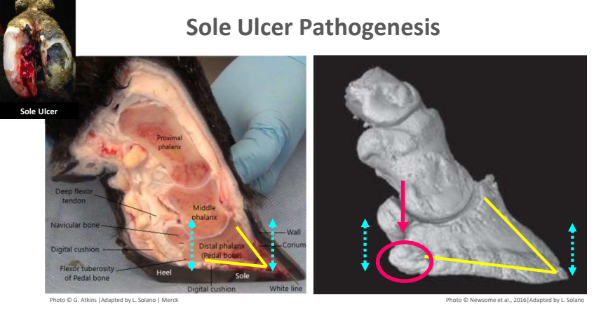
sole ulcer — pathogenesis
mechanical, hormonal, and/or metabolic changes weaken structures that suspend & support P3 → P3 “sinks” & its flexor tuberosity presses the corium against the sole
at first, compressed corium leaks blood into keratinocytes at the dermal-epidermal interface, which shows up as sole hemorrhage
extended periods of pressure damage the corium and interrupt horn growth, causing the corium to protrude through the defect, forming a sole ulcer
pressure also sets off inflammation that causes lasting anatomical changes to P3 (exostosis or new bone growth), which makes affected cows prone to relapse
when is the highest risk period for sole ulcers?
transition period (± 3 weeks around calving)
suspensory apparatus loosens & P3 sinks more easily
sole ulcer — prevention
reduce pressure on corium by reducing:
daily standing time on hard surfaces
manage risk factors associated with: stall comfort, time out of pen, heat abatement, stocking density, lock up time in headlocks
loss of body condition
thinning of digital cushion (fat pad) → ↓ cushioning & support below P3
hoof overgrowth & pressure on sole ulcer site
timely, correctly performed hoof trimming
what is an appropriate substrate for lying surfaces?
deep sand → provides cushion, traction, and support to facilitate rising & lying movements

white line disease — pathogenesis
starts at the white line, the junction between the sole and wall; weakest part of the claw
thought to be similar to sole ulcers → when P3’s suspensory mechanisms are compromised, the keratinocytes that grow white line horn are damaged
internal & external traumatic forces create a shearing action at the white line, which can separate the hoof wall
external forces include sharp turns, rough or slipper floors, and pushing during handling
these gaps can allow the entry of bacteria & foreign bodies, resulting in damage to the corium & inflammatory changes to P3

white line disease — prevention
minimize shear forces on the white line by improving:
flooring
good traction without causing trauma: non-slippery, non-abrasive concrete with proper grooving, strategic use of rubber flooring, and well-maintained pasture tracks
cattle handling
let cows walk at their own pace!
hoof (claw) balance and horn strength
timely, correctly performed hoof trimming to balance weight between claw, plus balanced diet that meets mineral & vitamin needs
planned pasture access
some cows, some of the time
how to fix a poor floor
get cows off it
regroove it
recommend adding oblique groove (diamond pattern) on high-risk areas
texture it
cover it up
concrete — minimum 3” (8 cm) depth
rubber
grooving pattern must provide enough flat stable standing surface & enough grip to prevent slipping, while not being so rough, resulting in excessive wear
what are strategic locations to use rubber floor covers?
high traffic area
transfer lanes — where cows walk long distances
holding areas — holding times >1 hr
parlor return lanes — where excessive slopes >2% may enhance wear
sharp turns (90-180°) — parlor platforms & exit areas with sharp turns
treatment of non-infectious hoof lesions — sole ulcers & white line disease
therapeutic trimming
remove loose horn (not tissue! leave corium alone)
for feet with typical lesions, preserve toe triangle & remove only wall in the heel
successful weight transfer from affected claw to unaffected claw
thin horns around the margins of the lesion, exposing healthy corium
elevate the affected claw by applying a hoof block to the unaffected claw
consider NSAIDs
when should you recheck sole ulcer / white line disease lesion?
recheck at 30 days
remove or adjust hoof block
explore lesion for healing and remove any persistent loose horn
establish repeat trimming schedule — “90 day cow”
thin sole, toe ulcer / necrosis — pathogenesis
usually one continuum
thin sole develops when sole horn wears away faster than it grows, until the sole flexes under thumb pressure but the corium is not yet exposed
if thin sole goes undetected, or wear is severe, the corium at the toe is exposed and a toe ulcer forms
bacteria then invade exposed corium → infection + original trauma cause widespread damage that can extend into P3 & cause toe necrosis
thin sole & toe ulcer — prevention
balance horn growth and wear by avoiding:
abrasive walking surface and/or bedding
over-trimming of sole & toe
long walking distances
rough handling that makes cows slip or turn sharply
abrupt changes in walking surface (ex. from pasture or dirt lots to concrete)
thin sole — treatment
apply thin plastic block to affected claw (ensure that there is no exposed corium) + management
toe ulcer — treatment
remove sole & wall horn around the lesion and block the unaffected claw
toe necrosis — treatment
block the healthy claw, plus remove infected bone and soft tissue → partial resection of P3
IV regional anesthesia
follow-up NSAIDs for pain relief
recovery: deeply bedded lying area
recheck in 2-4 wks: check healing & block position
what causes deep digital sepsis?
delayed or inappropriate treatment of sole ulcers, white line disease, sole punctures, and/or foot rot can lead to deep digital sepsis
how is deep digital sepsis different from foot rot?
deep digital sepsis does NOT respond to systemic antibiotic treatment (vs. foot rot — responds to antibiotic treatment)
deep digital sepsis — prevention
cows with DDS represent complete failure of herd lameness prevention & lameness identification program
presence of such a cow should trigger a full review of all lameness-related procedures on the farm
deep digital sepsis — treatment considerations
most cows should be euthanized on animal welfare grounds
severely lame cows should not be shipped for slaughter!
factors to consider when choosing treatment:
age, value, productivity, stage of gestation, footing, and ability to provide post-op care
restrain cow & examine foot after thorough cleaning. determine cause and extent of sepsis & discuss options with farmer — is this cow best culled?
deep digital sepsis — treatment
treatment should be implemented by a veterinarian
various surgical procedures exist:
involves drainage of infected area and ankylosis (fusion) of distal interphalangeal joint or amputation
apply local anesthesia & block sound claw. give NSAIDs before procedure
perform correct procedure
consider parenteral antibiotics & continue NSAIDs