ABDOMEN 2 ALL EXAMS
 Call Kai
 Learn
 Practice Test
 Spaced Repetition
 Match
 Flashcards
 Knowt Play
1/280

There's no tags or description
Looks like no tags are added yet.
281 Terms
At what diameter does a AAA have an increased risk of rupture? a. 5.5cm b. 3.0cm c. 7.0cm d. 6.0cm
c. 7.0cm
The development of which condition is strongly linked with renal artery stenosis? a. Hypotension b. Pancreatitis c. Osteoporosis d. Hypertension
d. Hypertension
The most common type of AAA is what? a. Fusiform b. Saccular c. Dissecting d. Pseudoaneurysm
a. Fusiform
Which layer of the arterial wall is changed by the presence of atherosclerosis? a. Intima b. Adventitia c. Media d. Albuginea
a. Intima
All BUT which of the following are correct regarding diaphragmatic hernia: a. Can allow stomach, small bowel or spleen into the thoracic cavity b. May result from a congenital malformation or post-surgical or other trauma c. Is often associated with chromosomal birth defects d. Mostly occurs on medial right
d. Mostly occurs on medial right
All BUT which of the following is true regarding Umbilical hernias? a. Most often contain large intestine and omentum b. Most common type of ventral hernia c. Usually present as small but gradually increase in size d. Most often occurs in males
d. Most often occurs in males
Paracentesis: a. can be done for diagnostic or therapeutic purposes b. is when a small, flexible catheter is inserted into an abscess pocket c. is a sterile procedure used to aspirate ascitic fluid from the thoracic cavity d. is only done when there is 6 liters of fluid or more identified on ultrasound
a. can be done for diagnostic or therapeutic purposes
Pleural effusion is: a. also known as hydrothorax b. defined as accumulation of fluid within the thoracic cavity c. seen sonographically as uni- or bilateral fluid collections superior to the diaphragm d. all are correct
d. all are correct
Sarcoma can be defined as: a. a mass of ectopic endometrial tissue planted on the abdominal wall b. a cancerous mass related to extension of primary carcinoma of a nearby structure c. a hypoechoic cancerous lesion arising from the abdominal muscle wall d. a collection of serum resulting from surgical incision or hematoma resolution
c. a hypoechoic cancerous lesion arising from the abdominal muscle wall
Strangulation can be a complication of a hernia, and: a. is called a "non-reducible" hernia b. is defined as compromised blood flow which can result in ischemia and progress to necrosis c. is defined as when contents of the hernia cannot be pushed back into the abdomen d. is expected to resolve on its own
b. is defined as compromised blood flow which can result in ischemia and progress to necrosis
What abdominal wall mass can be described as forming at the damaged end of a nerve, causing pain? a. Lipoma b. Neuroma c. Sarcoma d. Desmoid tumor
b. Neuroma
What condition is defined as partial or complete, uni- or bilateral abnormal elevation of the dome of the diaphragm: a. Diaphragmatic Hernia b. Diaphragmatic Eventration c. Diaphragmatic Rupture d. Diaphragmatic Inversion
b. Diaphragmatic Eventration
What condition presents with anechoic areas demonstrated on one or both sides of the chest superior to the diaphragm? a. Paralysis b. Eventration c. Inversion d. Pleural effusion
d. Pleural effusion
When epithelial cells spill into the peritoneal cavity, most commonly from appendiceal or ovarian rupture, which borderline malignant
condition can develop? a. Hematoma b. Peritoneal Mesothelioma c. Lymphocele d. Pseudomyxoma Peritonei d. Pseudomyxoma Peritonei
Which ascites is characterized by a high concentration of protein, cells, or solid material derived from blood cells? a. Hydremic b. Chylous c. Exudative d. Transudative
c. Exudative
Often seen sonographically as a result from a biliary tree ischemia, rupture, or cholecystectomy? a. Urinoma b. Biloma c. Seroma d. Hematoma
b. Biloma
Which of the following abnormalities of the diaphragm can inhibit respiration? a. Paralysis b. Eventration c. Hernia d. All are correct
d. All are correct
Which of the following findings is most suggestive of a hematoma? a. decreased hematocrit b. Increased appetite c. Clear skin without changes d. Improved mobility
a. decreased hematocrit
Which of the following is NOT true regarding Omental Caking? a. It is also called peritoneal carcinomatosis b. It is defined as thickening of the greater omentum due to malignant inflitration c. It is an indicatior for peritoneal mets d. All are correct
d. All are correct
Which of the following would be a clinical indicator of a suspected abscess? a. Redness b. Pain c. Swelling d. All are correct
d. All are correct
On transverse scans, what is the course of the left renal vein? a. Posterior to the Aorta b. Posterior to the inferior vena cava c. Anterior to the superior mesenteric artery and posterior to the inferior vena cava d. Between the superior mesenteric artery and the aorta
d. Between the superior mesenteric artery and the aorta
Precise determination of the measurement of aortic diameter should be taken: a. Perpendicular to the aorta b. Perpendicular to the skin surface c. In a coronal plane d. Sagittal to the skin surface
a. Perpendicular to the aorta
The superior mesenteric vein empties into which vein? a. Splenic b. Portal c. Right renal d. Inferior vena cava
b. Portal
Which vessel lies posterior to the bile duct and anterior to the portal vein? a. Hepatic vein b. Hepatic artery c. Cystic duct artery d. Gastroduodenal artery
b. Hepatic artery

You see this incidental finding on the ultrasound. What is your diagnosis? a. A pseudocyst b. cholangiocarcinoma c. polyp d. cholesterolosis
c. polyp
What happens to the normal aorta as it progresses inferiorly? a. Plasticity increases b. Diameter decreases c. Walls become thickened d. Joins other arteries and becomes larger
b. Diameter decreases
Which type of hernia has a higher incidence in women? a. femoral b. inguinal c. umbilical d. epigastric
a. femoral
c. umbilical

What type of aneurysm is in this transverse image of the aorta? a. dissecting b. saccular c. fusiform d. inguinal
a. dissecting
Which type of aneurysm is a gradual and progressive dilation of the complete circumference of the vessel? a. saccular b. fusiform c. dissecting d. pseudoaneurysm
b. fusiform
Which of the following is NOT a type of aneurysm? a. saccular b. fusiform c. dissecting d. inguinal
d. inguinal
Which of the following aneurysm is being described: Separation of the layers of the arterial wall. a. saccular b. inguinal c. dissecting d. fusiform
c. dissecting
Which of the following is a risk factor for aneurysms? a. atherosclerotic disease b. munchausen syndrome c. liver cirrhosis d. endometriosis
a. atherosclerotic disease
What is the most common cause of IVC obstruction? a. left sided heart failure b. right sided heart failure c. cardiomegaly d. pleural effusion
b. right sided heart failure

The findings are characteristic of: a. pleural effusion b. pleural abscess c. ascites d. dissecting aneurysm e. fatty liver
a. pleural effusion

What is this fluid finding called? (Ascites or pleural effusion?) Why?
Pleural effusion / Above the diaphragm
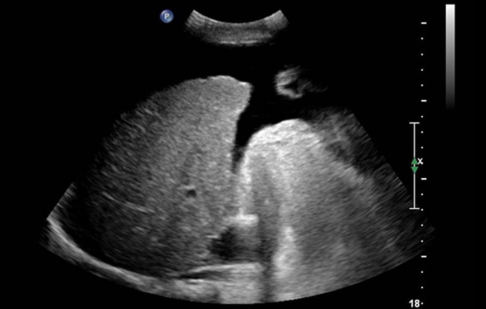
Where is this fluid located? What is the fluid called?
Peritoneum
Ascites
review manually with image).

Explain this image by first stating the plane of the image. Where is the fluid located? What is the fluid called?
located superior to diaphragm in the pleural cavity
pleural effusion
instructor did not correct plane). Fluid: pleural effusion.

What are your findings on this patient? (Must state type) What is the typical appearance that helps with the diagnoses? List the conditions most likely associated with this type of fluid.
Transudative Ascites (translucent)
-lack of protein and cellular materials in fluid
Typically has a simple appearance
Often associated with PHTN and congestive cardiac disease
characterized by a lack of protein and cellular materials in the fluid. 2. Typically has a simple appearance. 3. Often associated with portal hypertension and congestive cardiac disease.

What type of fluid is this finding? (must state type) What is the typical appearance that helps with the definitive diagnosis of this fluid? What are the most likely causes of this fluid?
Exudative Ascites - contains large amount of protein + cellular material
Typically results in more complex and echogenic appearance to fluid.
Associated with renal failure, inflammatory or ischemic bowel disease, peritonitis, and malignancy
contains a large amount of protein and cellular material. 2. Typically results in more complex and echogenic appearance to fluid. 3. Associated with renal failure, inflammatory or ischemic bowel disease, peritonitis, and malignancy.
List the five cardinal signs for inflammation: (0.5 pt for each)
Redness
heat
swelling
pain
loss of function
What instrumentation adjustment can help remove artifact echoes that may appear in the lumen of the abdominal aorta? a. Decrease time gain compensation (TGC) b. Decrease sensitivity c. Use a longer focused transducer d. Use a higher frequency transducer
a. Decrease time gain compensation (TGC)
What is often described as having a homogeneous ultrasound appearance with smooth borders and is found within the sac of an aneurysm? a. Calcification b. Plaque c. Intimal disruption d. Thrombus
d. Thrombus
What is the most common location of aneurysms associated with the continuation of an abdominal aortic aneurysm? a. Celiac axis b. Iliac artery c. Splenic artery d. Superior mesenteric artery
b. Iliac artery
What should be included in a possible list of imaging differential diagnosis when acoustic shadows are presented behind the aorta and the
sonographer is having difficulty visualizing both the anterior and posterior wall? a. Lymphadenopathy b. Pseudoaneurysm c. Arteriosclerosis d. Arteriovenous fistula c. Arteriosclerosis
What is the most common type of splanchnic artery aneurysm? a. Hepatic b. Renal c. Splenic d. Superior mesenteric artery
c. Splenic
Which of the following conditions is caused by portal hypertension? a. splenomegaly b. Courvoisier's GB c. hepatoma d. Aortic Aneurysm
a. splenomegaly
Which of the following is NOT an ultrasound finding for chronic hepatitis? a. hyperechoic parenchyma b. hypoechoic parenchyma c. decrease in liver size d. decreased echogenicity of hepatic vein walls
b. hypoechoic parenchyma
What is another term for the word echinococcal cyst? a. hydatid disease b. pseudocyst c. bacterial abscess d. fungal abscess
a. hydatid disease
Which of the following is NOT an ultrasound presentation of Budd Chiari? a. ascites b. splenomegaly c. hepatomegaly d. cholecystitis
d. cholecystitis

This patient has no known underlying conditions or symptoms other than RUQ pain due to gallstones. What is this isolated incidental finding on your patient abdominal ultrasound patient? a. Budd Chiari b. cavernous hemangioma c. metastasis disease d. echinococcal cyst
b. cavernous hemangioma

What is your diagnosis? a. steatosis b. Budd Chiari c. HCC d. normal
a. steatosis
Budd-Chiari syndrome is____________. a. Obstruction of the hepatic veins b. Obstruction of the portal veins c. Seen only with IVC tumor present d. none of the above
a. Obstruction of the hepatic veins
Hepatitis is an inflammation of the liver caused by____. a. Virus or toxins b. Hepatomegaly c. Budd Chiari d. Metastatic Disease
a. Virus or toxins
When suspecting liver metastases, which is least likely to be seen? a. Hypoechoic Lesion b. Hyperechoic Lesion c. Avascular Lesion d. Isoechoic lesion
c. Avascular Lesion
Which of these are not benign solid neoplasms? a. Liver Cell Adenoma b. Lipomas c. HCC d. Cavernous Hemangiomas
c. HCC
Splenomegaly is a consistent finding in which of the following liver pathologies? a. Polycystic disease b. hemangioma c. Portal hypertension d. Hepatocellular CA
c. Portal hypertension
All of the following are features of portal hypertension EXCEPT? a. splenomegaly b. collaterals c. hepatic artery thrombosis d. Ascites
c. hepatic artery thrombosis

What is the most likely diagnosis for this patient: a. Budd Chiari b. steatosis c. HCC d. metastatic disease
a. Budd Chiari
The type of cyst caused by a parasitic tapeworm is? a. Hematoma b. Pyogenic abscess c. Amebic Abscess d. Echinococcal Cyst
d. Echinococcal Cyst
This condition is an inherited disorder usually associated with autosomal dominant polycystic kidney disease? a. Focal nodular Hyperplasia b. Polycystic liver disease c. Fatty infiltration d. Echinococcal cyst
b. Polycystic liver disease
What is the most common predisposing factor for developing hepatocellular carcinoma? a. Cirrhosis b. Hepatitis c. Cavernous Hemangioma d. Polycystic liver disease
a. Cirrhosis
What is the typical liver echogenicity seen in chronic hepatitis? a. Isoechoic b. hyperechoic, similar to fatty infiltration c. Hypoechoic d. No change in liver parenchyma is seen
b. hyperechoic, similar to fatty infiltration
Which of the following is considered the most common benign liver tumor? a. Hepatocellular Carcinoma b. adenoma c. Cavernous Hemangioma d. Epitheliod hemangioendothelioma
c. Cavernous Hemangioma
Which condition is associated with an elevated alpha-fetoprotein? a. Hepatocellular carcinoma b. Fatty Infiltration of the liver c. Focal nodular hyperplasia d. budd-chiari syndrome
a. Hepatocellular carcinoma
One can image all of the following in a case of end-stage liver disease EXCEPT: a. ascites b. small atrophied liver c. biliary dilation d. portal hypertension e. nodular liver disease
c. biliary dilation
During an abdominal sonogram, recannalization of the umbilical vein is identified. This may be associated with: a. ascites b. an abscess c. hematoma d. hepatoma e. portal hypertension
e. portal hypertension
Chronic active hepatitis is a progressive destructive liver disease that eventually leads to: a. liver cysts b. hepatoma c. cirrhosis d. pancreatitis e. liver metastases
c. cirrhosis

Describe your incidental finding of this common liver mass that you found on your patient. What plane is this image taken in? What is your diagnosis?
RT Liver 5.2 x 6.5 cm well circumscribed, hyperechoic mass
Long
Cavernous hemangioma

Your 68-year-old male has a history of lymphoma and presents with an elevated AFP. Describe your findings. What is the most likely diagnosis?
multiple hypoechoic lesions throughout the liver
metastatic disease (lymphoma)

A patient with a history of alcohol abuse (ETOH) presents with these findings during their abdominal scan. The spleen measures 17cm. Describe all of your findings. What is your diagnosis?
Liver nodularity
Ascites
Enlarged spleen
GB stones
cirrhosis causing portal hypertension
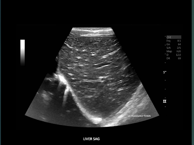
A 57-year-old male presents with elevated AST and ALT lab values as well as a fever, abdominal pain. Please describe your finding. What is the most likely diagnosis? What is the term for this appearance?
multiple tiny echogenic foci throughout the liver
"starry sky" appearance of the liver
Acute hepatitis

This image of the liver correlates with which parasitic process? a. Hydatid Cyst b. Abscess c. Von Gierke's disease d. Schistosomiasis
d. Schistosomiasis

This incidental finding in a patient referred for GB assessment is most likely?
Hemangioma

This benign liver lesion that presents with a spoke - wheel pattern is associated with what pathology?
FNH
Metastasis can have varied sonographic appearances? True False
True

A 66 year old man presents with a history of alcohol abuse and cirrhosis. What is your dx? a. HCC b. Abscess c. Metastasis d. Hemangioma
a. HCC
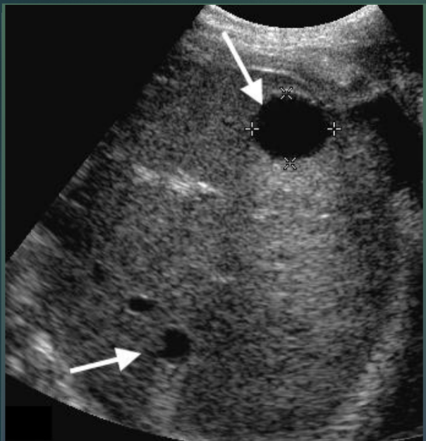
This patient has multiple findings. Describe using ultrasound terms. DX?
round, well-defined, smooth borders, anechoic, posterior acoustic enhancement DX:
simple cyst
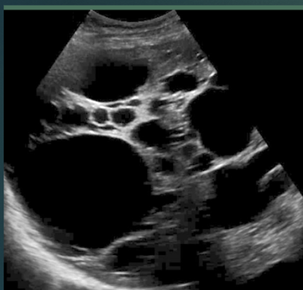
In this image of the liver. The patient most likely has which disease? a. FNH b. Hemangioma c. Polycystic Liver disease d. Metastasis
c. Polycystic Liver disease
Increase in liver echogenicity, hepatomegaly, increased attenuation, impaired visualization of intrahepatic vessels, and impaired visualization of diaphragm. These findings are all associated with fatty liver? True False
True
A 52 year old male with known liver cirrhosis presents for an abdominal ultrasound. You will carefully evaluate the liver to rule out the presence of any focal mass because of which TRUE statement below? a. Patients with liver cirrhosis are at increased risk for hepatocellular carcinoma b. Patients with liver cirrhosis tend to develop multiple cysts in their liver and pancreas c. The presence of regenerative nodules rules out cirrhosis d. All the above are correct
a. Patients with liver cirrhosis are at increased risk for hepatocellular carcinoma
You are scanning a patient with suspected liver cirrhosis. All of the following are sonographic features of cirrhosis EXCEPT: a. Surface nodularity b. Shrunken caudate lobe c. Altered echo texture d. Ascites e. Regenerative nodules
b. Shrunken caudate lobe
A patient is referred for a liver ultrasound with the clinical history of a raised serum alpha-fetoprotein level. What should you look for? a. HCC b. Fatty liver c. FNH
a. HCC
A patient is referred with right upper quadrant pain and tenderness. Patient has a history of oral contraceptive use. A solid, hypoechoic mass is identified in the right lobe of the liver. Color doppler reveals hypervascularity surrounding the mass. Which of the following scenarios is most likely? a. Hepatic lipoma b. Hepatic adenoma c. Hepatic abscess d. all of the above
b. Hepatic adenoma
A liver ultrasound on a 49 year old obese male demonstrates diffuse increased echogenicity with a focal hypoechoic area anterior to the portal vein. This most likely represents a. Liver cirrhosis with HCC b. Fatty metamorphosis of the liver with focal sparing c. Metastatic disease most likely due to a colon primary d. all of the above
b. Fatty metamorphosis of the liver with focal sparing
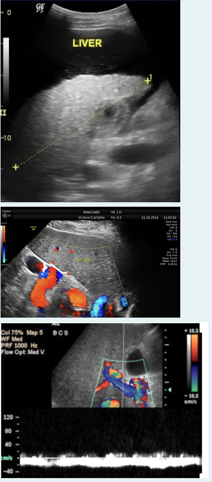
Describe all of the pathology seen in the following images. What is your DX?
Ascites
Nodular liver
splenic varices
hepatofugal flow
cirrhosis/portal hypertension

Patient has intense pain on the right side and is very ill. What is the most likely diagnosis of this image: a. Couvoisier's Gallbladder b. WES sign c. acute acalculous cholecystitis d. adenomyomatosis
c. acute acalculous cholecystitis
What symptom describes tenderness over the gallbladder with probe pressure? a. Morrison's sign b. Murphy's sign c. Douglas's sign d. Courvoisier's sign
b. Murphy's sign

You are scanning a patient with generalized abdominal pain and image the below. Upon interrogation there is no movement or flow seen. What is the most likely diagnosis? a. gangrenous cholecystitis b. adenomyomatosis c. acute cholecystitis d. sludge
b. adenomyomatosis

Patient had acute cholecystitis 8 days ago and now presents with this. What is the most likely diagnosis of this galllbladder image: a. empyema b. polyps c. emphysematous d. chronic cholecystitis
c. emphysematous

What is the most likely diagnosis for this Gallbladder image: a. WIAS sign b. adenomyomatosis c. Chronic cholecystitis d. acute cholecystitis
c. Chronic cholecystitis
If the patient has a known history of a porcelain gallbladder, what will the sonographer observe? a. enlarged and tender gallbladder b. the gallbladder contains multiple small cholesterol polyps c. varying amounts of calcification in the gallbladder wall d. asymmetrically thickened gallbladder wall
c. varying amounts of calcification in the gallbladder wall