Reproductive System Module Flashcard Set
 Call Kai
 Learn
 Practice Test
 Spaced Repetition
 Match
 Flashcards
 Knowt Play
1/61

Earn XP
Description and Tags
All Lectures combined
62 Terms
Control of body functions
The nervous and endocrine system act together to coordinate functions of all body systems. The nervous system acts through nerve impulses (action potentials) conducted along axons of neurons. At synapses, nerve impulses trigger the release of neurotransmitters. The endocrine system act through hormones, a mediator molecule that is released in one part of the body but regulates the activity of cells in other parts of the body.
Function of hormones
These processes help maintain internal homeostasis by regulating the chemical composition and volume of body fluids, metabolism and energy balance, muscle contraction, glandular secretion, and aspects of immune function. They also play key roles in controlling growth and development, regulating reproductive systems.
Hypothalamus:
is a neuroendocrine organ that maintains homeostasis by integrating neural and hormonal signals to regulate functions such as reproduction, stress, body temperature, hunger, thirst, and sleep.
Posterior pituitary
stores and releases the peptide hormones oxytocin and antidiuretic hormone (ADH), which are synthesised in the hypothalamus. Oxytocin stimulates smooth muscle contraction, particularly during childbirth and milk ejection.
Anterior pituitary
Hypothalamic neurosecretory neurons produce releasing and inhibiting hormones, transported to the anterior pituitary via the hypophyseal portal system (between capillary beds). These hormones regulate anterior pituitary cell secretion.
Gonadotrophs produce FSH and LH.
Features of hormone regulation
Hormone levels are regulated by complex homeostatic mechanisms involving positive and negative feedback loops that amplify and coordinate endocrine activity. Negative feedback is the most important of these, commonly operating through signalling pathways between the hypothalamus, pituitary, and target organs.
Pituitary hormones that regulate gonadal activity
Follicle Stimulating Hormone (FSH) and Luteinising Hormone (LH). Bind to receptors in the ovary and testis. Promote the synthesis of sex steroid hormones and gametogenesis.
The principle sex steroid hormones
Androgens - Testosterone is the main secretory product of the testis, and associated with the development and maintenance of male characteristics and fertility. Testosterone & 5α Dihydrotestosterone
Some key properties
• Male sex development
• Secondary sex characteristics
• Spermatogenesis
• Libido (sexual drive)
• Muscle development
Oestrogens - Main role is in development and maintenance of female characteristics and fertility. Oestradiol, Oestrone, Oestriol.
The main site of oestrogen production is the granulosa cells of the growing follicle.
Some key properties
• Female sex development
• Secondary sex characteristics
• Regulation of the menstrual cycle
• Growth of the endometrium
• Bone growth
Progestagens - the major steroidal hormone of the corpus luteum and of the placenta. Associated with the preparations for pregnancy and its maintenance.
Sex Determination & Differentiation
Determination - Commitment of the bipotential gonad to a testis or an ovary. The Sex-determining Region on the Y
chromosome (known as the SRY gene) provides the pathway for testes to develop. The presence of a testis determines the sexual fate of the embryo, against the basic feminine trend. In the absence of SRY, the embryo develops into a female.
Differentiation - The phenotypic development of genital structures due to the action of hormones produced following gonadal development.
The internal genitalia
Male differentiation - Müllerian duct regression occurs under the control of Anti-müllerian hormone (AMH). Testosterone secreted by the testis actively maintain the Wolan ducts. The Wolffian ducts develop into the epididymis, vas deferens and seminal vesicles. The testis descends from its internal position to the scrotum, usually after the 7th month.
Female differentiation - Female differentiation of the genital tract lags behind male organogenesis. The Wolffian ducts begin to regress slowly from about 10 weeks. The Müllerian ducts persist and develop to give rise to the fallopian tubes, uterus, cervix, and upper vagina.
The external genitalia
The male and female external genitalia develop from a single bipotential precursor.
Male differentiation - Fusion of the urethral folds enclosing the urethral tube forming the shaft of the penis. Labioscrotal swellings fuse in the midline forming the scrotum. The genital tubercle (glans area) expands forming the glans penis.
Female differentiationThe urethral folds and labioscrotal swellings remain separate, thus forming the labia minora and majora. The genital tubercle (glans area) forms the clitoris.
Puberty
The physical, emotional, and sexual transition from childhood to adulthood, occurring gradually but marked by distinct milestones. After the reproductive system develops in the embryo, it becomes relatively inactive during childhood, and puberty represents its reactivation, leading to secondary sexual maturation and reproductive capacity. Gonadotrophin levels remain very low until puberty begins.
Endocrine changes during puberty
An increase in plasma LH levels is the first endocrine sign of puberty and is the result of an increase in GnRH release. Gonadotrophin secretion (both LH and FSH) occurs in early puberty at night during sleep. In late puberty, daytime LH pulses also increase. Sex steroids rise in response to the increase in plasma LH.
Secondary sexual characteristics
Characteristics develop at different chronological ages in different individuals the sequence in which the changes occur are quite characteristic for each sex staging criteria (Tanner stage) allows abnormalities to be detected, and comparisons made between individuals.
Menopause
The menopause is the consequence of the ovaries running out of follicles and occurs between 50 and 52 years of age. The last episode of natural menstrual bleeding signifies the end of her reproductive life and is referred to as the menopause.
Postmenopausal oestrogen production. By ~ one year after the menopause the ovary has essentially ceased producing hormones (ovarian senescence). Oestrogen production reduces to less than one-tenth of previous. Oestrogen (oestrone) arises mainly from production in the stromal cells of adipose tissue. Oestrone is a weak oestrogen.
Clinical Symptoms Women Experience during Perimenopause
Vasomotor - hot flushes, night sweats
Genitourinary symptoms - atrophic changes, vaginal dryness
Bone metabolism - osteoporosis
Behavioural/Psychological changes - depression, tension, anxiety, mental confusion, loss of libido
Most of these symptoms of the menopause may be prevented or arrested by oestrogen treatment (known as menopausal hormone therapy)
Sperm Production: Spermatogenesis
Takes place in the seminiferous tubules of the testes
• Only occurs after puberty
- Huge numbers of sperm are produced constantly by the mature male
- 300-600 sperm/gram of testis tissue/second
• 3 phases to spermatogenesis:
1. Mitotic division
2. Meiotic division
3. Cytodifferentiation
• At puberty the primary gem cells are reactivated
- spermatogonial stem cells
• The spermatogonia divide by mitosis. 1 daughter cell remains undifferentiated (to maintain the stem cell population). The other daughter cell continues to divide by mitosis, forming spermatogonia. The spermatogonia continue to divide by mitosis.
• These mitotic divisions occur in the basal compartment of the seminiferous tubules.
• Spermatogonia have 46 chromosomes.
• When the mitotic divisions are complete the spermatogonia move between adjacent sertoli to the adluminal compartment of the seminiferous tubules.
• In the adluminal compartment the cells are called primary spermatocytes - they undergo meiosis.
• Each of these primary spermatocytes starts with
n = 46
The primary spermatocytes undergo meiosis I. The DNA content doubles: each of the spermatocytes still has 46 chromosomes.
(meiosis I) The primary spermatocytes then divide to produce secondary spermatocytes.
• Secondary spermatocytes have 23 chromosomes, each chromosome has two chromatids.
• Secondary spermatocytes divide very rapidly (meiosis II)
- to give four spermatids each with 23
chromosomes.

Spermiogenosis
• Spermatids are still round cells and have a round morphology. Round spermatids.
• The final process in spermatogenesis is spermiogenesis in which the round spermatids differentiate their shape and become spermatozoa (Sperm).
• Sperm move into the lumen of the seminiferous tubules. The round spermatids form a tail, a mid piece and a head
• The midpiece is packed with mitochondria to produce energy.
• The head contains the DNA and is covered by the acrosome a compartment filled with enymes that are reuired for egg penetration.
• The excess cytoplasm of the spermatid is also lost into a structure called the residual body that is phagocytosed by the Sertoli cells after the sperm leaves.

Summary
• Spermatogonialstem cells (n=2)
• Spermatogonia (n=2 divide by mitosis, basal compartment of the tubules)
• Primary spermatocytes (n=2, divide by meiosis I, adluminal compartment)
• Secondary spermatocytes (two chromatids, meiosis II)
• Spermatids (n=1, begin to cytodifferentiate)
• Sperm (n=1, move to lumen of tubules and exit the tubules)
Male infertility
Male infertility can have many causes. A common feature of infertile men is a reduced sperm count

The Testes and ducts
Sperm are produced in the seminiferous tubules then move:
--> the rete testis
--> the epididymis
--> the vas deferens

The Ducts
• Sperm acuire the ability to be motile and to fertilise while in the epididymis cuiring these abilities takes 10-14 days. The epididymis also reabsorbs liquid from around the sperm making it more concentrated
• Sperm then move to the vas deferens where sperm may be stored for several months. The vas deferens are about 45 cm long. Run from the epididymis up and around the
bladder --> then back down to join the ejaculatory duct. The seminal vesicles empty into the ejaculatory duct which joins the urethra at the prostate.
• The urethra is about 20 cm long and runs from the bladder through the prostate to the end of the penis.

Accessory glands
Seminal vesicles: Secretory glands (not storage areas) that secrete a mucoid (sticky) substance containing:
- Alkaline
- Fructose - energy source for sperm
- prostaglandins - may induce contractions in the female reproductive tract
- clotting proteins
The contents of the seminal vesicles are emptied into the ejaculatory duct directly after the sperm is ejected from the vas deferens and washes sperm down the ejaculatory duct.
The prostate
• A doughnut shaped organ about the size of a golf ball
• The urethra passes through the prostate which secretes prostatic fluid into the urethra ahead of sperm during ejaculation.
The prostate secretes:
- slightly acidic (pH 6.5)
- the acidity of prostatic fluid is neutralised in semen by seminal vesicle fluid.
- contains citrate (for ATP)
- a milky colour
- phosphate and calcium
- prostate specific antigen (PS) and other enymes (breaks down post ejaculation coagulum).
Semen: Ejaculated Fluid
Semen consists of (by volume):
1. sperm - 10%
2. seminal vesicle fluid - 60%
3. prostatic fluid - 30%
4. other secretions - small amounts
The pH of semen is approx. 7.5 with prostatic fluid and seminal fluid neutralising each other.
Semen varies greatly in its volume and content between species
- Volume (human ejaculate): 2-5 mls
- Volume (boar ejaculate): 500 mls
A normal human ejaculate contains at least 20 million sperm/ml
The aging man and his prostate
Benign prostatic hyperplasia
Excess growth of the prostate - occludes the urethra
Causes difficulty in voiding the bladder
Eventually causes weakening of the bladder
Can cause urinary infections and kidney problems.
90% of men over 85
Over 300,000 corrective operations/year
- second most common surgical procedure in men (after cataract)
- costs > 2 billion dollars/year
Treatments include:
• Selective -reductase inhibitor(s)
- Finasteride
- dutasteride
Stop the prostate enlarging or shrink it. Reuires long-term therapy
• Surgery
• others
• Complicated by autopsy cancer
- 30% of men > 50 yrs old have prostate cancer at autopsy
- But only 9.5% have been diagnosed with prostate cancer.
• In other words: 66% of the men with prostate cancer didn't know about it.
• Men can be screened for elevated levels of Prostate Specific Antigen (PSA).
• This may allow early detection UT also "over-diagnosis"
- What to do about the positive screen?
- Prostate biopsy or treat cancer - 2/3rds of men with prostate cancer don't even know they have it (i.e., it isn't causing any problems)
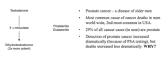
Prostate cancer
• Watchful waiting
• Androgen depletion
- 5a-reductase inhibitors (finasteride)
- castration
- Inhibitors of androgen synthesis
• Inhibition of testosterone action-block the androgen receptor.
• Surgery - prostatectomy
• others
PSA screening
- 136/1000 men → false positive.
- Prostatectomy risks: 1% death, 20-80% erectile dysfunction, 4-21% urinary incontinence.
- Mainly affects older men.
- Large trial (76,693 men) → no mortality reduction after 9 years.
- USPSTF initially recommended against routine screening.
- Later → individualised screening decisions for men aged 55-69.
The penis
1. Corpora cavernosa (x2) - the main erectile tissues.
2. Corpus spongiosum - surrounds the penile urethra and prevents occlusion
during erection.
3. penile urethra - conducts semen (and urine)
Erection occurs following sexual stimulation.
• Release of NO and Prostaglandin E1 causes the smooth muscle of the corpora cavernosa to relax.
• Blood fills the cavernous spaces of the corpora cavernosa.
- The erect penis contains about 8 times the blood volume of the flaccid penis.
• The engorgement of the corpora reduces venous outflow adding to the engorgement.
Viagra - Sildenafil
• Relaxation of the smooth muscle in the corpora cavernosa requires Guanosine Monophosphate (cGMP) (2messenger reduces intracellular calcium)
• The enzyme phosphodiesterase breaks down cGMP
• Viagra inhibits phosphodiesterase (type 5) leading to increased cGMP, relaxation of the arteries suppling the corpora cavernosa and erection.
Male reproductive hormones
Sertoli cells: Produce androgen-binding protein (ABP) and inhibin.
Interstitial (Leydig) cells: Produce testosterone.
LH: Stimulates testosterone production by Leydig cells.
Inhibin: Provides negative feedback to reduce FSH production.
3. Puberty and oocytes
First visible sign of male puberty: Enlargement of the testes.
Greatest number of oocytes: Before birth, during fetal development.
Vagina
An elastic muscular 7.5-9.0 cm tube extending from the cervix to the exterior of the body with three main functions. It serves as a passageway for the elimination of menstrual fluids. It receives the penis during sexual intercourse and holds spermatozoa before they pass into the uterus. It forms the lower portion of the birth canal through which the fetus passes during delivery.
Uterus
- Is a small, pear-shaped organ, that weighs ~ 30-40 g.
- Pathway for sperm transport. Provides mechanical protection, nutritional support, and waste removal for the developing embryo and fetus.
- Contractions in the muscular wall (myometrium) of the uterus are important in ejecting the fetus at the time of birth. Source of the menstrual flow.
- The endometrium can be subdivided into the inner functional zone (stratum functionalis), that contains most of the uterine glands and the outer basilar zone (stratum basalis; adjacent to the myometrium).
- The basilar zone attaches the endometrium to the myometrium.
Uterine (fallopian) tubes
Fertilisation typically occurs in the ampulla. The uterine tube provides a rich, nutritive environment containing lipids and glycogen, for spermatozoa, oocyte and the developing embryo. The epithelium lining of the uterine tube has both ciliated and nonciliated secretory columnar cells. The mucosa is surrounded by concentric layers of smooth muscle. Transport along the tube involves a combination of both ciliary movement and peristaltic contractions.
Ovaries
Adult human ovaries are oval, and weigh approximately 5-10 g. The ovary is comprised of three distinct regions. The outer ovarian cortex containing the ovarian follicles, the central ovarian medulla consisting of ovarian stroma and steroid producing cells, and the inner hilum (hilus) which acts as a point of entry for nerves and blood vessels.
Types of Follicle
Primordial follicle: The oocyte, once surrounded by a single layer of flat follicular cells (which develop into granulosa cells), form the primordial follicle.
Primary follicle: As the follicles grow, they are called primary or pre-antral follicles. Immature primary follicles consist of only one layer of granulosa cells. In response to FSH, some follicles get larger producing many layers of granulosa cells surrounding the oocyte.
Secondary follicle: As granulosa cells proliferate they produce a viscous follicular fluid that
coalesces to form a single follicular antrum. These are called secondary or antral follicles. The theca develops to become the inner glandular, highly vascular theca interna, and the surrounding fibrous capsule, the theca externa.
Mature (Graafian or pre-ovulatory) Follicle: As the follicular antrum grows, the oocyte becomes suspended in fluid. It is connected to the rim of peripheral granulosa cells by a thin stalk of cells.
Ovulation
The increasing size of the follicle and its position in the cortex of the ovarian stroma causes it to bulge out from the ovarian surface. The follicle ruptures, carrying with it the oocyte and its surrounding mass of cumulus cells. The oocyte is collected by cilia on the fimbria, which sweep the cumulus mass into the uterine tube.
Corpus luteum
The antrum breaks down, the basement membrane between the granulosa and thecal layers breaks down and blood vessels invade. The granulosa cells form large lutein (yellow pigment) cells (Corpus luteum = yellow body). This transformation is referred to as luteinisation and is associated with an increasing secretion of progestagens. The whitish scar tissue remaining, the corpus albicans (white body), is absorbed back into the stromal tissue of the ovary over weeks to months.
Fertilisation
If the oocyte is fertilised and begins to divide, the corpus luteum persists past its normal 2 week life span. It is rescued from degeneration by human chorionic gonadotropin (hCG). The hormone is produced by the chorion of the embryo beginning about 8 days after fertilisation.
The female reproductive cycle
The menstrual cycle: The average menstrual cycle is about 28 days. Two phases in the ovarian cycle: follicular phase (day 1 to ovulation) and luteal phase (ovulation to menstruation). Three phases in the endometrial cycle; menstrual, proliferative and secretory.
Main Events of Female Reproductive Cycle
1. Corpus luteum regresses, oestrogen and progesterone levels are low, see increased FSH.
2. FSH stimulation leads to increased follicular growth.
3. About day 6-7, see selection of the dominant follicle, with increased oestrogen.
4. Oestradiol suppress FSH (and LH) production in the pituitary.
5. Oestrogen levels rise, by ~day 12, a threshold concentration of oestradiol is exceeded. If this
is maintained for ~36 hours, there is a temporary switch from a negative to positive feedback.
6. Oestrogen-mediated positive feedback triggers a rise in GnRH leading to an LH surge.
7. LH surge induces ovulation.
8. Corpus luteum develops, see increased progesterone.
9. Elevated progesterone levels inhibit GnRH, lead to decreased FSH and LH.
10. Demise of the corpus luteum.

Female reproductive system
Zona pellucida: The glycoprotein layer between the oocyte and granulosa cells.
Oogenesis: Begins before birth.
Theca interna: Contains hormone-producing secretory cells.
Endometrium: Is thickest late in the postovulatory phase.
Progesterone: Reaches its highest levels late in the postovulatory phase and helps thicken and maintain the endometrium.