cell bio exam 3 flashcards
 Call Kai
 Learn
 Practice Test
 Spaced Repetition
 Match
 Flashcards
 Knowt Play
1/73

Earn XP
Description and Tags
the cytoskeleton, cell signaling, transcriptional regulators, the cell cycle
74 Terms
3 families of cytoskeleton
actin
microtubules
intermediate filaments
actin
shape, locomotion, pinching
microtubules
organelle position, intracellular transport, mitotic spindles
intermediate filaments
mechanical strength
microtubules
stiff, hollow cylinders, not good at bending
monomers —> heterodimer —> protofilament—>(13) form cylinder, a hollow microtubule polymer
microtubule monomer
alpha and beta tubulin
join to create heterodimer

microtubule heterodimer
link together end to end to form a protofilament
microtubule protofilament
order is alpha-beta, alpha-beta, alpha-beta
creates polarity
2 ends differ
NEGATIVE END: ALPHA TUBULIN
POSITIVE END: BETA TUBULIN

protofilament negative end
alpha tubulin
protofilament positive end
beta tubulin
has a GDP/GTP binding site (E-site)
hollow microtubule polymer cylinder
formed by 13 parallel protofilaments
microtubule instability
-heavily influenced by GTP
-can bind to beta tubulin subunit and is hydrolyzed to GDP (sometimes)
-when hydrolyzed, linkage between heterodimers weakens. protofilaments curve. instable. microtubule disassembles

solution to microtubule instability
MAINTAIN GTP CAP
-ensures all protofilament ends are straight and stable
-promotes growth, mostly at positive end
-loss of GTP results in catastrophe.
alternative: regain results in rescue where it regains GTP cap

microtubule organizing center
where nucleation occurs
in animals: centrosome

centrosome
2 centrioles surrounded by pericentriolar material
centriole helps extend mitotic spindles
requires gamma-tubulin for gamma tubulin ring complex (yTuRC)
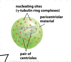
yTuRC
microtubule extends from each one
negative end is nucleating center

primary cillium/mother centriole?

organization in cell
varies by species, cell type, etc

microtubule dissociated proteins (MAP)
MAP2
tau

MAP2 and tau
-enriched in neurons
-stability/bundle spacing
-distance between microtubules depends on arm length of MAP2 or tau

MAP2
-more dispersed
-more dendrites

tau
-less spacing
-more axons

kinesin-13 protein
-rips away heterodimers
-speeds up depolymerization
-alters state of GTP cap plus end
CATASTROPHE
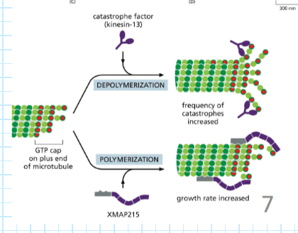
XMAP215 protein
GROWTH
-speeds up polymerization
-recruits heterodimers to growing end
-catalyst
-alters state of GTP cap

accessory protein Augmin
-facilitates branching
-binds to side of existing microtubule while recruiting y-TuRC complex to nucleate a new microtubule
-lack of augmin results in stunted plant growth

motor proteins (2)
kinesin
dynein
both provide bidirectional movement

kinesin motor protein
walk to POSITIVE end of microtubule
structural similarity to myosin
can influence neurogenesis in developing brains

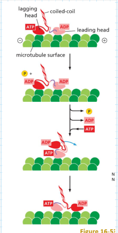
kinesin structure
large globular motor domains (2) at N-terminus, cargo connected at C terminus
lagging and leading head connected by a coiled stalk

kinesin movement steps
stepwise walk
2 ‘feet’
leading head ADP
lagging head ATP
1.ATP binds to leading head, lagging head moves forward
2.movement only occurs when one is hydrolyzed and the other has ADP
dynein
walk to NEGATIVE end of microtubule
uses its tail domain and connects to dynactin with 4Arp1 and then complex attaches to the adapter protein on the vesicle

dynein structure and movement (power stroke cycle)
-protein stalks with microtubule binding domains
-ATPase motor head
-hydrolysis of ATP rotates ATPase domain to cause movement. stalks lift, extend, ATP hydrolysis rotates head, causing power stroke

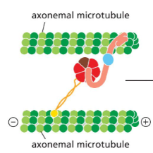
microtubule movement in flagella
-they line the length of flagella tails, which are highly conserved across evolution
-movement caused by bending of axoneme via motor proteins (dynamin=bending) and ATP (sliding/walking)
9 pairs of outer doublet microtubules (A and B) connected via nexin (linker protein)
intermediate filaments
-nonpolar
-do not bind or have a binding site for any nucleotides
-rope-like structure
-coiled
-easy to bend but difficult to break because fibers are staggered on eachother

intermediate filament monomer
alpha helices
-combine to form dimers—>tetramers—>full filament is 8 tetramers interwoven
1 tetramer=4 monomers
tetramer is overlapped coil-coil dimers
individual monomers twist together and form a coiled-coil dimer and 2 of these come together in a staggered orientation and form tetramer. 8 tetramers form bundle

different types of intermediate filaments
all help protect from mechanical stress and damage
ex. epithelial keratins
epithelial keratins
mutations in keratin can lead to condition epidermolysis bullosa simplex
(Weakening of ability of epithelial cells to stay anchored to basement membrane)
cells rupture and blister easily

cell signaling
the process by which cells are stimulated or inhibited by extracellular signal causing change! (main goal)
cell signaling components (3)
1.reception
2.transduction
3.response
cell signaling RECEPTION requirements (2)
-extracellular signal: could be molecule or protein**
-receptor protein: binding of signaling molecule=activation**
transduction requirements
intracellular signaling proteins**
-cascade and can translocate to nucleus of the cell
-ex: second messengers
response requirements
effector proteins
-alter cell function, metabolism and development
types of intracellular signaling (5)
-contact dependent/juxtracrine
-paracrine
-synaptic
-endocrine
-autocrine
contact-dependent/juxtracrine signaling
2 cells must be very close together, each cell expresses protein on their surface that can link together
paracrine signaling
1 signal producing cell creates and secretes ligand into ECF (extracellular fluid) around it. molecules bind to receptors on nearby cells
-close proximity still. binds to receptors on target cell
synaptic signaling
neuron specific & form of paracrine
-end of axon becomes synapse and releases molecules aka neurotransmitters into synaptic gap near target cell receptors
-caused by action potential
endocrine
-long distance
-signal producing cell secretes hormone signaling molecule
-hormone uses bloodstream to move to target cell and bind
autocrine signaling
-cell releases molecules that bind to its own receptors
-self regulation
-local/short distance
receptor location varies—> what are some things that can act as a signaling molecule on a receptor?
location of receptor is indicative of the binding signal molecule properties
ex. signal molecule can be proteins, small peptides, amino acids, nucleotides, steroids, or even dissolved gases
receptors on surface
hydrophilic signal molecule
large polar molecules
intracellular receptor
hydrophobic signal molecule
simple diffusion via carrier protein
translating signals overview —> what prevents cells from doing what they want?
-different combos of signals create different responses and presence: survive, grow, divide, differentiate, or death. require combination
-ABSENCE of signal: initiates cell death APOPTOSIS
acetylcholine effects in different cell types
1.heart pacemaker: decreased rate of firing
2.salivary gland: secretion
3.skeletal muscle: contraction
advantage: creates efficiency. less NRG investment into making/using diff transmitter
transduction relay race: messages relayed via intracellular signaling molecules (3 steps) —> 3 possible mechanisms for effector protein activation
1.phosphorylation
2.signaling by GTP binding
3.inhibitor protein can bind to transcription regulator
phosphorylation in transduction (step 1)
-add phosphates to protein, activating it
-phosphorylation cascade
-kinases add phosphates to proteins when active
-phosphatases remove phosphate and stop process
-travels to effector protein
molecular switch
-kinases add phosphates to proteins when active. protein kinases ultimately activate effector proteins
-phosphatases remove phosphate and stop process
signaling by GTP binding (step 2 in transduction)
-inactive GDP bound protein can be swapped out for GTP—>now active
-can be hydrolyzed again
-GEF and GAP exchange factors
GEF
adds GTP to protein and signal goes out
GAP
removes phosphate, hydrolyzes ADP
step 3 of transduction: inhibitor protein can bind to transcription regulator
kinase inhibits inhibitor, leading to gene expression
weakens the polymer of GTP-tubulin dimers
GTP hydrolysis
what grows from y-tubulin ring
microtubules at + end that has a microtubule seam and alpha-beta alternating subunits
centrosome cycle
1) pericentriolar center and centrioles
2) centrioles undergo duplication, form mitotic spindle
3) cell splits and mother and daughter cells are separated
4) cycle restarts
fibroblast microtubules
centralized star-like structure where minus ends are concentrated near the center and plus ends extend outward toward the cell edge
plant cell microtubules
arranged in parallel structure along the cortex
intestinal/epithelial cell microtubules
ordered, vertical array from apical to basal surface
neuronal cell microtubules
organized distinctly across its regions. microtubules along dendrite and axon that dynein and kinesin move along
state of dispersed melanophore and pigments along microtubules
increase in cAMP causes pigment to disperse on microtubule, cell looks darker
state of aggregated melanophore and the pigment along their microtubules
decrease in cAMP causes pigment to aggregate on microtubule making cell look lighter
axoneme
internal core microtubule of cilia and flagella
is transcription regulatory protein always on?
no, has inhibitor protein that deactivates it
when signaling protein kinase enters, it inhibits inhibitor, freeing the protein by adding ATP and starting gene expression
explain assemble of signaling complex on phosphoinositide (second messenger) docking sites
signal molecule binds to receptor
phosphoinositides get a phosphate, becoming hyperphosphorylated
allows intracellular signaling proteins to bind and send signals (activated)
explain assembly of signaling complex
signal molecule binds to receptor
phosphates are attached (act as docking sites for inactive in