Cellular Immunity
 Call Kai
 Learn
 Practice Test
 Spaced Repetition
 Match
 Flashcards
 Knowt Play
1/65

There's no tags or description
Looks like no tags are added yet.
66 Terms
Major histocompatibility complex (MHC)
important in self vs non-self recognition
T cell recognition of Ag includes recognizing self-MHC
non-self MHC=foreign, these cells are destroyed
HLA
HLA=human leukocyte antigens=human MHC
3 classes of MHC gene locus
class I MHC, class II MHC, and class III MHC
Class I MHC
-on most nucleated cells
-HLA-A, HLA-B, and HLA-C (in humans)
Class II MHC
-mostly expressed by professional antigen presenting cells (APCS=macrophages, B cell, DC)
HLA-DP, HLA-DQ, HLA-DR (in humans)
Class III MHC
-secreted proteins that are found in serum
-complement proteins, TNF, proteins in Ag processing
MHC Class I Protein
-single chain associated with another protein NOT encoded for by MHC locus (ex: beta2 microglobulin)
-CD8+ T cells (CTLs)
MHC Class II Proteins
-heterodimers encoded for by alpha and beta genes in MHC locus
-CD4+ T cells (helper T)
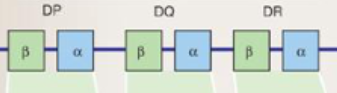
Class II MHC genes

Class I MHC genes

Class III MHC genes
Co-dominant
both alleles are expressed at once, both give us the haplotype
MHC genes are:
polygenic
have multiple similar genes (A,B,C)
(in humans) 6 from class I MHC, 12 from class II MHC
Allotype
genetic sequence of a gene within a species
HLA locus genes are:
Classical and non-classical
Classical MHC genes
classical=T cell recognition of Ag
ex: HLA-A,B,C and HLA-DP,DQ,DR
Non-classical MHC genes
NK cells and others
Heterodimer of MHC class II proteins
-alpha and beta chains
-protein is divided into alpha1/alpha2 and beta1/beta2
CD4 contacts the class II molecule
MHC class I vs MHC class II antigenic peptides
class I: closed-ended structure accommodates 8-9 AA
class II: open-ended structure accommodates 18-20 AA
Endogenous intracellular peptides
ER is where class I MHC and Ag peptide associate
Nonamers (9 AA) are preferred peptide length and bind the strongest
How peptides associate with MHC class I
proteins originate in cell
proteasome complexes degrade proteins
degraded proteins go to rER (TAP proteins) and associate with MHC class I
Exogenous peptides
peptides degraded by endocytic pathway
MHC class II expressed on: B cells, macrophages, DCs
How peptides associate with MHC class II
proteins/cells are endocytosed
endosome fuses with lysosome
once CLIP is release, Ag peptide can sit in MHC class II
MHC class II/Ag then moves to cell surface
Human MHC genes are:
polymorphic=allow population to respond to the largest possible number of antigens
Antigen recognition for T cells
T-cell receptor (TCR)
T cells
TCR is always cell bound
TCRs recognize Ag in association to MHC

MHC class I T cell activation and function
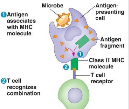
MHC class II T cell activation and function
TCR gene rearrangement
occurs in thymus
controlled by: RAG1 and RAG2
2 possibilities: alpha-beta or gamma-beta
TCR structure
Heterodimer
alpha-beta and gamma-beta chains are similar to Fab of Igs
anchored by disulfide bonds
variable and constant regions
3 CDRs (complimentary determining regions)
TCR gene rearrangement
similar to Ig gene rearrangement
CD3 complex
Signaling function (similar to immunoglobulin Ig-alpha/Ig-beta)
TCRs are members of:
Ig gene superfamily, bc of Ig fold (intrachain disulfide bond)
T cell maturation and differentiation
progenitor T cells originate in bone marrow and move to thymus
thymus undergoes ‘education‘ process for alpha-beta T cells
Lack of thymus
causes T cell defects
thymectomy
DiGeorge syndrome (deletion of part of chromosome 22=thymic aplasia)
Positive thymic selection of T cells
Select T cells that can interact with self-MHC
cells that bind get a protective signal and survive
Negative thymic selection of T cells
T cells with high affinity for self-MHC and T cells are reactive to self-peptides=die with apoptosis
cells NOT reacting=selected to survive and leave thymus
Diversity of TCR (5)
combinatorial joining (3×10^6 possibilities)
alternative joining of D segments (beta chain)
junctional flexibility (imprecise cutting/joining of genes)
P nucleotides (palindromic bases added)
N nucleotides: Tdt enzyme addition
estimates 10^13 possibilities
Major CD4+ T helper cell subsets (5)
Th0=naive T cells
Th1= CMI, intracellular microbes
Th2= humoral, extracellular microbes, worms
Treg(Th3)= regulate autoimmunity
Th17= extracellular bacteria, fungi, associated with autoimmunity
Th cells are divided into two major subsets:
Th1=CMI (IL-2, IFN-gamma, TNF-beta)
Th2= (IL-4,5,6,10,13)
Th1/Th2 subsets
Th0 to Th1/Th2 subset is dependent on APC stimulation
IL-12 from DC stimulates Th1 production
IL-4 from Th2 stimulates Th2 production
Cytokine activity
cytokines must be bound to specific cytokine receptor
once bound, signaling cascade exerts biological activity
act locally, because short in vivo half life
cytokine receptors have 2-3 chains
Cytokine receptor redundancy
in a cytokine receptor family, often share same signaling chain
get higher affinity with binding of 2-3 chains
after cell activation=increased cytokine receptor expression
SCID (X-inked severed combined immunodeficiency)
defect in gamma chain gene
Th1
stimulate CMI/inflammation, IL-2, IFN-gamma
Th2
stimulate humoral responses, IL-4,5,6,10
Th3 (Tregs)
regulatory cells, make IL-10, TGF-beta
Th17
extracellular bacteria and fungi
IL-2 receptor family
shares common gamma chain
CD4 and CD8
CD4=class II=beta2, CD8=class I=alpha3
T cell activation/co-stimulation
T cells need 2 signals to be stimulated
Signal 1: TCR-Ag (pMHC)
Signal 2: CD28/B7
inhibitory signal:CTLA-4 binds to B7 over CD28 and sends suppression signal
Lack of 2 signals causes T cells to:
T cells become anergic (suppressed) or undergo apoptosis
How to killa microbe
innate defenses: phagocytosis
adaptive responses: antibodies, cytotoxic T lymphocytes (CTLs), NK cells, ADCC
Effector cell
effector cell is a cell with a ‘job‘
effector cells are not naive (increased expression of adhesion molecules)
Cytotoxic T cells
important in eliminating altered self cells (like virally infected cells or tumor cells)
B and T cell ‘jobs‘
B cell= is the plasma cell
T cell= is the CTL, Th1 and Th2 cell
CTL-mediated killing of target cells
is the release of cytotoxic mediators at interface between Tc (target cell)
perforin
pore forming
granzymes
enter target cell through pores
FasL on CTL and Fas on target cell
cascade events= apoptosis
TNF-alpha binds to Fas =apoptosis

Perforin activity
NK cell-mediated killing of altered-self cells
similar to CTL but no AG specific recognition
NK cells active before CTLs
produce cytokines to increase macrophage activity
produce perforin and granzymes
NK cells detect alterations in expression on surface of normal proteins
Antibody-dependent cellular cytotoxicity (ADCC)
Ab serves the specificity to deliver Ag to non-specific cells to target'
FcR binding
NK cells, neutrophils, eosinophils, macrophages
release of granules=cell death

impact of HIV on T helper cells
NK cells kill altered cells via:
reduction in MHC class I and presence of stress proteins
ADCC
when antibody binds to target cell, Fc region can interact with FcRs on surface of cells (neutrophils, NK, macrophages, eosinophils), these cells release toxic chemicals that kill the target cell